Abstract
Although transgenic crops expressing Bacillus thuringiensis (Bt) toxins have been used successfully for management of lepidopteran and coleopteran pest species, the sap-sucking insects (Hemiptera) are not particularly susceptible to Bt toxins. To overcome this limitation, we demonstrate that addition of a short peptide sequence selected for binding to the gut of the targeted pest species serves to increase toxicity against said pest. Insertion of a 12-aa pea aphid gut-binding peptide by adding to or replacing amino acids in one of three loops of the Bt cytolytic toxin, Cyt2Aa, resulted in enhanced binding and toxicity against both the pea aphid, Acyrthosiphon pisum, and the green peach aphid, Myzus persicae. This strategy may allow for transgenic plant-mediated suppression of other hemipteran pests, which include some of the most important pests of global agriculture.
Keywords: aphid management, biotechnology, insect resistance
A significant proportion of world food production is lost to arthropod pests (1). Although the application of chemical insecticides has reduced crop losses, problems associated with the development of resistance in the targeted pest and unintended impacts on nontarget organisms require the deployment of alternative pest management approaches. Insect resistant transgenic crops that express insect-specific toxins derived from the bacterium Bacillus thuringiensis (Bt), have provided effective suppression of lepidopteran (moth) and coleopteran (beetle) pests and have been widely adopted in the United States and elsewhere (2, 3). However, the sap-sucking hemipteran pests are not particularly susceptible to the Bt-derived toxins (4–6). Indeed, in some cases, chemical insecticides have to be applied to curtail damage caused by hemipteran pests on Bt transgenic crops, such that the benefit of using the insect resistant transgenic varieties is lost (7–9).
Among the Hemiptera, aphids are pervasive pests of temperate agriculture (10). Aphid activities that result in yield loss include feeding and consequent weakening of the plant, production of honey dew that promotes the growth of harmful sooty molds on plant surfaces, and the vectoring of plant viruses. Some 275 plant viruses from 19 different genera are transmitted by aphids, representing half of the insect-vectored plant viruses (11). Aphid management relies almost exclusively on the application of chemical insecticides. A few toxins derived from Bt have low levels of toxicity against aphids but are insufficiently toxic for use in aphid management (6).
We isolated a pea aphid gut-binding peptide, GBP3.1, which impedes uptake of a plant virus by its vector, the pea aphid (12). GBP3.1 (amino acid sequence: TCSKKYPRSPCM) binds to the midgut and hindgut epithelia of the pea aphid, and to a lesser extent, to the gut epithelia of the green peach aphid and the soybean aphid under in vitro conditions (12). We determined that GBP3.1 binds to membrane alanyl aminopeptidase-N on the surface of the aphid gut epithelium. We then addressed whether addition of this peptide sequence to a Bt toxin would enhance binding and increase toxicity against aphids. To test this concept, we used the Bt cytolytic toxin Cyt2Aa on the bases that (i) the mode of action of Cyt toxins is less complex than that of Bt crystal (Cry) toxins (13, 14), (ii) Cyt2Aa is toxic to mosquito larvae (13), allowing for assessment of whether toxin modifications impair toxicity, and (iii) Cyt2Aa has low toxicity against the pea aphid.
Results
GBP3.1 Binds to Pea Aphid Aminopeptidase.
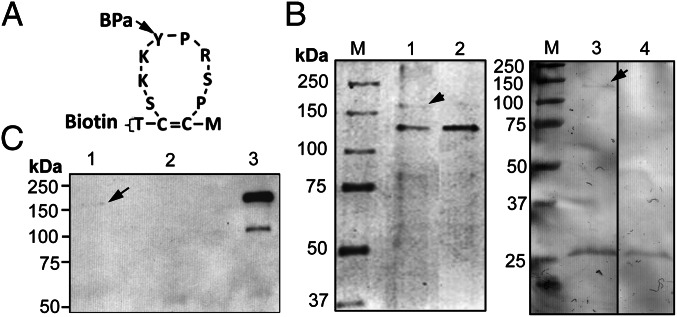
Following UV-cross-linking to interacting partners, GBP3.1 consistently bound to a ∼180-kDa pea aphid gut protein in pull-down assays (Fig.1 A and B). The GBP3.1-interacting protein was identified by liquid chromatography–tandem mass spectrometry (LC MS/MS) analysis as pea aphid membrane alanyl aminopeptidase-N (APN; Uniprot accession no. J9JIN6). A total of 35 unique peptides representing 42% of the APN sequence were identified. Western blot detection of this protein using polyclonal anti-pea aphid APN antiserum provided additional confirmation of the identity of the ∼180-kDa protein (Fig. 1C).
Fig. 1.
Identification of APN as the GBP3.1 receptor. (A) A double derivatized synthetic GBP3.1, with a biotin residue at the N terminus and a UV-cross-linking residue (pbenzoyl-l- phenylalanine, BPa) replacing the tyrosine residue in the loop was used for receptor identification. (B) Coomassie Blue-stained SDS-polyacrylamide gel of proteins interacting with synthetic GBP3.1 in a pull-down experiment. The GBP3.1–receptor protein complex was pulled down by streptavidin agarose beads. Lane 1, GBP3.1 incubated with pea aphid gut and UV-cross-linked to interacting proteins. The ∼180-kDa protein (arrow) was consistently pulled down following UV-cross-linking to GBP3.1. Lane 2, GBP3.1 incubated with pea aphid gut, without UV-cross-linking. Lane 3, streptavidin beads with pea aphid gut only, showing the ∼140-kDa gut protein (arrow) pulled down by the streptavidin beads. Lane 4, streptavidin beads only. Molecular mass standards (M) are indicated. (C) Western blot analysis with anti-pea aphid APN antiserum confirmed the identity of the GBP3.1-interacting protein from pull-down experiments as APN. Lane 1, pea aphid gut proteins UV-cross-linked to GBP3.1. Lane 2, pea aphid gut proteins incubated with GBP3.1 without UV-cross-linking before pull-down. Lane 3, pea aphid gut proteins (positive control).
Binding Specificity of GBP3.1.
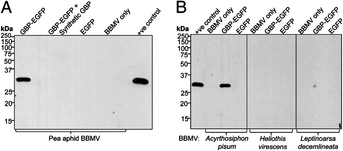
GBP3.1-EGFP binding to pea aphid brush border membrane vesicles (BBMVs) was outcompeted by addition of synthetic GBP3.1, indicating specificity of binding (Fig. 2). Although GBP3.1-EGFP bound to pea aphid BBMV, no binding was detected to BBMV from the cotton budworm, Heliothis virescens or the Colorado potato beetle, Leptinotarsa decemlineata, indicating that H. virescens and L. decemlineata BBMV lack binding sites (Fig. 2B). The aminopeptidases from H. virescens and L. decemlineata do not appear to function as receptors for GBP3.1, possibly due to their low homology with pea aphid APN (Table S1).
Fig. 2.
GBP3.1 specifically binds pea aphid BBMV. (A) Binding of GBP3.1-EGFP (GBP-EGFP; 50 nM) to pea aphid BBMV was out-competed by addition of synthetic GBP3.1 (50 µM). (B) GBP3.1 did not bind to BBMV proteins (10 µg) from H. virescens or L. decemlineata. Binding of EGFP and BBMV only were used as negative controls for both experiments. Positive control, GBP3.1-EGFP.
Production of Modified Cyt2Aa.
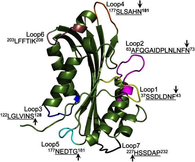
The amino acid sequences of all Cyt toxins show a high degree of identity and have similar crystal structures (14) with a single α-β domain composed of two outer layers of α-helix hairpins wrapped around a sheet (15). A homology-based model of Cyt2Aa has been developed (16), and seven loops on the surface of Cyt2Aa have been identified as potential sites for GBP3.1 insertion (Fig. 3, Fig. S1). Loop 6 was not selected for modification due to its role in potentiation of Cry toxin action (17) whereas the remaining six loops are not involved in protein production and folding (18). Two sets of modified Cyt2Aa were constructed, by addition of GBP3.1 to each Cyt2Aa loop [Cyt2Aa-GBP3.1-addition-Loopn (CGALn)] or by substitution of amino acids in each Cyt2Aa loop with GBP3.1 (CGSLn). Homology-based models of all 12 modified toxins showed surface-exposed GBP3.1, with no apparent changes to the core structure of Cyt2Aa (Fig. S2). Comparison of CGALn and CGSLn models with that of Cyt2Aa showed nonsignificant variation in the average distance between the α-carbon atoms of superimposed models (19) and similar overall topologies (Table S2) (20).
Fig. 3.
Ribbon structure of Cyt2Aa homology model showing the amino acid sequence of loops predicted to be on the surface of the molecule. The core Cyt2Aa structure is shown in green with loops of nonmodified Cyt2Aa shown in various colors. The aphid gut binding peptide GBP3.1 was inserted into each of these loops except for loop 6, which is implicated in Cyt toxin action (17). Arrows indicate sites of addition of GBP3.1 for CGALn toxins. Underlined amino acids were replaced with GBP3.1 in CGSLn toxins.
The expression levels of the modified Cyt2Aa protoxins in Escherichia coli were low relative to those of Cyt2Aa protoxin. As the method used for purification (21) resulted in low amounts of partially purified protoxins, further purification on Q-Sepharose FF was used (Fig. S3). However, CGSL3 was unstable, and insufficient quantities were obtained for inclusion in all analyses (Fig. S4).
Among the modified toxins, trypsin treatment resulted in mobility shifts for CGAL1 and CGAL3 only, suggestive of protoxin activation during production and purification of the other modified toxins (22, 23). Excluding the unstable CGSL3, and CGAL2 and CGAL5, which showed partial proteolytic instability, the remaining nine activated CGALn and CGSLn were stable following trypsin treatment (Fig. S4). Insertion of GBP3.1 into Cyt2Aa resulted in an increase in molecular mass from the 22 kDa of activated Cyt2Aa to ∼25 kDa.
Impact of Modifications on Cyt2Aa Toxicity.
To assess the impact of GBP3.1 insertions on Cyt2Aa toxicity, bioassays of activated CGALn and CGSLn were conducted with 3-d-old Aedes aegypti larvae. Only 5 (CGAL1, CGAL3, CGAL4, CGASL1, and CGSL4) of the 11 modified toxins tested retained toxicity at levels comparable with that of Cyt2Aa (Table 1). All toxins with GBP3.1 insertions (by addition or substitution) at loops 2, 5, and 7 showed reduced toxicity against Ae. aegypti relative to Cyt2Aa.
Table 1.
Aphid toxicity of Cyt2Aa, CGALn, and CGSLn
| Toxin | LC50 ± SE, μg/mL (CL 95%) |
||||||
| Mosquito larvicidal activity |
Aphicidal activity |
||||||
| Ae. aegypti | Relative LC50 | A. pisum | M. persicae | ||||
| Cyt2Aa | 0.37 ± 0.07 | (0.21–0.90) | 1 | >>150 ± 0.00 | (NA) | >>150 ± 0.00 | (NA) |
| CGAL1 | 0.22 ± 0.04 | (0.06–0.43) | 0.58 | 19.71b ± 5.74 | (2.51–21.00) | 58.04 ± 2.08 | (35.01–65.73) |
| CGAL3 | 0.62 ± 0.04 | (0.24–1.30) | 1.68 | 9.55a ± 2.54 | (0.65–12.23) | 42.68 ± 0.49 | (17.18–83.04) |
| CGAL4 | 0.18 ± 0.05 | (0.01–0.82) | 0.49 | 11.92a ± 1.99 | (0.83–22.43) | 92.75 ± 2.54 | (34.67–152.96) |
| CGSL1 | 0.36 ± 0.06 | (0.19–0.79) | 0.97 | 28.74b ± 2.92 | (6.40–93.40) | ND | |
| CGSL4 | 0.40 ± 0.05 | (0.12–0.91) | 1.09 | 15.13a ± 0.23 | (4.3–25.60) | ND | |
Mortality resulting from aphid ingestion of the five modified toxins that retained toxicity against mosquito larvae was significantly higher than mortality following Cyt2Aa ingestion for A. pisum and three (CGAL1, CGAL3, and CGAL4) for M. persicae (one-way ANOVA, P < 0.001). Significant differences in modified toxin LC50 values for A. pisum are indicated by different letters (a and b). CGAL1, CGAL3, and CGAL4 were significantly more toxic to A. pisum than to M. persicae (one-way ANOVA, P < 0.005). A total of 360 second instar aphids were used for each toxin, with mortality scored after 4 d. LC50 values presented are representative of data from three replicate bioassays. CGSL3 was not included in the aphid feeding assays due to instability of this toxin. ND, not determined.
GBP3.1-Mediated Binding of Modified Toxins to Aphid Gut BBMV.
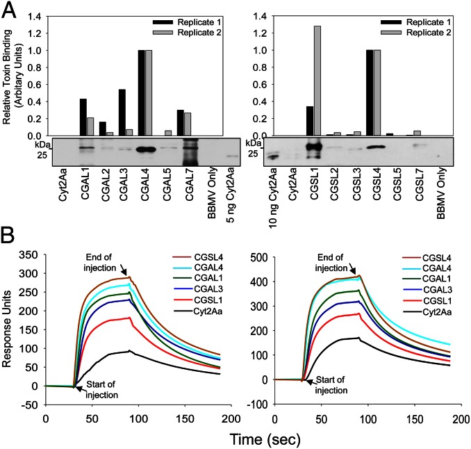
Pull-down assays (17) were used to demonstrate increased binding of all 12 CGALn and CGSLn to pea aphid gut BBMVs compared with Cyt2Aa (Fig. 4A). Binding of Cyt2Aa to pea aphid gut BBMV was not detected in these assays. Toxin constructs with loop 4 modifications (CGAL4 and CGSL4) along with CGAL3 and CGSL1 showed the highest levels of binding in pull-down assays (Fig. 4A, Table S3). It is notable that all of the modified toxins, including those with reduced toxicity relative to Cyt2Aa, bound aphid gut BBMV in these assays, demonstrating that toxin binding does not always correlate with toxicity.
Fig. 4.
CGALn and CGSLn bind to pea aphid gut BBMV more strongly than Cyt2Aa. (A) Pull-down assays were conducted following incubation of activated Cyt2Aa, CGALn, or CGSLn and pea aphid gut BBMV. Membrane bound toxin was detected by Western blot with anti-Cyt2Aa antiserum. Western blot images (Lower) were scanned and processed using ImageJ to estimate the relative amount of activated toxin associated with pea aphid BBMV (histogram Upper). Relative toxin binding is shown for two pull-down assays with Western blot images shown for Replicate 1 in each case. (B) BIAcore surface plasmon resonance analysis of toxin binding to small unilamellar vesicles (SUV). Sensorgram showing the real-time interaction between 6 µM Cyt2Aa, CGAL1, CGAL3, CGAL4, CGSL1, CGSL4, and immobilized pea aphid gut membrane SUV. L1 chip surfaces were prepared with 4,000 RU of ligands. Data are shown for two independent experiments.
Surface plasmon resonance (SPR) was used to quantify the relative binding of Cyt2Aa and five of the modified toxins (CGAL1, CGAL3, CGAL4, CGSL1, and CGSL4) to small unilamellar vesicles (SUV) prepared from pea aphid gut BBMV (Fig. 4B). Association and dissociation profiles were similar for all six toxins. The extent of binding at the end of injection of CGAL1, CGAL3, CGAL4, and CGSL4 was significantly higher than that of Cyt2Aa (one-way ANOVA, P < 0.05; Table S3). The extent of binding of Cyt2Aa at the end of injection was not significantly different from that of CGSL1 (P = 0.070).
Aphid Toxicity and Impact of Modified Toxins on Aphid Gut Epithelial Membrane.
Membrane feeding assays were used to assess the aphicidal activity of Cyt2Aa, CGALn, and CGSLn against second instar Acyrthosiphon pisum and Myzus persicae (Table 1). The six toxins with reduced toxicity against Ae. aegypti relative to Cyt2Aa (CGAL2, CGAL5, CGAL7, CGSL2, CGSL5, and CGSL7) also lacked toxicity against aphids. The five toxins that retained toxicity against Ae. aegypti (CGAL1, CGAL3, CGAL4, CGSL1, and CGSL4) showed increased toxicity against A. pisum, and three (CGAL1, CGAL3, and CGAL4) showed increased toxicity against M. persicae (one-way ANOVA, P < 0.001; Table 1). CGAL3, CGAL4, and CGSL4 were significantly more toxic to A. pisum than CGAL1 and CGSL1 (one-way ANOVA, P < 0.01; Table S4). CGAL1, CGAL3, and CGAL4 were significantly more toxic to A. pisum than to M. persicae (one-way ANOVA, P < 0.005), reflecting selection of GBP3.1 for binding to the gut of A. pisum (12).
Ingestion of CGAL1 or CGSL4 resulted in extensive damage to the pea aphid midgut epithelium as seen in light and transmission electron micrographs (Fig. 5). Damage was consistent in all six aphid midguts examined per treatment (Fig. 5). Midgut epithelial cells had completely disintegrated in some regions. Disruption of the gut epithelium was also apparent to a lesser degree following ingestion of Cyt2Aa, consistent with the low toxicity of Cyt2Aa against A. pisum (Table 1).
Fig. 5.
CGAL1 and CGSL4 cause extensive damage to second instar pea aphid midgut epithelium. (A) Light micrographs of midgut cross sections, and (B) transmission electron micrographs, showing the impact of CGAL1 and CGSL4 on the midgut epithelium relative to that of control and Cyt2Aa-fed aphids. Micrographs show the intact gut epithelial cell (EC), intact apical surface of the gut epithelial membrane with microvilli (M) projecting into the gut lumen (L) in aphids fed on control diet (Control). The microvilli of Cyt2Aa-fed aphids showed some damage, consistent with the low level of toxicity seen again A. pisum with this toxin. The integrity of the gut epithelia of aphids fed on CGAL1 and CGSL4 was severely compromised. LEC, lysed epithelial cells.
Discussion
The relatively low toxicity of Bt toxins against hemipteran pests has thus far prevented their application for management of these sap-sucking pests (4) although a Cry toxin with potential for use against Lygus hesperus has now been identified (24). We have demonstrated the use of a synthetic peptide to increase toxin binding to the aphid gut. Binding of a Bt toxin to the gut of the target insect is an important step for toxicity (25). In the case of aphids, the physiological bases for low levels of toxicity vary (5, 6, 26, 27) but include toxin instability in the aphid gut and low levels of binding (5). We selected the cytolytic toxin Cyt2Aa to test the concept that addition of an aphid gut binding peptide to the toxin would increase toxin binding and associated toxicity.
Importance of the Site and Type of Peptide Insertion.
Insertion of the aphid gut binding peptide GBP3.1 at six different loops of Cyt2Aa by either addition to, or substitution of, native loop amino acids resulted in protein instability in the case of CGSL3, and reduced Cyt2Aa toxicity for loops 2, 5, and 7. Based on the model for Cyt2Aa action at the gut epithelium (28), loops 5 and 7 are inserted through the membrane to the cytoplasmic side whereas loop 2 remains on the surface. Insertion of GBP3.1 into loops 5 and 7 likely affected the ability of the modified toxins to insert into the membrane. Loop 2 of Cyt2Aa has been reported to function in toxin stability (15), and addition of GBP3.1 to loop 2 did result in partial degradation on trypsin treatment. Loops 1, 3, and 4 of the membrane-inserted toxin are predicted to bind to the cell membrane surface, exposing toxin helices αA, αB, αC, and αD to form an oligomer with other Cyt2Aa monomers (28). Our results for increased toxin binding on insertion of GBP3.1 into loops 1, 3, and 4 are consistent with this prediction based on the model for Cyt2Aa membrane insertion (28).
Increased binding of all of the modified toxins relative to Cyt2Aa binding was demonstrated by pull-down assay (Fig. 4A) whereas increased binding was demonstrated by SPR analysis for CGAL1, CGAL3, CGAL4, and CGSL4, but not CGSL1 (Fig. 4B). Both methods indicate particularly strong binding of toxins with loop 4 modifications (CGAL4 and CGSL4). All five modified toxins that retained activity against mosquito larvae showed increased aphid toxicity in membrane feeding bioassays (Table 1), demonstrating a correlation between increased aphid gut binding and toxicity for the active toxins.
Our data indicate that loop 4 provides the optimal site for insertion of insect gut binding peptides into Cyt2Aa. Modification by addition to, or substitution of, native amino acids in loop 4 did not affect toxin stability (Fig. S4) or toxicity to mosquito larvae (Table 1), but significantly increased binding to pea aphid gut BBMV (Fig. 4, Table S3) and resulted in increased toxicity against A. pisum relative to other toxins (with the exception of CGAL3). In contrast, the outcome of peptide insertions into loop 3 varied according to the type of insertion, with CGSL3 being highly unstable, and loop 1 insertions exhibiting significantly less toxicity against A. pisum relative to toxins with loop 4 modifications.
Specificity of Modified Bt Cyt2Aa Toxins.
The greater toxicity of the modified toxins against the pea aphid compared with toxicity against the green peach aphid reflects the selection of GBP3.1 for binding to the pea aphid gut, and stronger binding to the gut of this species relative to that of the green peach aphid (12). In contrast, GBP3.1 did not bind to BBMV derived from species with relatively low APN homology (Fig. 2, Table S1). The fact that GBP3.1 did not bind to BBMV from representative lepidopteran or coleopteran species indicates that toxins modified with GBP3.1 are unlikely to interfere with other APN-binding toxins that are used for management of lepidopteran and coleopteran pests.
For optimal efficacy of a modified toxin against a given pest species, a gut-binding peptide selected for binding to the gut of that species should be used. Related species may be impacted by such modified toxins, however, if the peptide binding partners in the insect gut are conserved across species, as is the case for GBP3.1 (12). Modified toxins with lower potency against species related to the target pest could result in selection for resistance in the nontarget species. Plant expression of multiple toxins modified with different peptides to target different pests in the cropping system could be used to avoid this outcome.
Toxin modification can affect toxin specificity: Mutations S108C and V109A in helices αA and αC of Cyt2Aa2 resulted in significantly reduced toxicity against Culex quinquefasciatus, but not Ae. aegypti. These alpha helices bind to glycoprotein or lipoprotein components on the cell membrane (29). It is unknown whether modification of toxin loop sequences could similarly affect host specificity.
Domain III of Bt-derived crystal (Cry) toxins is implicated in insect specificity. Swapping of this domain between different Cry toxins resulted in hybrid toxins with improved toxicity against some insect pests (30). In addition, incorporation of the nontoxic galactose-binding domain of ricin at the C terminus of Cry1Ac enhanced binding and toxicity against several insect pests, including the leafhopper, Cicadulina mbila (Hemiptera) (31). The advantage of peptide-mediated toxin modification as described in this paper over these alternative approaches is that modifications are tailored to target a specific pest insect and have a more predictable outcome.
The median lethal dose (LC50) values for modified Cyt2A against the pea aphid (9.55–28.74 µg/mL) and against the green peach aphid (42.68–92.75 µg/mL) are considerably higher than those for Cry toxins used in transgenic plants for resistance to lepidopteran pests (32), but are more similar to the potency of Cry3 proteins against coleopteran pests (33, 34). That said, given the disparate feeding strategies (ingestion of phloem versus leaf material) and volumes ingested, direct comparison of LC50 values may be inappropriate. The question of whether the modified Cyt2Aa toxins are sufficiently toxic for practical use for aphid-resistant transgenic plants will require empirical determination.
Taken together, this research proves the concept that gut-binding peptides can be used for effective retargeting of the Bt toxin Cyt2Aa to pest species that are otherwise refractory to toxin action. This strategy will provide a valuable approach for management of hemipteran pests of agriculture.
Materials and Methods
Derivatized GBP3.1 Peptide Synthesis.
A double-derivatized GBP3.1 peptide with biotin at the N terminus and a UV-cross-linker attached phenylalanine (Bpa; pbenzoyl-l- phenylalanine) in place of the tyrosine (Y) within the 8-aa loop (Fig. 1A) was synthesized by Genemed Synthesis.
Peptide Pull-Down Assays.
Pea aphid guts were dissected at 4 °C in PBS containing protease inhibitor (PI) mixture (Sigma; P8340) and stored on ice. Dissected guts were washed thoroughly with several changes of PBS containing PI to remove cell debris and other contaminating materials. The guts were then incubated with the double-derivatized GBP3.1 peptide (Fig. 1A) in PBS (10 μg/mL) for 1 h at 4 °C in the dark. Guts were washed to remove unbound peptide and exposed to UV light for 30 min in a laminar hood (240 nm wave length) at a distance of ∼1 cm between the guts and the UV lamp. The guts were then centrifuged briefly, resuspended in lysis buffer (250 mM KAc, 10 mM MgAc2, 50 mM Hepes, pH 7.4, 0.1% Nonidet P-40) and homogenized in the presence of PI mixture. Homogenates were incubated at 4 °C for 20 min. The lysate was clarified by centrifugation at 25,000 × g for 1 h, and the supernatant was collected. Clear supernatant was mixed with ∼100 mL of streptavidin-agarose bead suspension (Molecular Probes; S-951) preequilibrated in binding buffer (lysis buffer without Nonidet P-40) and incubated on a rotary shaker at room temperature for 1 h. The beads were then washed several times by centrifugation to remove unbound proteins, followed by addition of SDS/PAGE sample buffer and boiling for 3 min. Boiled beads were centrifuged briefly, and clear supernatant was separated on 8% (wt/vol) SDS/PAGE and stained with Coomassie Blue R250. The ∼180-kDa protein band was excised from the gel and submitted to the University of Iowa Proteomics Facility for identification. Small aliquots from the pull-down assay were used for Western blot detection of GBP3.1-interacting aminopeptidase. Polyclonal anti-APN antibodies against E.coli-expressed 67-kDa pea aphid APN (GenBank accession no. ABD96614) were generated at the Iowa State University Hybridoma Facility.
Protein Identification by LC MS/MS.
The GBP3.1-interacting protein was analyzed by LC-MS/MS on the Dionex 3000 nanoRSLC series HPLC system (Thermo-Electron). LC effluent was directed to the electrospray source of a linear ion-trap mass spectrometer (LTQ/XL; Thermo-Electron). MS/MS spectra were acquired in a data-dependent acquisition mode that automatically selected and fragmented the five most abundant peaks from each MS spectrum. All MS/MS samples were analyzed using Mascot (Matrix Science; version 2.4.0), Spectrum Mill (Agilent; version Unknown) and X! Tandem [The GPM (thegpm.org); version CYCLONE (2010.12.01.1)] with the PeaAphid_20130306 database (unknown version; 33,591 entries) assuming digestion with the enzyme trypsin. Scaffold (version Scaffold_4.0.0; Proteome Software) was used to validate MS/MS-based peptide and protein identifications. Protein probabilities were assigned by the Protein Prophet algorithm.
Production of Peptide–EGFP Fusion and EGFP.
The GBP3.1-EGFP fusion protein and EGFP expression constructs in pBAD/His B (Invitrogen) were prepared as described previously (12). Protein induction was carried out at room temperature overnight by adding 0.02% L arabinose. The His-tagged fusion proteins were purified using Ni-NTA agarose resin (QIAGEN) according to the manufacturer's directions. Purification was conducted under native conditions using a batch purification method at 4 °C. Purified protein fractions were separated by SDS/PAGE [12% (wt/vol) gel] and stained with Coomassie Blue to check protein purity. The purest fractions were pooled and dialyzed at 4 °C against PBS with three buffer changes. The concentration of the dialyzed protein was determined using the Coomassie Plus Protein Assay (Bio-Rad; 500–0006EDU) kit with BSA as a standard. Aliquots of 500 µl in 1.5-mL Eppendorf tubes were snap frozen in liquid N2 and stored at −80 °C until further use.
Preparation of Brush Border Membrane Vesicles.
Pea aphid (n = 5,000), late instar Helicoverpa zea and L. decemlineata larval guts were dissected for preparation of BBMV by differential precipitation using MgCl2 (35). Leucine aminopeptidase assays on the crude homogenate and resulting BBMV were conducted to assess the level of enrichment (36). Protease inhibitor mixture (Sigma; P8340) was added to the BBMV preparation to a dilution of 1:100, and protein concentration was determined using BSA as standard (37). BBMV were stored at −80 °C until use.
Specificity of GBP3.1-EGFP Binding.
Pull-down binding assays were carried out as described previously (17) to investigate the binding of GBP3.1-EGFP in competition assays, and the relative binding to pea aphid, H. zea, and L. decemlineata BBMV. EGFP and BBMV only negative control reactions were included in both assays. Equal concentrations of BBMV protein (10 μg) were used for competition and relative binding assays. For the relative binding assay, equimolar concentrations (50 nM) of GBP3.1-EGFP were incubated with BBMV (pea aphid, H. zea, or L. decemlineata) in 100 μL of binding buffer [PBS; 0.1% (wt/vol) BSA/ 0.1% (vol/vol) Tween 20, pH 7.6]. Postincubation, BBMV, along with bound GBP3.1-EGFP or EGFP, was pelleted by centrifugation at 11,000 × g for 10 min at 4 °C, and pelleted BBMV were washed several times to remove unbound GBP3.1-EGFP or EGFP. The final pellet was boiled in SDS-sample buffer for 5 min and centrifuged briefly, and proteins in the supernatant were separated by SDS/PAGE [12% (wt/vol) gel]. The proteins were then transferred to nitrocellulose membrane for Western blot detection of BBMV-bound GBP3.1-EGFP with anti-EGFP antibodies (Sigma; G1544). For competition assays, the same protocol was followed, except GBP3.1-EGFP (50 nM) was incubated with pea aphid BBMV in the presence of 1,000-fold molar excess of synthetic GBP3.1 (50 μm). GBP3.1-EGFP (50 nM) with pea aphid BBMV only served as a positive control reaction.
Homology-Based Models and Expression of CGALn and CGSLn.
The Cyt2Aa1 structure (PDB ID code 1cby) (15) used for homology modeling differs by one amino acid (N230 rather than D230) from Cyt2Aa. The structure was analyzed with PyMol (PyMOL Molecular Graphics System, version 1.5.0.4; Schrödinger, LLC.) to identify surface exposed toxin loops. Six of the seven surface loops were selected for modification with the aphid gut binding peptide GBP3.1. Two sets of homology-based models of Cyt2Aa-GBP3.1 were developed using the online program LOMETS (16), with one set for addition of the GBP3.1 peptide sequence (CGALn) and the second set for substitution of Cyt2Aa loop sequence with GBP3.1 (CGSLn). Structural comparisons of Cyt2Aa with all 12 modified toxin models was carried out using ProCKSI (38), and differences in the root mean square deviation (rmsd) and TM-score were estimated by Dalilite and TM-align programs, respectively (19, 20).
The plasmid Cyt2Aa/pGEM-Teasy (39) was used for production of Cyt2Aa and for generation of modified toxins. CGALn and CGSLn constructs were exact copies of the Cyt2Aa construct except for the addition or substitution of GBP3.1 into the desired loop by overlapping and extension PCR (40) using primers listed in Table S5, as indicated in Fig. 3 and Fig. S1. Expression constructs were transfected into BL21(DE3)pLysE cells (Life Technologies).
Toxin Preparation and Analysis of Proteolytic Processing.
Toxin inclusions were extracted from E. coli as described (21). Extracted toxin inclusion bodies were solubilized in 50 mM Na2CO3, pH 10.5, 5 mM DTT. Toxin purity was assessed by SDS/PAGE followed by Coomassie Blue R250 staining. Concentrations of solubilized proteins were determined (37) using BSA as standard or by using ImageJ (http://rsb.info.nih.gov/ij; developed by Wayne Rasband, National Institutes of Health) to quantify toxin bands on Western blots. Modified toxins were further purified on a Q-Sepharose FF ion exchange column. The ion exchange column was run in 50 mM Tris⋅HCl, pH 8.5, and bound toxin was eluted with an NaCl step gradient. Toxin was eluted at 0.4 M NaCl. Eluted protein was dialyzed against 50 mM Tris⋅HCl, pH 8.5, at 4 °C and stored at 4 °C until further use.
For analysis of toxin proteolytic activation, wild-type Cyt2Aa, CGALn, and CGSLn were incubated with trypsin at a final concentration of 1% of the toxin concentration at 37 °C for 1 h. Proteolysis was stopped by adding 1 mM phenylmethylsulfonyl fluoride. The samples were boiled in denaturing SDS-sample buffer for 5 min and separated by 12% (wt/vol) SDS/PAGE, and Western blot was conducted using polyclonal Cyt2Aa antiserum.
Relative Binding of Cyt2Aa, CGALn, and CGSLn to Pea Aphid Gut BBMV.
For pull-down binding assays, equimolar concentrations (50 nM) of activated Cyt2Aa, CGALn, or CGSLn were incubated with 10 μg of pea aphid gut BBMV in 100 μL of binding buffer [PBS; 0.1% (wt/vol) BSA/ 0.1% (vol/vol) Tween 20, pH 7.6]. The control reaction consisted of BBMV alone, without addition of toxin. The assays were carried out as described previously (17) and as described above.
The BBMV-associated toxins were detected by overnight incubation of nitrocellulose membranes with anti-Cyt2Aa antiserum (1:5,000 dilution). The relative intensities of the BBMV bound toxins visualized by Western blot were estimated by analysis of the image using ImageJ. The extent of toxin binding to BBMV is represented in arbitrary units relative to the binding of toxins with loop 4 modifications (CGAL4, CGSL4), which were assigned the value of 1.0. Pull-down assays that included Cyt2Aa were conducted separately for CGALn and CGSLn.
Surface plasmon resonance (SPR) assays using a BIAcore T100 (BIAcore) (41) were also used for quantification of relative toxin binding. Small unilamellar vesicles (SUV) were prepared from pea aphid gut BBMV (42). The buffer HBS-N (BIAcore) was used for all experiments. Preparations of Cyt2Aa, CGAL1, CGAL3, CGAL4, CGSL1, and CGSL4 were dialyzed in HBS-N to a final concentration of 6 µM. SUV were immobilized on to the surface of the L1 chip (BIAcore) with an immobilization target level of 4000 resonance units (RU). The vesicle surface was stabilized by injecting a short pulse (1 min) of 10 mM NaOH at 30 µL/min. The SUV surface was blocked with BSA (0.1 mg/mL) at 30 µL/min until a stable baseline was obtained. Analysis of the relative binding of Cyt2Aa, CGAL1, CGAL3, CGAL4, CGSL1, and CGSL4 was performed by injecting activated toxins (6 µM in HBS-N) at 30 µL/min for 60 s, dissociation for 100 s, regeneration with 10 mM NaOH for 100 s, and stabilization for 100 s. Reference flow cells without immobilization of SUV were included in the experiments. Pull-down assays and SPR assays were conducted twice with two different pea aphid gut BBMV preparations.
Impact of GBP3.1 Insertions on Cyt2Aa Toxicity.
Toxicity assays using 3-d-old larvae of A. ageypti were conducted in 24-well culture plates with 2 mL of protein solution in distilled water per well. Toxin dilutions ranged from 100 µg/mL to 0.195 µg/mL in serial twofold dilutions. Three different groups (control, Cyt2Aa, and CGALn/CGSLn) were set up in duplicate with 10–15 larvae per well. Plates were incubated at 28 °C with 75% humidity and an 18:6 light:dark photoperiod. Mortality of larvae was recorded every 24 h, and the assay was run for 2 d. The assay was replicated twice on different dates.
Assessment of Toxicity to Aphids.
The toxicity of CGALn and CGSLn was tested against second instars of the pea aphid, A. pisum, and the green peach aphid, M. persicae. Although late instar and adult aphids were also sensitive to the modified toxins, better survival of second instar aphids was noted on artificial diet. Membrane-feeding assays were conducted with six concentrations (100, 50, 25, 12.5, 6.25, and 3.12 μg/mL) of Cyt2Aa, CGALn, and CGSLn in complete artificial liquid diet (43). Treatments of E. coli plus vector protein (100 μg/mL) and BSA (100 μg/mL) were included in bioassays to control for the effects of contaminating proteins and high protein concentration, respectively. Aphids were maintained in a growth chamber at 24 °C with an 18:6 light:dark photoperiod. Assays were set up in duplicate with 10 aphids per replicate. Mortality was recorded every 24 h, and diet was replaced every third day, with assays run for a period of 7 d. Three independent replicates were conducted.
Impact of CGAL1 and CGSL4 on the Pea Aphid Gut.
Second instar pea aphids were fed on a single concentration (75 μg/mL) of Cyt2Aa, CGAL1, or CGSL4 in complete artificial diet by membrane feeding, with control aphids fed on diet alone. Ten aphids per treatment were fed for 72 h in a growth chamber at 24 °C with an 18:6 light:dark photoperiod, with three replicate assays. Aphids from all replicates were pooled. The distal abdominal segments and head were cut, and aphids were immediately fixed [2% (vol/vol) paraformaldehyde, 2.5% (vol/vol) glutaraldehyde, 0.05 M cacoldylate buffer, pH 7.1]. Whole aphids from each treatment group were sectioned longitudinally for examination by transmission electron microscopy at the ISU Microscopy and NanoImaging Facility, with the midguts of six aphids per treatment examined in detail.
Statistical Analysis.
One-way ANOVA was carried out to identify statistically significant differences between the binding of modified toxins and Cyt2Aa to BBMV in pull-down and SPR assays. Probit analysis of mortality data to estimate the concentration required to kill 50% of larvae (LC50) and 95% confidence limits (CL) was carried out by PoloPlus (44).
Supplementary Material
Acknowledgments
We thank Dr. Lyric Bartholomay (Iowa State University) for provision of Ae. aegypti larvae and Dr. Boonhiang Promdonkoy (National Science and Technology Development Agency, Bangkok) for provision of the plasmid Cyt2Aa/pGEM-Teasy and polyclonal Cyt2Aa antiserum. This work was supported by the Iowa State University Plant Sciences Institute, the Consortium for Plant Biotechnology Research Inc., Dow AgroSciences, and the Hatch Act and State of Iowa funds.
Footnotes
Conflict of interest statement: H.L., K.E.N., and T.M. were employed by Dow AgroSciences while the research described in this publication was undertaken.
This article is a PNAS Direct Submission.
This article contains supporting information online at www.pnas.org/lookup/suppl/doi:10.1073/pnas.1222144110/-/DCSupplemental.
References
- 1.Edwards MG, Gatehouse AMR. 2007. Biotechnology in crop protection: Towards sustainable insect control. Novel Biotechnologies for Biocontrol Agent Enhancement and Management., eds Vurro M, Gressel J (Springer, New York), pp 1–24. [Google Scholar]
- 2.Gatehouse JA. Biotechnological prospects for engineering insect-resistant plants. Plant Physiol. 2008;146(3):881–887. doi: 10.1104/pp.107.111096. [DOI] [PMC free article] [PubMed] [Google Scholar]
- 3.Sanahuja G, Banakar R, Twyman RM, Capell T, Christou P. Bacillus thuringiensis: A century of research, development and commercial applications. Plant Biotechnol J. 2011;9(3):283–300. doi: 10.1111/j.1467-7652.2011.00595.x. [DOI] [PubMed] [Google Scholar]
- 4.Chougule NP, Bonning BC. Toxins for transgenic resistance to hemipteran pests. Toxins (Basel) 2012;4(6):405–429. doi: 10.3390/toxins4060405. [DOI] [PMC free article] [PubMed] [Google Scholar]
- 5.Li H, Chougule NP, Bonning BC. Interaction of the Bacillus thuringiensis delta endotoxins Cry1Ac and Cry3Aa with the gut of the pea aphid, Acyrthosiphon pisum (Harris) J Invertebr Pathol. 2011;107(1):69–78. doi: 10.1016/j.jip.2011.02.001. [DOI] [PubMed] [Google Scholar]
- 6.Porcar M, Grenier AM, Federici B, Rahbé Y. Effects of Bacillus thuringiensis delta-endotoxins on the pea aphid (Acyrthosiphon pisum) Appl Environ Microbiol. 2009;75(14):4897–4900. doi: 10.1128/AEM.00686-09. [DOI] [PMC free article] [PubMed] [Google Scholar]
- 7.Greene JK, Turnipseed SG, Sullivan MJ, May OL. Treatment thresholds for stink bugs (Hemiptera: Pentatomidae) in cotton. J Econ Entomol. 2001;94(2):403–409. doi: 10.1603/0022-0493-94.2.403. [DOI] [PubMed] [Google Scholar]
- 8.Lu Y, et al. Mirid bug outbreaks in multiple crops correlated with wide-scale adoption of Bt cotton in China. Science. 2010;328(5982):1151–1154. doi: 10.1126/science.1187881. [DOI] [PubMed] [Google Scholar]
- 9.Li G, et al. Effects of transgenic Bt cotton on the population density, oviposition behavior, development, and reproduction of a nontarget pest, Adelphocoris suturalis (Hemiptera: Miridae) Environ Entomol. 2010;39(4):1378–1387. doi: 10.1603/EN09223. [DOI] [PubMed] [Google Scholar]
- 10.van Emden H, Harrington R. 2007. Aphids as Crop Pests (CABI Publishing, London), p 717.
- 11. Sylvester ES (1989) Viruses transmitted by aphids. Aphids: Their Biology, Natural Enemies and Control, eds Minks AK, Harrewijn P (Elsevier, Amsterdam), Vol C, pp 65–88.
- 12.Liu S, Sivakumar S, Sparks WO, Miller WA, Bonning BC. A peptide that binds the pea aphid gut impedes entry of Pea enation mosaic virus into the aphid hemocoel. Virology. 2010;401(1):107–116. doi: 10.1016/j.virol.2010.02.009. [DOI] [PubMed] [Google Scholar]
- 13.Butko P. Cytolytic toxin Cyt1A and its mechanism of membrane damage: Data and hypotheses. Appl Environ Microbiol. 2003;69(5):2415–2422. doi: 10.1128/AEM.69.5.2415-2422.2003. [DOI] [PMC free article] [PubMed] [Google Scholar]
- 14.Thomas WE, Ellar DJ. Bacillus thuringiensis var israelensis crystal delta-endotoxin: Effects on insect and mammalian cells in vitro and in vivo. J Cell Sci. 1983;60:181–197. doi: 10.1242/jcs.60.1.181. [DOI] [PubMed] [Google Scholar]
- 15.Li J, Koni PA, Ellar DJ. Structure of the mosquitocidal delta-endotoxin CytB from Bacillus thuringiensis sp. kyushuensis and implications for membrane pore formation. J Mol Biol. 1996;257(1):129–152. doi: 10.1006/jmbi.1996.0152. [DOI] [PubMed] [Google Scholar]
- 16.Wu S, Zhang Y. LOMETS: A local meta-threading-server for protein structure prediction. Nucleic Acids Res. 2007;35(10):3375–3382. doi: 10.1093/nar/gkm251. [DOI] [PMC free article] [PubMed] [Google Scholar]
- 17.Pérez C, et al. Bacillus thuringiensis subsp. israelensis Cyt1Aa synergizes Cry11Aa toxin by functioning as a membrane-bound receptor. Proc Natl Acad Sci USA. 2005;102(51):18303–18308. doi: 10.1073/pnas.0505494102. [DOI] [PMC free article] [PubMed] [Google Scholar]
- 18.Thammachat S, Pathaichindachote W, Krittanai C, Promdonkoy B. Amino acids at N- and C-termini are required for the efficient production and folding of a cytolytic delta-endotoxin from Bacillus thuringiensis. BMB Rep. 2008;41(11):820–825. doi: 10.5483/bmbrep.2008.41.11.820. [DOI] [PubMed] [Google Scholar]
- 19.Holm L, Park J. DaliLite workbench for protein structure comparison. Bioinformatics. 2000;16(6):566–567. doi: 10.1093/bioinformatics/16.6.566. [DOI] [PubMed] [Google Scholar]
- 20.Zhang Y, Skolnick J. TM-align: A protein structure alignment algorithm based on the TM-score. Nucleic Acids Res. 2005;33(7):2302–2309. doi: 10.1093/nar/gki524. [DOI] [PMC free article] [PubMed] [Google Scholar]
- 21.Promdonkoy B, Ellar DJ. Membrane pore architecture of a cytolytic toxin from Bacillus thuringiensis. Biochem J. 2000;350(Pt 1):275–282. [PMC free article] [PubMed] [Google Scholar]
- 22.Koni PA, Ellar DJ. Biochemical characterization of Bacillus thuringiensis cytolytic delta-endotoxins. Microbiology. 1994;140(Pt 8):1869–1880. doi: 10.1099/13500872-140-8-1869. [DOI] [PubMed] [Google Scholar]
- 23.Alyahyaee SAS, Ellar DJ. Maximal toxicity of cloned CytA delta-endotoxin from Bacillus thuringiensis subsp israelensis requires proteolytic processing from both the N- and C-termini. Microbiol-Uk. 1995;141:3141–3148. [Google Scholar]
- 24.Baum JA, et al. Cotton plants expressing a hemipteran-active Bacillus thuringiensis crystal protein impact the development and survival of Lygus hesperus (Hemiptera: Miridae) nymphs. J Econ Entomol. 2012;105(2):616–624. doi: 10.1603/ec11207. [DOI] [PubMed] [Google Scholar]
- 25.Soberón M, et al. Engineering modified Bt toxins to counter insect resistance. Science. 2007;318(5856):1640–1642. doi: 10.1126/science.1146453. [DOI] [PubMed] [Google Scholar]
- 26.Walters FS, English LH. Toxicity of Bacillus thuringiensis delta-endotoxins toward the potato aphid in an artificial diet bioassay. Entomol Exp Appl. 1995;77(2):211–216. [Google Scholar]
- 27.Walters FS, Kulesza CA, Phillips AT, English LH. A stable oligomer of Bacillus thuringiensis delta-endotoxin, CryIIIA. Insect Biochem Mol Biol. 1994;24(10):963–968. [Google Scholar]
- 28.Promdonkoy B, Ellar DJ. Structure-function relationships of a membrane pore forming toxin revealed by reversion mutagenesis. Mol Membr Biol. 2005;22(4):327–337. doi: 10.1080/09687860500166192. [DOI] [PubMed] [Google Scholar]
- 29.Promdonkoy B, et al. Amino acid substitutions in alphaA and alphaC of Cyt2Aa2 alter hemolytic activity and mosquito-larvicidal specificity. J Biotechnol. 2008;133(3):287–293. doi: 10.1016/j.jbiotec.2007.10.007. [DOI] [PubMed] [Google Scholar]
- 30.Bravo A, et al. Evolution of Bacillus thuringiensis Cry toxins insecticidal activity. Microb Biotechnol. 2013;6(1):17–26. doi: 10.1111/j.1751-7915.2012.00342.x. [DOI] [PMC free article] [PubMed] [Google Scholar]
- 31.Mehlo L, et al. An alternative strategy for sustainable pest resistance in genetically enhanced crops. Proc Natl Acad Sci USA. 2005;102(22):7812–7816. doi: 10.1073/pnas.0502871102. [DOI] [PMC free article] [PubMed] [Google Scholar]
- 32.van Frankenhuyzen K. Insecticidal activity of Bacillus thuringiensis crystal proteins. J Invertebr Pathol. 2009;101(1):1–16. doi: 10.1016/j.jip.2009.02.009. [DOI] [PubMed] [Google Scholar]
- 33.Park Y, Abdullah MA, Taylor MD, Rahman K, Adang MJ. Enhancement of Bacillus thuringiensis Cry3Aa and Cry3Bb toxicities to coleopteran larvae by a toxin-binding fragment of an insect cadherin. Appl Environ Microbiol. 2009;75(10):3086–3092. doi: 10.1128/AEM.00268-09. [DOI] [PMC free article] [PubMed] [Google Scholar]
- 34.Walters FS, Stacy CM, Lee MK, Palekar N, Chen JS. An engineered chymotrypsin/cathepsin G site in domain I renders Bacillus thuringiensis Cry3A active against Western corn rootworm larvae. Appl Environ Microbiol. 2008;74(2):367–374. doi: 10.1128/AEM.02165-07. [DOI] [PMC free article] [PubMed] [Google Scholar]
- 35.Wolfersberger MG. Enzymology of plasma membranes of insect intestinal cells. Am Zool. 1984;24(1):187–197. [Google Scholar]
- 36.Garczynski SF, Crim JW, Adang MJ. Identification of putative insect brush border membrane-binding molecules specific to Bacillus thuringiensis delta-endotoxin by protein blot analysis. Appl Environ Microbiol. 1991;57(10):2816–2820. doi: 10.1128/aem.57.10.2816-2820.1991. [DOI] [PMC free article] [PubMed] [Google Scholar]
- 37.Bradford MM. A rapid and sensitive method for the quantitation of microgram quantities of protein utilizing the principle of protein-dye binding. Anal Biochem. 1976;72(1/2):248–254. doi: 10.1006/abio.1976.9999. [DOI] [PubMed] [Google Scholar]
- 38.Barthel D, Hirst JD, Błazewicz J, Burke EK, Krasnogor N. ProCKSI: A decision support system for Protein (structure) Comparison, Knowledge, Similarity and Information. BMC Bioinformatics. 2007;8:416. doi: 10.1186/1471-2105-8-416. [DOI] [PMC free article] [PubMed] [Google Scholar]
- 39.Promdonkoy B, et al. Efficient expression of the mosquito larvicidal binary toxin gene from Bacillus sphaericus in Escherichia coli. Curr Microbiol. 2003;47(5):383–387. doi: 10.1007/s00284-003-4035-3. [DOI] [PubMed] [Google Scholar]
- 40.Ho SN, Hunt HD, Horton RM, Pullen JK, Pease LR. Site-directed mutagenesis by overlap extension using the polymerase chain reaction. Gene. 1989;77(1):51–59. doi: 10.1016/0378-1119(89)90358-2. [DOI] [PubMed] [Google Scholar]
- 41.Masson L, Mazza A, Brousseau R, Tabashnik B. Kinetics of Bacillus thuringiensis toxin binding with brush border membrane vesicles from susceptible and resistant larvae of Plutella xylostella. J Biol Chem. 1995;270(20):11887–11896. doi: 10.1074/jbc.270.20.11887. [DOI] [PubMed] [Google Scholar]
- 42.Pereira EJG, Siqueira HAA, Zhuang M, Storer NP, Siegfried BD. Measurements of Cry1F binding and activity of luminal gut proteases in susceptible and Cry1F resistant Ostrinia nubilalis larvae (Lepidoptera: Crambidae) J Invertebr Pathol. 2010;103(1):1–7. doi: 10.1016/j.jip.2009.08.014. [DOI] [PubMed] [Google Scholar]
- 43.Febvay G, Delobel B, Rahbe Y. Influence of the amino-acid balance on the improvement of an artificial diet for a biotype of Acyrthosiphon pisum (Homoptera, Aphididae) Can J Zool. 1988;66(11):2449–2453. [Google Scholar]
- 44. LeOra-Software (1987) POLO-PC, A User's Guide to Probit and Logit Analysis (LeOra Software, Berkeley, CA)
Associated Data
This section collects any data citations, data availability statements, or supplementary materials included in this article.


