Abstract
Background:
Clinical outcome of high-risk melanoma patients is not reliably predicted from histopathological analyses of primary tumours and is often adjusted during disease progression. Our study aimed at extending our previous findings in skin metastases to evaluate the prognostic value of tyrosinase-related protein 1 (TYRP1) in lymph node metastases of stages III and IV melanoma patients.
Methods:
TYRP1 mRNA expression in 104 lymph node metastases was quantified by real-time PCR and normalised to S100 calcium-binding protein B (S100B) mRNA expression to correct for tumour load. TYRP1/S100B ratios were calculated and median was used as cutoff value. TYRP1/S100B mRNA values were correlated to clinical follow-up and histopathological characteristics of the primary lesion.
Results:
A high TYRP1/S100B mRNA ratio significantly correlated with reduced disease-free (DFS) and overall survival (OS; Cox regression analysis, P=0.005 and 0.01, respectively), increased Breslow thickness (Spearman's rho test, P<0.001) and the presence of ulceration (Mann–Whitney test, P=0.02) of the primaries. Moreover, high TYRP1/S100B was of better prognostic value (lower P-value) for OS than Breslow thickness and ulceration. Finally, it was well conserved during disease progression with respect to high/low TYRP1 groups.
Conclusion:
High TYRP1/S100B mRNA expression in lymph node metastases from melanoma patients is associated with unfavourable clinical outcome. Its evaluation in lymph node metastases may refine initial prognosis for metastatic patients, may define prognosis for those with unknown or non-evaluable primary lesions and may allow different management of the two groups of patients.
Keywords: TYRP1, Breslow, ulceration, melanoma, survival
Cutaneous melanoma is the less common (4%) and the deadliest form of skin cancer; it causes the majority of deaths related to skin cancer (75%), with an increasing incidence in the last decades (Berwick et al, 2009). Early detection and complete surgical excision of melanoma are the best strategies to reduce mortality. However, about 15% of patients diagnosed with primary melanoma develop distant metastases. Treatment options for advanced melanoma are limited and rarely curative with survival rates often below 15% (Miller and Mihm, 2006). Importantly, although long-term survival for patients with advanced melanoma is low, it is highly variable (Balch et al, 2010). Many efforts are done to identify metastatic melanoma patients with a longer life expectancy. Thus, it would potentially be useful to classify melanoma that has already metastasised into categories that more accurately predict patient survival. Indeed, in the absence of effective therapy for the late-stage melanoma, the early identification of patients of better prognostic is critical.
The current melanoma AJCC staging system proposed clinical and pathological prognostic factors to estimate the survival of melanoma patients (Balch et al, 2009). However, a recent multivariate analysis of prognostic factors among 2313 patients with stage III melanoma demonstrated remarkable heterogeneity of prognosis (Balch et al, 2010). Such results support the evaluation of additional marker(s) in metastases of melanoma patients in order to refine the initial prognosis and reclassify patients at the time of melanoma progression. In this context, various groups identified genes associated with survival (Mandruzzato et al, 2006) or suggested the use of serum and tissue markers in order to refine the prognosis of patients with high-risk melanoma, to ensure adequate follow-up, and to predict the possible benefit from a therapy. These markers, associated with a significant poorer prognosis and a shorter survival, included high level of the autophagosome marker LC3B (Lazova et al, 2012), elevated cytoplasmic p27 levels (Chen et al, 2011), high p-proteasome levels (Henry et al, 2010), both elevated S100B and MIA (melanoma inhibitory activity; Díaz-Lagares et al, 2011), high vascular endothelial growth factor-C and its receptor VEGFR-3 (Mouawad et al, 2009), high percentage of protease inhibitor 9-positive tumour cells (van Houdt et al, 2005), strong nuclear ATF2 expression (Berger et al, 2003), high ℒ-Dopa/ℒ-Tyrosine ratio and high Ki-67-labeling index (Stoitchkov et al, 2003). However, most studies consist in small series requiring additional evaluation in larger populations and thus, their results have not been translated into a meaningful prognostic tool in clinics up-to-now.
In our previous study combining microarray analysis and real-time PCR in skin melanoma metastases, we found significant correlation between tyrosinase-related protein 1 (TYRP1) expression and distant metastasis-free survival, overall survival (OS) and Breslow thickness, suggesting that TYRP1 can be of a prognostic value particularly useful when pathology parameters at the primary lesion are lacking (about 20% of all cases of both unknown and some ulcerated primaries; Journe et al, 2011).
In the current study, we extend our initial observations by evaluating TYRP1 mRNA expression in 104 lymph node metastases from melanoma patients by real-time PCR. As lymph node biopsies may contain variable amounts of stroma and tumour tissue, we related TYRP1 values to those of S100 calcium-binding protein B (S100B) mRNA, a gene whose expression is specific of melanocyte lineage, thus accounting for tumour load. We calculated correlations between TYRP1/S100B mRNA expression and disease-free survival (DFS), OS and conventional histopathological parameters, such as Breslow thickness, ulceration and lymph node involvement. Then, we compared the prognostic value of TYRP1/S100B to those of Breslow thickness and ulceration.
Materials and methods
Patients and tissue collection
Lymph node metastases were collected from patients with stage III (n=86) and IV (n=18) melanoma undergoing surgery as a part of the diagnostic work-up or therapeutic strategy at Institut Jules Bordet (Brussels, Belgium) from 1998 to 2009. Samples were collected randomly with no inclusion or exclusion criteria. All were palpable lymph node macrometastases confirmed by sonography and pathology examination. Immediately after surgery, specimens were snap-frozen in liquid nitrogen and stored at −80 °C until use. This study was approved by the ethic committee of Institut Jules Bordet and performed in accordance with the REMARK guidelines (Alonzo, 2005; McShane et al, 2005). The clinical characteristics of the patients are outlined in Table 1.
Table 1. Characteristics of lymph node metastasis samples and melanoma patients.
| Median (range) | Number of samples in category | Frequency (%) | |
|---|---|---|---|
| Histology of primary |
|
104 |
|
| Unknown | 14 | 13.5 | |
| Unclassable | 10 | 9.6 | |
| SSM | 46 | 44.2 | |
| NM | 20 | 19.2 | |
| ALM | 8 | 7.7 | |
| LMM | 1 | 1.0 | |
| Mucosal |
|
5 |
4.8 |
| Gender |
|
104 |
|
| Male | 47 | 45.2 | |
| Female |
|
57 |
54.8 |
| Age (years)a |
52 (25–87) |
104 |
|
| 15–39 | 26 | 25.0 | |
| 40–64 | 49 | 47.1 | |
| ⩾65 |
|
29 |
27.9 |
|
Survival data (years) | |||
| DFSb | 1.2 (0.1–25.7) | 104 | |
| OSc |
5.1 (0.8–29.3) |
100 |
|
| Breslowd |
2.2 (0.7–45.0) |
94 |
|
| ≤1.00 | 10 | 10.6 | |
| 1.01–2.00 | 30 | 31.9 | |
| 2.01–4.00 | 39 | 41.5 | |
| >4.00 |
|
15 |
16.0 |
| Ulceratione |
|
54 |
|
| Yes | 33 | 61.1 | |
| No |
|
21 |
38.9 |
| Lymph node involvementf |
|
79 |
|
| 0 | 50 | 63.3 | |
| ⩾1 |
|
29 |
36.7 |
| Treatment |
|
74 |
71.2 |
| Chemotherapy | 32 | 30.8 | |
| Immunotherapy | 20 | 19.2 | |
| Chemo/immunotherapy |
|
22 |
21.2 |
| No treatment |
|
30 |
28.8 |
| TYRP1/S100Bg | 8.7 × 10⁻³ (5.2 × 10⁻⁶–63.6) | 104 | |
Abbreviations: ALM=acral lentiginous melanoma; DFS=disease-free survival; LMM=lentigo maligna melanoma; NM=nodular melanoma; OS= overall survival; Primary=primary lesion; S100B=S100 calcium-binding protein B; SSM=superficial spreading melanoma; TYRP1=tyrosinase-related protein 1.
Age at the diagnosis of primary (years).
DFS (years): time between primary and the first (loco)-regional metastasis.
OS (years): time between primary and patient death.
Breslow is the thickness (mm) of primary tumours as determined by histopathological examination.
Ulceration of primary.
Number of positive lymph node at primary.
mRNA ratio, real-time PCR determination.
RNA extraction
Frozen samples were homogenised using the FastPrep-24 homogeniser system with lysing matrix D (MP Biomedicals, Illkirch, France) in RLT buffer supplemented with β-mercaptoethanol (RNeasy Mini Kit, Qiagen, Venlo, The Netherlands) at 4 °C, and then centrifuged with RNeasy spin column for the separation of melanin from the total RNA. After washing steps, RNA was collected in RNase-free water and subjected to DNase treatment as described by the manufacturer. RNA concentration was evaluated using a NanoDrop 1000 spectrophotometer (Thermo Scientific, Wilmington, DE, USA). RNA quality was assessed based on the RNA profile generated by a Bioanalyzer 2100 (Agilent Technologies, Santa Clara, CA, USA); all samples used in this study had RNA integrity number values >6.
Real-time PCR
TYRP1, S100B and β-actin mRNA expression were quantified by real-time PCR. cDNA was synthesised using a standard reverse transcription method (qScript cDNA SuperMix, Quanta Biosciences, Gaithersburg, MD, USA). Real-time PCR reactions were performed using the SYBR Green PCR Master Mix (Applied Biosystems, Foster City, CA, USA) and sequence-specific primer sets designed from PrimerBank (http://pga.mgh.harvard.edu/primerbank/) for TYRP1 (forward: 5′-AGCCCTCAGTATCCCCATGAT-3′, reverse: 5′-CCCGGACAAAGTGGTTCTTTT-3′, amplicon size: 214), for S100B (forward: 5′-TGGCCCTCATCGACGTTTTC-3′, reverse: 5′-ATGTTCAAAGAACTCGTGGCA-3′, amplicon size: 248) and for β-actin (forward: 5′-CATGTACGTTGCTATCCAGGC-3′, reverse: 5′-CTCCTTAATGTCACGCACGAT-3′, amplicon size: 250; Life Technologies, Gent, Belgium). The amplification was performed on a LightCycler 480 System (Roche Diagnostics GmbH, Mannheim, Germany) using an initial activation step (95 °C for 10 min) followed by 40 cycles of amplification (95 °C for 15 s and 60 °C for 60 s). Melting curves from 60 °C to 99 °C were assessed to evaluate PCR specificity. A preliminary analysis demonstrated linear and similar amplification efficacies. Relative quantification was determined by normalising the crossing threshold (CT) of TYRP1 and S100B with the CT of β-actin (loading control) using the 2−ΔCT method. Both relative mRNA expressions were then used to calculate TYRP1/S100B ratio.
Statistical analysis
Statistical correlation between two variables was assessed using Spearman's rho test. Statistical significance between two independent groups was examined using the Mann–Whitney test. Disease-free survival and OS were estimated using the Kaplan–Meier method. Univariate and multivariate analyses were performed by Cox regression model to estimate hazard ratios (HRs) and 95% confidence intervals. Significance of the positive predictive value was determined by Fisher's exact test. P-values <0.05 were considered as statistically significant. All statistical analyses were performed using SPSS 15.0 Inc. (Chicago, IL, USA).
Results
Characteristics of melanoma patients
The clinical and pathological characteristics of melanoma patients whose lymph node metastases have been evaluated in this study are reported in Table 1. Samples were obtained from patients diagnosed between 1981 and 2007. The majority of the melanomas were of superficial spreading or nodular histological subtypes. Eighty-nine percent of patients had primary melanoma with Breslow thickness >1 mm. Data for ulceration were known for about half of the patients (52%). Disease-free survival and OS ranged from 0.1 to 25.7 and from 0.8 to 29.3 years with median of 1.2 and 5.1 years, respectively. Of note, OS refers to disease-specific survival. Four patients were lost for follow-up for OS. In all, 81% of patients were deceased at the time of the study, 19% were still alive. About one-third of patients were only treated by surgery, whereas other received additional chemotherapy (DTIC, cisplatin, etc.), immunotherapy (interferon α-2b or vaccine) or both therapies (Table 1). These treatments did not affect OS (Mann–Whitney test, P>0.270). No patient included in the present study has been treated with the recent anti-CTLA4 antibodies or the BRAF inhibitors.
Evaluation of TYRP1/S100B ratio in lymph node metastases
To rule out any possible bias due to variations in tumour tissue content of lymph node samples, we calculated the specific melanocyte marker S100B/β-actin ratio and corrected the TYRP1/β-actin value by calculating a TYRP1/S100B ratio (Table 1). Of note, S100B/β-actin ranged from 0.001 to 8.815 (median=0.226) confirming a high variation in tumour load among lymph node samples. In addition, we checked that TYRP1/β-actin did not correlate with S100B/β-actin (r=0.050, P=0.615, Spearman's rho test), suggesting no direct relationship between the expression of both genes.
Correlation of TYRP1/S100B ratio with pathological features of the primary lesion
TYRP1/S100B mRNA expression (as a continuous variable) in lymph node metastases significantly correlated with Breslow thickness (P<0.001) and ulceration (P=0.023) of the primary lesions of melanoma patients (Table 2), demonstrated that TYRP1/S100B is not an independent factor. However, it did not correlate with the number of positive lymph nodes detected at the time of excision of the primary (P=0.504). Moreover, we also found significant correlations between TYRP1/S100B ratio and both DFS (P=0.017) and OS (P=0.008; Table 2). Of note, although TYRP1/β-actin showed excellent correlation with TYRP1/S100B (ρ=0.888, P<0.001, Spearman's rho test) and OS (ρ=−279, P=0.005), it did not significantly correlate with DFS (P=0.123), supporting the fact that normalisation of TYRP1 with S100B improved significance. Of note, S100B/β-actin did not correlate with neither DFS (P=0.07, Cox regression analysis) nor OS (P=0.39).
Table 2. Association of TYRP1 mRNA expression in lymph node metastases with pathological parameters of primaries.
| TYRP1/S100B | |
|---|---|
|
DFSa | |
| N | 104 |
| ρ | −0.235 |
|
P |
0.017 |
|
OSa | |
| N | 100 |
| ρ | −0.265 |
|
P |
0.008 |
|
Breslowa | |
| N | 94 |
| ρ | 0.410 |
|
P |
<0.001 |
|
Lymph nodea,b | |
| N | 79 |
| ρ | 0.076 |
|
P |
0.504 |
|
Ulceration (yes/no)c | |
| N | 54 |
| P | 0.023 |
Abbreviations: DFS=disease-free survival; OS= overall survival; S100B=S100 calcium-binding protein B; TYRP1=tyrosinase-related protein 1
Correlation test (Spearman's rho test).
Number of positive lymph nodes at primary.
Non-parametric test (Mann–Whitney test).
Comparison of the prognostic values of TYRP1/S100B ratio, Breslow thickness and ulceration
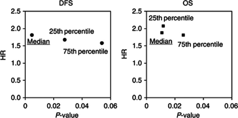
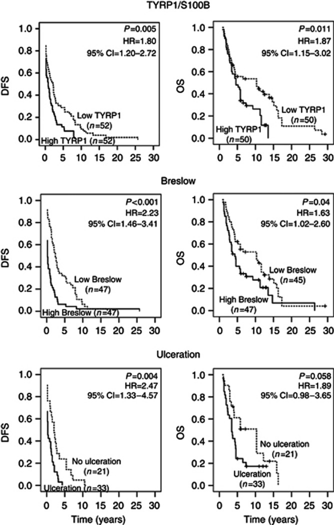
To find out the most relevant cutoff point for TYRP1/S100B ratio, we divided the population into two groups according to the median, the 25th and the 75th percentiles. We found that the median cutoff point gave a lower P-value than other cutoff points with a comparable HR for DFS and OS (Cox regression analysis; Figure 1). Then, using the median cutoff point, we defined a group with ‘low TYRP1' and a group with ‘high TYRP1' and showed the corresponding Kaplan–Meier curves in Figure 2 (top panel). High TYRP1 was significantly associated with a shorter DFS and a shorter OS in univariate analyses.
Figure 1.
Identification of a cutoff point for TYRP1/S100B ratio. Population (n=104) was classified with regard to ascending TYRP1/S100B ratio that was divided into two groups according to the median, the 25th percentile and the 75th percentile. P-values and hazard ratios (HR) were calculated (Cox regression) for DFS and OS for each cutoff point.
Figure 2.
Survival curves for TYRP1/S100B ratio, Breslow and ulceration. DFS and OS curves (Kaplan–Meier analysis) were determined according to high/low TYRP1, high/low Breslow and yes/no ulceration. Cox regressions were used to calculate P-values, hazard ratios (HRs) and 95% confidence intervals (CIs). ‘+' symbol indicates patients alive at the time of analysis.
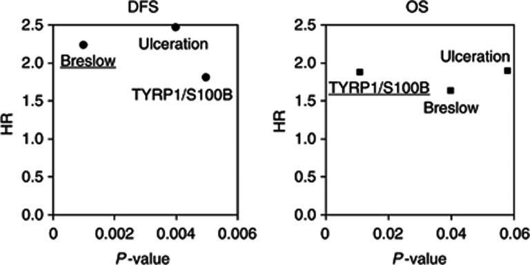
The prognostic power of TYRP1/S100B ratio was then compared with those of Breslow and ulceration by considering a median Breslow thickness of 2.3 mm and the presence/absence of ulceration as cutoff points. Kaplan–Meier analyses and Cox regressions showed that high Breslow thickness was significantly correlated with a shorter DFS and a shorter OS (Figure 2, medium panel). Similarly, ulceration of primaries was also significantly associated with a shorter DFS and tended to be significant with a shorter OS (P=0.058) (Figure 2, bottom panel). Altogether, comparing P-values and HRs for TYRP1, Breslow and ulceration, we found that all markers gave similar HRs but Breslow thickness showed a lower P-value for DFS and TYRP1 demonstrated a lower P-value for OS (Figure 3). These data suggest that TYRP1 could be a more valuable marker to refine prognosis of metastatic patients.
Figure 3.
Comparison of the prognostic value of each of TYRP1/S100B ratio, Breslow and ulceration. P-values and hazard ratios (HRs) were calculated (Cox regression) for each prognostic markers and were compared according to DFS and OS.
Moreover, high TYRP1, high Breslow and ulceration predicted, respectively, 88% (P=0.049, Fisher's exact test), 94% (P=0.003) and 100% (P=0.006) of first relapse before 5 years and 82% (P=0.003), 81% (P=0.007) and 91% (P=0.035) of mortality before 10 years. Globally, TYRP1 evaluated in lymph node metastases and Breslow/ulceration at the diagnosis showed similar predictive performances, especially for OS.
Multivariate analyses of TYRP1, Breslow and ulceration (as dichotomous variables) vs DFS and OS showed a significant correlation only between Breslow and DFS (P=0.003). Neither TYRP1, Breslow thickness nor ulceration was significant with regard to OS. This is most probably due to the small number of residual samples in this test (n=53, 42 events for OS) lowering significance. Indeed, univariate analysis of the same 53 samples did not show any significance neither (data not shown).
Assessment of TYRP1/S100B ratio in thin melanoma
The current study included the analyses of lymph node metastases from 10 patients with thin melanomas. As expected, we found that these patients had significantly longer DFS (median: 4.6 years) and OS (14.6 years) compared with patients with thick lesions (Breslow >1 mm, n=84; median DFS: 1.1 and median OS: 4.6 years, P=0.011 and 0.009 for DFS and OS, respectively, Mann–Whitney test). Moreover, the lymph node metastases of thin melanomas have a significant lower TYRP1/S100B ratio as compared to thick melanomas (n=84; median 1.1 × 10⁻⁴ vs 260 × 10−4, P<0.001, Mann–Whitney test), and belonged to the group of ‘low TYRP1' (ratio<cutoff) associated with a better prognosis. This finding further supports the prognostic value of TYRP1/S100B and its association with Breslow.
TYRP1/S100B ratio in two recurrent lymph node metastases of the same patient
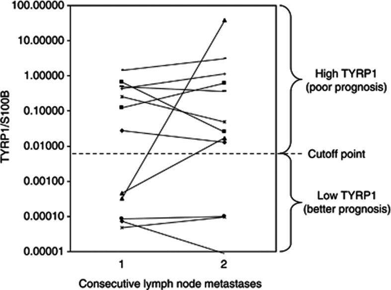
TYRP1/S100B was measured in two metastases obtained at different points in time (median: 2.1 years, range: 0.1–25.8) from each of 12 patients (Figure 4). We found that the TYRP1/S100B ratio remained within the same group of poor or good prognosis for 10 out of the 12 patients. Out of these 10 patients, 3 progressed from stage III to IV without any significant impact on TYRP1/S100B ratios. This further supports that TYRP1/S100B ratio has a prognostic value regardless of the time of metastases occurrence and suggests a well-conserved ratio during tumour progression.
Figure 4.
Variation of TYRP1/S100B ratio in two recurrent lymph node metastases within same patient. TYRP1/S100B mRNA expression were evaluated in triplicates by real-time PCR in two different melanoma lymph node metastasis obtained over years from each of 12 patients. The median TYRP1/S100B ratio calculated in Table 1 (cutoff point) is plotted.
Discussion
Our current study aimed at evaluating the prognostic value of TYRP1/S100B mRNA expression in lymph node metastases and to compare its predictive performance to those of pathological parameters from the primary melanoma (Breslow, lymph node involvement, ulceration). We demonstrated that high TYRP1/S100B ratio significantly correlates with shorter survival (DFS and OS) as well as with high Breslow thickness and the presence of ulceration in primary lesions. Moreover, it was well conserved during melanoma progression according to high/low TYRP1 groups. Our findings, linking TYRP1 and melanoma progression, are in line with recent studies reporting that TYRP1 variants were associated with melanoma risk (Nan et al, 2009; Chatzinasiou et al, 2011).
Importantly and unlike in skin metastases, tumour cells within lymph nodes can be subcapsular, parenchymal or multifocal (Dewar et al, 2004), so that corresponding snap-frozen tissue samples are likely to contain various amounts of stroma. Of note, the wide distribution range of S100B/β-actin values that we observed among our samples confirms and underlines a high variation in tumour load among lymph node metastases. Therefore and to account for tumour load, we normalised TYRP1 transcript against the one of S100B. Indeed, S100B is a gene whose expression is specific of melanocyte lineage and glial cells. It has many cell functions and is mainly involved in cytoskeleton integrity, cell cycle regulation (Millward et al, 1998) and apoptosis through its interaction with p53 (Markowitz et al, 2005). Importantly, serum S100B provides a good indication of tumour burden, disease progression as well as the response to chemotherapy in stage IV melanoma patients (Hamberg et al, 2003), through a release mechanism related to cell damage or cell death (Ghanem et al, 2001). More recently, it was reported that serial determination of serum S100B in stage IIb–III melanoma patients is a strong independent prognostic marker, even stronger compared with stage and number of positive lymph nodes (Bouwhuis et al, 2011). Our data confirmed the valuable use of S100B as an indicator of tumour load (Kruijff and Hoekstra, 2012).
In our study, univariate analyses demonstrated that TYRP1/S100B ratio (P=0.01), Breslow thickness (P=0.04) and ulceration (P=0.06) were predictive of OS with comparable HRs (range 1.6–1.9). Unfortunately, multivariate analyses were limited by the relatively small sample size of patients with available information for all of TYRP1/S100B, Breslow and ulceration (n=42). Our data are partly in accordance with a recent multivariate analysis of prognostic factors showing that the number of tumour-containing nodes, primary ulceration and patient age, but not Breslow thickness, independently predicted survival (P<0.01) in a population of 268 stage III melanoma patients with nodal macrometastases (Balch et al, 2010). Of note, our population contained 18% of stage IV melanoma patients, which may impact on our observations.
It is unclear how TYRP1 expression alters cell behaviour. TYRP1 is involved in maintenance of melanosome structure and may affect melanocyte proliferation and death (Sarangarajan and Boissy, 2001). However, the link between TYRP1 and patient survival is not known yet. Actually, TYRP1 could affect melanoma progression by stabilising tyrosinase (Kobayashi et al, 1998) and protecting melanocyte against toxic melanin intermediates produced by tyrosinase (Rad et al, 2004). In this context, an in vitro study from our group is on going to better understand the impact of TYRP1 on melanoma cell proliferation, migration and invasion (Mogha et al, 2011). The expression of TYRP1 is classically regulated by the microphthalmia-associated transcription factor, which is the master transcription factor involved in cell differentiation/melanogenesis, proliferation and apoptosis (Fang et al, 2002). However, additional regulatory processes could implicate microRNA. In particular, miR-155 (Li et al, 2012) and miR-137 (Dong et al, 2012) have been recently identified as downregulators of TYRP1 and microphthalmia-associated transcription factor, respectively, opening new perspectives for prognostic marker discovery. TYRP1 is also a melanocyte differentiation antigen that is important for both autoimmune destruction of melanocytes resulting in depigmentation or vitiligo, and anti-melanoma immune response. In this context, Liu et al (2009) reported that immunisation with a lentivector stimulated potent CD8 T cell responses against melanoma self-antigen TYRP1 and generated anti-tumour immunity in mice. In addition, active mouse immunisation against TYRP1-induced melanoma rejection and higher survival (Takechi et al, 1996). More recently, a new fully human anti-TYRP1 monoclonal antibody (20D7) has been developed, which recognises native TYRP1 in melanoma cells and significantly suppresses the growth of human melanoma xenografts in nude mice (Patel et al, 2007). These data allowed the activation of a phase I clinical trial that is still open using the 20D7 antibody (recently called flanvotumab, IMC-20D7S; http://clinicaltrials.gov/ct2/show/NCT01137006).
About 3–5% of patients with thin melanoma (≤1.0 mm) develop distant metastases (Kalady et al, 2003). Additional prognostic markers could be valuable to enable more accurate stratification of such patients with regard to survival, clinical management and follow-up regimens (Murali et al, 2012). Our data showed that 10 patients with thin primary melanoma had low TYRP1/S100B mRNA expression in lymph node metastasis, which is consistent with a better prognosis, and that these patients have effectively a longer survival, as expected with a low Breslow. These observations suggest that TYRP1 marker could be also of value for patients with thin melanomas, confirming a better prognosis.
In addition, by examining 12 patients with two lymph node metastases occurring at different dates, 10 remained within the same group of high or low TYRP1/S100B. First, this supports the correlations found with DFS and OS, as all samples have been collected randomly regardless of the time of their occurrence. Second, it indicates that such ratio can be used not only as a valuable prognostic marker but also as a potential target for therapy specially that TYRP1 expression is conserved during disease progression.
In conclusion, our present study, performed in melanoma lymph node metastases, extends and confirms our previous findings in skin metastases as to a significant association between TYRP1/S100B mRNA ratio and patient survival. Its measurement in lymph node metastases of stages III and IV melanoma patients may have important clinical implications to refine initial prognosis, to establish prognosis for those with unknown or non-evaluable primaries, and possibly to impact the management of high-risk patients with TYRP1-positive lymph nodes.
Acknowledgments
This study received financial support from MEDIC Foundation, Les Amis de l'Institut Bordet, Fondation Lambeau-Marteaux, EORTC—Melanoma Group and Wallonie-Bruxelles International. Petra El Hajj is the recipient of a fellowship from the Lebanese National Council for Scientific Research and the Lebanese University.
Footnotes
This work is published under the standard license to publish agreement. After 12 months the work will become freely available and the license terms will switch to a Creative Commons Attribution-NonCommercial-Share Alike 3.0 Unported License.
References
- Alonzo TA. Standards for reporting prognostic tumor marker studies. J Clin Oncol. 2005;23:9053–9054. doi: 10.1200/JCO.2005.04.3778. [DOI] [PubMed] [Google Scholar]
- Balch CM, Gershenwald JE, Soong S-J, Thompson JF, Atkins MB, Byrd DR, Buzaid AC, Cochran AJ, Coit DG, Ding S, Eggermont AM, Flaherty KT, Gimotty PA, Kirkwood JM, McMasters KM, Mihm MC, Morton DL, Ross MI, Sober AJ, Sondak VK. Final version of 2009 AJCC melanoma staging and classification. J Clin Oncol. 2009;27:6199–6206. doi: 10.1200/JCO.2009.23.4799. [DOI] [PMC free article] [PubMed] [Google Scholar]
- Balch CM, Gershenwald JE, Soong S-J, Thompson JF, Ding S, Byrd DR, Cascinelli N, Cochran AJ, Coit DG, Eggermont AM, Johnson T, Kirkwood JM, Leong SP, McMasters KM, Mihm MC, Morton DL, Ross MI, Sondak VK. Multivariate analysis of prognostic factors among 2,313 patients with stage III melanoma: comparison of nodal micrometastases versus macrometastases. J Clin Oncol. 2010;28:2452–2459. doi: 10.1200/JCO.2009.27.1627. [DOI] [PMC free article] [PubMed] [Google Scholar]
- Berger AJ, Kluger HM, Li N, Kielhorn E, Halaban R, Ronai Z, Rimm DL. Subcellular localization of activating transcription factor 2 in melanoma specimens predicts patient survival. Cancer Res. 2003;63:8103–8107. [PubMed] [Google Scholar]
- Berwick M, Erdei E, Hay J.2009Melanoma epidemiology and public health Dermatol Clin 27205–214.viii. [DOI] [PMC free article] [PubMed] [Google Scholar]
- Bouwhuis MG, Suciu S, Kruit W, Salès F, Stoitchkov K, Patel P, Cocquyt V, Thomas J, Liénard D, Eggermont AMM, Ghanem G. Prognostic value of serial blood S100B determinations in stage IIB-III melanoma patients: a corollary study to EORTC trial 18952. Eur J Cancer. 2011;47:361–368. doi: 10.1016/j.ejca.2010.10.005. [DOI] [PubMed] [Google Scholar]
- Chatzinasiou F, Lill CM, Kypreou K, Stefanaki I, Nicolaou V, Spyrou G, Evangelou E, Roehr JT, Kodela E, Katsambas A, Tsao H, Ioannidis JPA, Bertram L, Stratigos AJ. Comprehensive field synopsis and systematic meta-analyses of genetic association studies in cutaneous melanoma. J Natl Cancer Inst. 2011;103:1227–1235. doi: 10.1093/jnci/djr219. [DOI] [PMC free article] [PubMed] [Google Scholar]
- Chen G, Cheng Y, Zhang Z, Martinka M, Li G. Prognostic significance of cytoplasmic p27 expression in human melanoma. Cancer Epidemiol Biomarkers Prev. 2011;20:2212–2221. doi: 10.1158/1055-9965.EPI-11-0472. [DOI] [PubMed] [Google Scholar]
- Dewar DJ, Newell B, Green MA, Topping AP, Powell BWe, Cook MG. The microanatomic location of metastatic melanoma in sentinel lymph nodes predicts nonsentinel lymph node involvement. J Clin Oncol. 2004;22:3345–3349. doi: 10.1200/JCO.2004.12.177. [DOI] [PubMed] [Google Scholar]
- Dong C, Wang H, Xue L, Dong Y, Yang L, Fan R, Yu X, Tian X, Ma S, Smith GW. Coat color determination by miR-137 mediated down-regulation of microphthalmia-associated transcription factor in a mouse model. RNA. 2012;18:1679–1686. doi: 10.1261/rna.033977.112. [DOI] [PMC free article] [PubMed] [Google Scholar]
- Díaz-Lagares A, Alegre E, Arroyo A, González-Cao M, Zudaire ME, Viteri S, Martín-Algarra S, González A. Evaluation of multiple serum markers in advanced melanoma. Tumour Biol. 2011;32:1155–1161. doi: 10.1007/s13277-011-0218-x. [DOI] [PubMed] [Google Scholar]
- Fang D, Tsuji Y, Setaluri V. Selective down-regulation of tyrosinase family gene TYRP1 by inhibition of the activity of melanocyte transcription factor, MITF. Nucleic Acids Res. 2002;30:3096–3106. doi: 10.1093/nar/gkf424. [DOI] [PMC free article] [PubMed] [Google Scholar]
- Ghanem G, Loir B, Morandini R, Sales F, Lienard D, Eggermont A, Lejeune F. On the release and half-life of S100B protein in the peripheral blood of melanoma patients. Int J Cancer. 2001;94:586–590. doi: 10.1002/ijc.1504. [DOI] [PubMed] [Google Scholar]
- Hamberg AP, Korse CM, Bonfrer JM, de Gast GC. Serum S100B is suitable for prediction and monitoring of response to chemoimmunotherapy in metastatic malignant melanoma. Melanoma Res. 2003;13:45–49. doi: 10.1097/00008390-200302000-00008. [DOI] [PubMed] [Google Scholar]
- Henry L, Lavabre-Bertrand T, Douche T, Uttenweiler-Joseph S, Fabbro-Peray P, Monsarrat B, Martinez J, Meunier L, Stoebner P-E. Diagnostic value and prognostic significance of plasmatic proteasome level in patients with melanoma. Exp Dermatol. 2010;19:1054–1059. doi: 10.1111/j.1600-0625.2010.01151.x. [DOI] [PubMed] [Google Scholar]
- Journe F, Id Boufker H, Van Kempen L, Galibert M-D, Wiedig M, Salès F, Theunis A, Nonclercq D, Frau A, Laurent G, Awada A, Ghanem G. TYRP1 mRNA expression in melanoma metastases correlates with clinical outcome. Br J Cancer. 2011;105:1726–1732. doi: 10.1038/bjc.2011.451. [DOI] [PMC free article] [PubMed] [Google Scholar]
- Kalady MF, White RR, Johnson JL, Tyler DS, Seigler HF.2003Thin melanomas: predictive lethal characteristics from a 30-year clinical experience Ann Surg 238528–535.discussion 535–537. [DOI] [PMC free article] [PubMed] [Google Scholar]
- Kobayashi T, Imokawa G, Bennett DC, Hearing VJ. Tyrosinase stabilization by Tyrp1 (the brown locus protein) J Biol Chem. 1998;273:31801–31805. doi: 10.1074/jbc.273.48.31801. [DOI] [PubMed] [Google Scholar]
- Kruijff S, Hoekstra HJ. The current status of S-100B as a biomarker in melanoma. Eur J Surg Oncol. 2012;38:281–285. doi: 10.1016/j.ejso.2011.12.005. [DOI] [PubMed] [Google Scholar]
- Lazova R, Camp RL, Klump V, Siddiqui SF, Amaravadi RK, Pawelek JM. Punctate LC3B expression is a common feature of solid tumors and associated with proliferation, metastasis, and poor outcome. Clin Cancer Res. 2012;18:370–379. doi: 10.1158/1078-0432.CCR-11-1282. [DOI] [PMC free article] [PubMed] [Google Scholar]
- Li J, Liu Y, Xin X, Kim TS, Cabeza EA, Ren J, Nielsen R, Wrana JL, Zhang Z. Evidence for positive selection on a number of microRNA regulatory interactions during recent human evolution. PLoS Genet. 2012;8:e1002578. doi: 10.1371/journal.pgen.1002578. [DOI] [PMC free article] [PubMed] [Google Scholar]
- Liu Y, Peng Y, Mi M, Guevara-Patino J, Munn DH, Fu N, He Y. Lentivector immunization stimulates potent CD8 T cell responses against melanoma self-antigen tyrosinase-related protein 1 and generates antitumor immunity in mice. J Immunol. 2009;182:5960–5969. doi: 10.4049/jimmunol.0900008. [DOI] [PMC free article] [PubMed] [Google Scholar]
- Mandruzzato S, Callegaro A, Turcatel G, Francescato S, Montesco MC, Chiarion-Sileni V, Mocellin S, Rossi CR, Bicciato S, Wang E, Marincola FM, Zanovello P. A gene expression signature associated with survival in metastatic melanoma. J Transl Med. 2006;4:50. doi: 10.1186/1479-5876-4-50. [DOI] [PMC free article] [PubMed] [Google Scholar]
- Markowitz J, Mackerell AD, Carrier F, Charpentier TH, Weber DJ. Design of Inhibitors for S100B. Curr Top Med Chem. 2005;5:1093–1108. doi: 10.2174/156802605774370865. [DOI] [PubMed] [Google Scholar]
- McShane LM, Altman DG, Sauerbrei W, Taube SE, Gion M, Clark GM. REporting recommendations for tumour MARKer prognostic studies (REMARK) Br J Cancer. 2005;93:387–391. doi: 10.1038/sj.bjc.6602678. [DOI] [PMC free article] [PubMed] [Google Scholar]
- Miller AJ, Mihm MC. Melanoma. N Engl J Med. 2006;355:51–65. doi: 10.1056/NEJMra052166. [DOI] [PubMed] [Google Scholar]
- Millward TA, Heizmann CW, Schäfer BW, Hemmings BA. Calcium regulation of Ndr protein kinase mediated by S100 calcium-binding proteins. EMBO J. 1998;17:5913–5922. doi: 10.1093/emboj/17.20.5913. [DOI] [PMC free article] [PubMed] [Google Scholar]
- Mogha A, Gilot D, Primot A, Debbache J, Journe F, Bennett DC, Dreno B, Napolitano A, Ghanem G, Galiber MD.2011Uncovered role of Tyrosinase-related Protein 1 (TYRP1) in melanoma cells aggressiveness Pig Cell Mel Res 24791abstract C44. [Google Scholar]
- Mouawad R, Spano J-P, Comperat E, Capron F, Khayat D. Tumoural expression and circulating level of VEGFR-3 (Flt-4) in metastatic melanoma patients: correlation with clinical parameters and outcome. Eur J Cancer. 2009;45:1407–1414. doi: 10.1016/j.ejca.2008.12.015. [DOI] [PubMed] [Google Scholar]
- Murali R, Haydu LE, Long GV, Quinn MJ, Saw RPM, Shannon K, Spillane AJ, Stretch JR, Kefford RF, Thompson JF, Scolyer RA. Clinical and pathologic factors associated with distant metastasis and survival in patients with thin primary cutaneous melanoma. Ann Surg Oncol. 2012;19:1782–1789. doi: 10.1245/s10434-012-2265-y. [DOI] [PubMed] [Google Scholar]
- Nan H, Kraft P, Hunter DJ, Han J. Genetic variants in pigmentation genes, pigmentary phenotypes, and risk of skin cancer in Caucasians. Int J Cancer. 2009;125:909–917. doi: 10.1002/ijc.24327. [DOI] [PMC free article] [PubMed] [Google Scholar]
- Patel D, Balderes P, Lahiji A, Melchior M, Ng S, Bassi R, Wu Y, Griffith H, Jimenez X, Ludwig DL, Hicklin DJ, Kang X. Generation and characterization of a therapeutic human antibody to melanoma antigen TYRP1. Hum Antibodies. 2007;16:127–136. [PubMed] [Google Scholar]
- Rad HH, Yamashita T, Jin H-Y, Hirosaki K, Wakamatsu K, Ito S, Jimbow K. Tyrosinase-related proteins suppress tyrosinase-mediated cell death of melanocytes and melanoma cells. Exp Cell Res. 2004;298:317–328. doi: 10.1016/j.yexcr.2004.04.045. [DOI] [PubMed] [Google Scholar]
- Sarangarajan R, Boissy RE. Tyrp1 and oculocutaneous albinism type 3. Pigment Cell Res. 2001;14:437–444. doi: 10.1034/j.1600-0749.2001.140603.x. [DOI] [PubMed] [Google Scholar]
- Stoitchkov K, Letellier S, Garnier J-P, Bousquet B, Tsankov N, Morel P, Ghanem G, Le Bricon T. Evaluation of the serum L-dopa/L-tyrosine ratio as a melanoma marker. Melanoma Res. 2003;13:587–593. doi: 10.1097/00008390-200312000-00008. [DOI] [PubMed] [Google Scholar]
- Takechi Y, Hara I, Naftzger C, Xu Y, Houghton AN. A melanosomal membrane protein is a cell surface target for melanoma therapy. Clin Cancer Res. 1996;2:1837–1842. [PubMed] [Google Scholar]
- van Houdt IS, Oudejans JJ, van den Eertwegh AJM, Baars A, Vos W, Bladergroen BA, Rimoldi D, Muris JJF, Hooijberg E, Gundy CM, Meijer CJLM, Kummer JA. Expression of the apoptosis inhibitor protease inhibitor 9 predicts clinical outcome in vaccinated patients with stage III and IV melanoma. Clin Cancer Res. 2005;11:6400–6407. doi: 10.1158/1078-0432.CCR-05-0306. [DOI] [PubMed] [Google Scholar]


