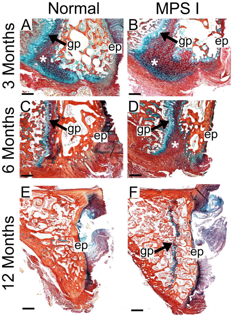
Figure 5. Cartilage to bone conversion in the C2 vertebral epiphysis.
Mid-sagittal histological sections double-stained with Alcian blue (glycosaminoglycans) and picrosirius red (collagen) illustrating increased cartilage in the ventral epiphyses (*) at 3 months-of-age (A and B) and at 6 months-of-age (C and D). At 12 months-of age, the epiphyseal growth plate in normal animals was absent (E); however, in MPS I animals, this growth plate persisted (F). Scale bars = 500 μm (A–D) and 1 mm (E and F); gp = growth plate and ep = caudal vertebral endplate. All images are oriented with the ventral side at the bottom.

