Abstract
Nucleophosmin, encoded by NPM1, is a haploinsufficient suppressor in hematologic malignancies. NPM1 mutations are mostly found in acute myeloid leukemia patients with normal karyotype and associated with favorable prognosis. A polymorphic nucleotide T deletion with unknown significance is present in the NPM1 3′-untranslated region. Here, we showed that the homozygous nucleotide T deletion was associated with adverse outcomes and could independently predict shortened survival in patients with de novo acute myeloid leukemia. Mechanistically, we demonstrated that the nucleotide T deletion created an illegitimate binding NPM1 for miR-337-5p, which was widely expressed in different acute myeloid leukemia subtypes and inhibited NPM1 expression. Accordingly, NPM1 levels were found to be significantly reduced and correlated with miR-337-5p levels in patients carrying a homozygous nucleotide T-deletion genotype. Together, our findings uncover a microRNA-mediated control of NPM1 expression that contributes to disease heterogeneity and suggest additional prognostic values of NPM1 in acute myeloid leukemia.
Introduction
The NPM1 gene is frequently altered in hematologic malignancies.1 In acute myeloid leukemia (AML), NPM1 is mainly disrupted by C-terminus mutations, causing aberrant cytoplasmic expression of the NPM1 protein.2NPM1-mutated AML has distinctive clinicopathological and molecular features. Most notably, the mutation is closely associated with normal karyotype and relatively favorable prognosis in the absence of FLT3-internal tandem duplication (ITD).3 While genotyping the NPM1 mutations, a polymorphic nucleotide T deletion (delT) at position 1,146 of the NPM1 3′-untranslated region (UTR) was reported in 60%–70% of AML patients.4,5 However, the significance of this polymorphism was unclear. Previous studies have shown that polymorphisms in other genes, including WT1 and IDH1, can affect AML prognosis in addition to mutations in the cognate genes.6,7 Here, we revealed that the homozygous state of the NPM1 delT polymorphism had important clinical and biological implications in AML involving an illegitimate microRNA (miRNA) regulation.
Design and Methods
Patients
We analyzed 149 adult and 70 childhood patients with de novo AML; therapy-related AML or AML arising from a prior myelodys-plastic syndrome were excluded. Patients’ characteristics are shown in Online Supplementary Table S1. All patients gave informed consent for the study, which was approved by the Joint CUHK-NTEC Clinical Research Ethics Committee and was conducted in accordance with the Declaration of Helsinki. Patients with acute promyelocytic leukemia (APL) were excluded for survival analysis. Overall, complete survival data were available from 93 adult and 61 childhood patients with non-APL who had received treatment. All the 93 adult patients were treated with the standard cytarabine plus daunorubicin ‘7+3’ induction chemotherapy regimen.8 Patients who achieved complete remission (CR) were then given consolidation treatment stratified by cytogenetics. Patients with non-favorable cytogenetics were referred for assessment for allogeneic stem cell transplantation. Patients who were not eligible for transplantation and those with favorable cytogenetics were given high-dose cytarabine-based chemotherapy for consolidation.9 Of the 61 pediatric patients, 41 were treated with a modified UK MRC AML 12 protocol10 and 20 with the NOPHO-AML 2004 protocol,11 as previously described. Two adult and 11 pediatric patients received transplantation as consolidation therapy. For these patients, survival data had been censored at the time of transplantation.
Normal bone marrow (BM) and peripheral blood samples were obtained from healthy donors who had no prior history of malignancy.
Cell culture
Cell lines were cultured in RPMI-1640 medium containing 10% fetal bovine serum.
Cytogenetic and mutational studies
Cytogenetics were classified into favorable, intermediate, and adverse according to the UK MRC classification.12,13 Intermediate/adverse cytogenetics are hereafter collectively referred to as non-favorable. Patients with unknown cytogenetics were screened for the presence (favorable) or absence (non-favorable) of RUNX1-ETO, CBFB-MYH11 and PML-RARA fusion transcripts.10
Mutation analysis of FLT3-ITD, KIT (exons 8 and 17), NPM1 (C-terminus and the delT polymorphism), and CEBPA was performed as previously described.10 Exon 4 of IDH1 (R132) and IDH2 (R140 and R172) were analyzed by direct sequencing. Primer sequences are provided in Online Supplementary Table S2.
Constructs, transfection, and luciferase reporter assays
Full-length NPM1 3′-UTR carrying the wild-type (pmirGLO-3′UTR-WT) or delT (pmirGLO-3′UTR-delT) genotype was cloned into the dual-luciferase pmirGLO vector (Promega), which co-expresses Renilla luciferase for normalization of transfection efficiency. pmirGLO-WT×3 and pmirGLO-delT×3 were prepared by cloning of the annealed oligonucleotides NPM1-WT×3-F/NPM1-WT×3-R and NPM1-delT×3-F/NPM1-delT×3-R (nucleotide sequences are provided in Online Supplementary Table S2) into pmirGLO. Pre-miR precursors were obtained from Life Technologies. Transfection was performed using Lipofectamine 2000 (Life Technologies). Luciferases were measured by Dual-Glo Luciferase Assay System (Promega) 24 h after transfection.
Quantitative RT-PCR (qRT-PCR)
RNA was extracted using TRIzol (Life Technologies). Quantitative real-time polymerase chain reaction (qRT-PCR) was performed using TaqMan assays (Life Technologies). NPM1 levels were normalized to GAPDH, and miRNAs normalized to RNU48. Relative expression was calculated by 2−ΔΔCt.
Western blotting
Western blotting was performed as previously described using NPM1 and β-tubulin antibodies (Cell Signaling).10
Statistical analysis
Overall survival (OS) and relapse-free survival (RFS) were defined as previously described.14 Kaplan-Meier curves were compared by log rank test. Multivariate Cox’s regression analysis was performed to test the significance of the delT polymorphism with adjustment for other potential prognostic factors. Pearson’s correlation was used to analyze correlations between continuous parameters. Two-sided P<0.05 was considered statistically significant. SPSS 13.0 was used for statistical analyses (SPSS).
Results and Discussion
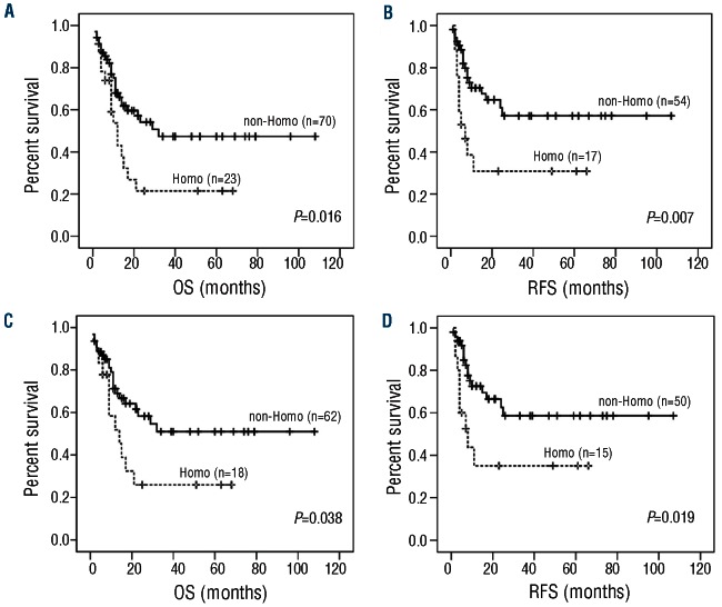
Of the 149 adult AML patients, 105 carried the delT polymorphism (32 homozygous and 73 heterozygous). The polymorphism was also determined in 211 healthy individuals (54% males; median age 50 years, range 21–74 years) and the genotype frequency was found to be similar between the normal (38 homozygous and 102 heterozygous) and adult patient (P=0.603) cohort, suggesting that the polymorphism might not affect susceptibility to leukemia development. Among 93 adults with non-APL with survival data, 44 (47%) died during a mean follow up of 21 months. Seventy-one patients (76%) achieved CR of whom 27 (38%) relapsed. Patients carrying a homozygous delT genotype had higher relapse rates (59% vs. 31%; P=0.051) and significantly shortened OS (median 9 months vs. 12 months, P=0.016) and RFS (median 5 months vs. 12 months; P=0.007) than patients carrying a non-homozygous genotype (Figure 1 A and B). CR rates were similar between the two groups (74% vs. 77%, P=0.781). In multivariate analysis, the homozygous delT genotype (P=0.035), age (P=0.013), white blood cell count (P=0.006), cytogenetics (P=0.044), and CEBPA double mutation (P=0.004) were significant prognostic factors for OS (Online Supplementary Table S3). For RFS, the homozygous delT genotype (P=0.018), age (P=0.005), cytogenetics (P=0.026), and CEBPA double mutation (P=0.002) were significant factors.
Figure 1.
The homozygous NPM1 3′-UTR delT genotype was associated with inferior outcomes in AML patients. Kaplan-Meier analysis of OS and RFS based on the delT genotype in the entire adult AML cohort (A and B) or younger adult patients aged 18 to 60 years (C and D). Patients failed to achieve CR were omitted in the RFS analysis. Homo, homozygous; non-Homo, non-homozygous.
Because elderly and young adult AML patients have different biological and clinical behaviors, we next restricted our analysis to younger adult patients who were 18–60 years old (n=80). Younger patients with homozygous delT had higher relapse rates (60% vs. 32%; P=0.07) and significantly reduced OS (median 10.5 months vs. 13 months; P=0.038) and RFS (median 7 months vs. 13 months; P=0.019) than patients with a non-homozygous genotype (Figure 1C and D). CR rates were similar between the two groups (83% vs. 81%, P=1.000). The homozygous delT genotype remained prognostic for poorer RFS (P=0.028) in multivariate analysis (Online Supplementary Table S3). Based on these findings, we further evaluated the impact of the homozygous delT genotype on younger adult patients with different cytogenetic and molecular features. Despite the limited sample size, a trend or significant association of homozygous delT with poorer RFS was observed in all the subgroups analyzed (Online Supplementary Figure S1). The prognostic impact of the delT polymorphism in elderly patients was not determined because of the small number of patients over the age of 60 years.
The delT polymorphism was detected in 44 (12 homozygous and 32 heterozygous) of 70 childhood AML patients. Among 61 non-APL patients with survival data, 12 (20%) died during a mean follow up of 54 months. Fifty-nine patients (97%) achieved CR of whom 16 (27%) relapsed. The homozygous delT genotype had no significant impacts on OS, RFS, and CR rates in the entire cohort. However, when patients with FLT3-ITD (n=7) and KIT mutations (n=5), the two molecular markers reported to correlate with high relapse risk in childhood AML,15 were excluded, a significant association between homozygous delT and poorer RFS (P=0.032) was noted; the association with OS was not statistically significant (P=0.106).
The delT polymorphism showed no significant correlation with any clinicopathological or molecular parameters in the adult and childhood patient cohorts (Online Supplementary Table S4).
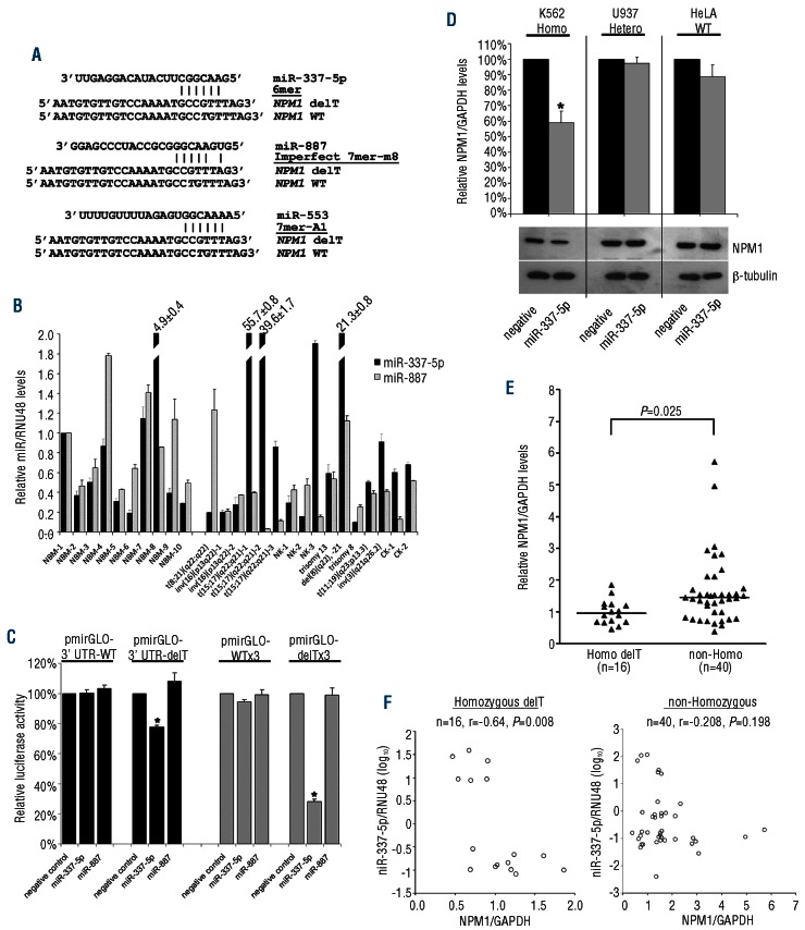
Polymorphisms within 3′-UTR might modify miRNA-mediated gene regulation. Using the PITA algorithm,16 we found that the delT created three putative binding sites for miR-337-5p, miR-887, and miR-553 (Figure 2A). To investigate the potential relevance of these miRNAs in regulating NPM1, we first examined their expression in BM from patients with different AML subtypes. All the 16 AML samples and 10 normal BM expressed detectable levels of miR-337-5p and miR-887 (Figure 2B). In contrast, none of the BM examined (16 patients and 11 normal) showed miR-553 expression (data not shown). Thus, miR-337-5p and miR-887 were selected for functional validation using luciferase reporter assays in HeLa which expressed barely detectable levels of the two miRNAs (data not shown). Co-transfection of pre-miR-337-5p exerted a dose-dependent repressive effect on the NPM1 3′-UTR harboring the delT (Figure 2C and Online Supplementary Figure S2). In contrast, no effect on the 3′-UTR-delT construct was observed when pre-miR-887 was used. Moreover, co-transfection of these two miRNA precursors had no effect on the wild-type 3′-UTR construct (Figure 2C). To validate this finding, we created two additional constructs, each containing three tandem copies of the delT (pmirGLO-delT×3) or wild-type (pmirGLO-WT×3) sequences. Co-transfection with pre-miR-337-5p but not pre-miR-887 drastically repressed the pmirGLO-delTx3 construct (Figure 2C). We next transfected K562, U937 and HeLa cells with pre-miR-337-5p to investigate the effect on endogenous NPM1 expression. These cell lines carried the homozygous delT, heterozygous delT and wild-type genotypes, respectively, and expressed barely detectable levels of miR-337-5p (data not shown). Transfection of pre-miR-337-5p reduced both NPM1 mRNA and protein levels in K562 cells harboring the homozygous delT polymorphism (Figure 2D). In contrast, no apparent effect on NPM1 expression was observed in U937 and HeLa cells. Collectively, these findings indicate that the delT caused illegitimate repression of NPM1 by miR-337-5p. We next compared NPM1 transcript levels in AML patients with or without homozygous delT. Since NPM1 is ubiquitously and abundantly expressed,1 only patients whose BM contained at least 80% of blasts were selected for analysis. As shown in Figure 2E, NPM1 levels were significantly reduced in the homozygous delT group as compared to the non-homozygous group. Importantly, those patients with lower NPM1 expression were also found to have poorer outcomes than those with higher NPM1 expression (Online Supplementary Figure S3). Notably, a significant inverse relationship between NPM1 and miR-337-5p levels was observed in patients carrying the homozygous delT genotype, but not in patients carrying the other genotypes (Figure 2F).
Figure 2.
The homozygous delT polymorphism in NPM1 3′-UTR caused illegitimate regulation by miR-337-5p. (A) Sequence alignment of miR-337-5p, miR-887, and miR-553 with the NPM1 3′-UTR delT sequence. The delT in the wild-type (WT) sequence is in italics. The type of microRNA seed match20 is indicated. (B) Expression of miR-337-5p and miR-887 in BM samples from 16 adult AML patients and 10 normal individuals. Relative expression was compared to NBM-1. The cytogenetics of each patient is indicated. NK: normal karyotype; CK: complex karyotype; NBM: normal BM. Results are expressed as mean±SE from triplicate assays. The column bar of four outliners was truncated and the relative levels are shown. (C) NPM1 3′-UTR luciferase constructs (0.2 μg) were co-transfected with pre-miR-337-5p or pre-miR-887 precursor at a final concentration of 100nM into HeLa cells. Transfection with the same amount of pre-miR negative control was done in parallel. Results are presented as relative luciferase activity by comparing the normalized firefly luciferase activity of the construct co-transfected with pre-miR precursor to that co-transfected with the negative control. Results are expressed as mean±SE from at least triplicate assays. *indicates P<0.05. (D) K562, U937, and HeLa cells were transfected with 100nM of pre-miR-337-5p or pre-miR negative control and NPM1 expression was determined 72 h after the transfection. Flow cytometric analysis of FAM-labeled pre-miR negative control revealed a transfection efficiency of 91–97% for the three cell lines (data not shown). NPM1 mRNA levels (upper panel) were compared between the pre-miR-337-5p and control transfection. qRT-PCR results are presented as mean±SE from triplicate assays. *P<0.05. Lower panel, NPM1 levels were examined by Western blot and β-tubulin was used as loading control. Representative blots from repeated experiments are shown The delT genotype in each cell line is indicated. Homo: homozygous delT; Hetero, heterozygous delT. (E) Comparison of NPM1 mRNA levels in adult AML patients with or without the homozygous delT polymorphism. Each triangle represents one patient and the number of patients in each group is shown. Horizontal lines indicate the mean NPM1/GAPDH ratio. (F) Correlation of NPM1 with miR-337-5p levels in adult AML patients with different NPM1 3′-UTR genotypes. Each circle represents one patient and the number of patients and the Pearson’s correlation coefficient (r) in each group is shown. For (E) and (F), all patients had at least 80% blast counts in their BM. The cohort includes 20 patients with PML-RARA and 36 patients with a non-favorable karyotype (31 intermediate, 3 adverse and 2 unknown). Relative expression levels in each patient were determined in triplicates and compared to U937 (NPM1) and NBM-1 (miR-337-5p).
NPM1-mutated AML has been found to have a distinctive miRNA expression profile,17–19 however, whether NPM1 is subjected to miRNA regulation is unclear. In this study, we demonstrated that a polymorphic delT in the NPM1 3′-UTR generated an illegitimate binding site for miR-337-5p. Analysis of the entire NPM1 mRNA sequence revealed no further miR-337-5p site, highlighting the significance of the polymorphism in miR-337-5p-mediated NPM1 regulation. Moreover, the use of other databases failed to reveal additional miRNAs targeting this delT site. 6-mer seed-matched sites typically have lower efficacy20 and this might explain the modest NPM1 repression by miR-337-5p. It is worth noting that the miR-337-5p-mediated NPM1 regulation might be cell context-dependent as we failed to observe similar correlations of NPM1 and miR-337-5p expression in normal peripheral blood samples (Online Supplementary Figure S4). Indeed, we noticed that the normal samples had markedly higher NPM1 levels (approx. 7.3-fold) than the AML patient samples while miR-337-5p expression was similar between the two groups. Since the endogenous level of the target mRNA is an important determinant of miRNA regulation,21 these findings implicate potential differential regulation of NPM1 expression in normal and AML leukemic cells. To date, very little is known about the role of miR-337-5p. It was shown that the miRNA might be involved in the regulation of the tyrosine kinase gene LYN in B-cell chronic lymphocytic leukemia.22
The similar frequencies of the delT polymorphism between adult AML patients and normal adults suggested that the polymorphism might not predispose to leukemia. Likewise, the cytoplasmic NPM1 mutant also failed to cause AML in transgenic mice, suggesting the need for cooperating mutations.23 On the other hand, our findings indicated that the delT in homozygous state had negative impacts on AML outcome. Although the mechanisms by which the delT impacts outcome are unclear, it is possible that the delT-associated reduction in NPM1 expression may cause centrosome amplification and genomic instability and promote tumorigenicity of the leukemic cells.1 Another potential mechanism is that the delT-harboring NPM1 mRNA may act as competing endogenous RNA (ceRNA) to compete with other mRNA targets for miR-337-5p binding, thereby regulating their function in trans and perturbing normal miRNA regulation.24 Larger prospective studies are needed to further evaluate the prognostic value of the delT polymorphism and NPM1 expression as well as their interaction with other mutations in AML patients.
In summary, we demonstrated the impact of genetic variations in an miRNA network involving fine control of a tumor suppressor and identified a potential poor-risk marker in AML.
Acknowledgments
We thank the Children’s Cancer Foundation for the support of patient care in leukemia and Yonna Leung for recruiting normal blood samples from healthy donors.
Footnotes
The online version of this article has a Supplementary Appendix.
Funding
This work was supported in part by a grant from the Research Council of the Hong Kong SAR, China (project n. CUHK CERG 4415/05M).
Authorship and Disclosures
Information on authorship, contributions, and financial & other disclosures was provided by the authors and is available with the online version of this article at www.haematologica.org.
References
- 1.Grisendi S, Mecucci C, Falini B, Pandolfi PP. Nucleophosmin and cancer. Nat Rev Cancer. 2006;6(7):493–505 [DOI] [PubMed] [Google Scholar]
- 2.Falini B, Mecucci C, Tiacci E, Alcalay M, Rosati R, Pasqualucci L, et al. Cytoplasmic nucleophosmin in acute myelogenous leukemia with a normal karyotype. N Engl J Med. 2005;352(3):254–66 [DOI] [PubMed] [Google Scholar]
- 3.Falini B, Martelli MP, Bolli N, Sportoletti P, Liso A, Tiacci E, et al. Acute myeloid leukemia with mutated nucleophosmin (NPM1): is it a distinct entity?. Blood. 2011;117(4):1109–20 [DOI] [PubMed] [Google Scholar]
- 4.Döhner K, Schlenk RF, Habdank M, Scholl C, Rücker FG, Corbacioglu A, et al. Mutant nucleophosmin (NPM1) predicts favorable prognosis in younger adults with acute myeloid leukemia and normal cytogenetics: interaction with other gene mutations. Blood. 2005;106(12):3740–6 [DOI] [PubMed] [Google Scholar]
- 5.Chou WC, Tang JL, Lin LI, Yao M, Tsay W, Chen CY, et al. Nucleophosmin mutations in de novo acute myeloid leukemia: the age-dependent incidences and the stability during disease evolution. Cancer Res. 2006;66(6):3310–6 [DOI] [PubMed] [Google Scholar]
- 6.Damm F, Heuser M, Morgan M, Yun H, Grosshennig A, Göhring G, et al. Single nucleotide polymorphism in the mutational hotspot of WT1 predicts a favorable outcome in patients with cytogenetically normal acute myeloid leukemia. J Clin Oncol. 2010;28(4):578–85 [DOI] [PubMed] [Google Scholar]
- 7.Wagner K, Damm F, Göhring G, Görlich K, Heuser M, Schäfer I, et al. Impact of IDH1 R132 mutations and an IDH1 single nucleotide polymorphism in cytogenetically normal acute myeloid leukemia: SNP rs11554137 is an adverse prognostic factor. J Clin Oncol. 2010;28(14):2356–64 [DOI] [PubMed] [Google Scholar]
- 8.Rai KR, Holland JF, Glidewell OJ, Weinberg V, Brunner K, Obrecht JP, et al. Treatment of acute myelocytic leukemia: a study by cancer and leukemia group B. Blood. 1981; 58(6): 1203–12 [PubMed] [Google Scholar]
- 9.Mayer RJ, Davis RB, Schiffer CA, Berg DT, Powell BL, Schulman P, et al. Intensive postremission chemotherapy in adults with acute myeloid leukemia. Cancer and Leukemia Group B. N Engl J Med. 1994;331 (14):896–903 [DOI] [PubMed] [Google Scholar]
- 10.Cheng CK, Li L, Cheng SH, Ng K, Chan NP, Ip RK, et al. Secreted-frizzled related protein 1 is a transcriptional repression target of the t(8;21) fusion protein in acute myeloid leukemia. Blood. 2011;118(25):6638–48 [DOI] [PubMed] [Google Scholar]
- 11.Abrahamsson J, Forestier E, Heldrup J, Jahnukainen K, Jónsson OG, Lausen B, et al. Response-guided induction therapy in pediatric acute myeloid leukemia with excellent remission rate. J Clin Oncol. 2011;29(3):310–5 [DOI] [PubMed] [Google Scholar]
- 12.Grimwade D, Hills RK, Moorman AV, Walker H, Chatters S, Goldstone AH, et al. Refinement of cytogenetic classification in acute myeloid leukemia: determination of prognostic significance of rare recurring chromosomal abnormalities among 5876 younger adult patients treated in the United Kingdom Medical Research Council trials. Blood. 2010;116(3):354–65 [DOI] [PubMed] [Google Scholar]
- 13.Harrison CJ, Hills RK, Moorman AV, Grimwade DJ, Hann I, Webb DK, et al. Cytogenetics of childhood acute myeloid leukemia: United Kingdom Medical Research Council Treatment trials AML 10 and 12. J Clin Oncol. 2010;28(16):2674–81 [DOI] [PubMed] [Google Scholar]
- 14.Döhner H, Estey EH, Amadori S, Appelbaum FR, Büchner T, Burnett AK, et al. Diagnosis and management of acute myeloid leukemia in adults: recommendations from an international expert panel, on behalf of the European LeukemiaNet. Blood. 2010;115(3):453–74 [DOI] [PubMed] [Google Scholar]
- 15.Meshinchi S, Arceci RJ. Prognostic factors and risk-based therapy in pediatric acute myeloid leukemia. Oncologist. 2007;12(3): 341–55 [DOI] [PubMed] [Google Scholar]
- 16.Kertesz M, Iovino N, Unnerstall U, Gaul U, Segal E. The role of site accessibility in microRNA target recognition. Nat Genet. 2007;39(10):1278–84 [DOI] [PubMed] [Google Scholar]
- 17.Garzon R, Garofalo M, Martelli MP, Briesewitz R, Wang L, Fernandez-Cymering C, et al. Distinctive microRNA signature of acute myeloid leukemia bearing cytoplasmic mutated nucleophosmin. Proc Natl Acad Sci USA. 2008;105(10):3945–50 [DOI] [PMC free article] [PubMed] [Google Scholar]
- 18.Becker H, Marcucci G, Maharry K, Radmacher MD, Mrózek K, Margeson D, et al. Favorable prognostic impact of NPM1 mutations in older patients with cytogenetically normal de novo acute myeloid leukemia and associated gene- and microRNA-expression signatures: a Cancer and Leukemia Group B study. J Clin Oncol. 2010;28(4):596–604 [DOI] [PMC free article] [PubMed] [Google Scholar]
- 19.Russ AC, Sander S, Lück SC, Lang KM, Bauer M, Rücker FG, et al. Integrative nucleophosmin mutation-associated microRNA and gene expression pattern analysis identifies novel microRNA - target gene interactions in acute myeloid leukemia. Haematologica. 2011;96(12):1783–91 [DOI] [PMC free article] [PubMed] [Google Scholar]
- 20.Friedman RC, Farh KK, Burge CB, Bartel DP. Most mammalian mRNAs are conserved targets of microRNAs. Genome Res. 2009;19(1):92–105 [DOI] [PMC free article] [PubMed] [Google Scholar]
- 21.Doench JG, Sharp PA. Specificity of microRNA target selection in translational repression. Genes Dev. 2004;18(5):504–11 [DOI] [PMC free article] [PubMed] [Google Scholar]
- 22.Hussein K, von Neuhoff N, Büsche G, Buhr T, Kreipe H, Bock O. Opposite expression pattern of Src kinase Lyn in acute and chronic haematological malignancies. Ann Hematol. 2009;88(11):1059–67 [DOI] [PubMed] [Google Scholar]
- 23.Cheng K, Sportoletti P, Ito K, Clohessy JG, Teruya-Feldstein J, Kutok JL, et al. The cytoplasmic NPM mutant induces myeloproliferation in a transgenic mouse model. Blood. 2010;115(16):3341–5 [DOI] [PMC free article] [PubMed] [Google Scholar]
- 24.Poliseno L, Salmena L, Zhang J, Carver B, Haveman WJ, Pandolfi PP. A coding-independent function of gene and pseudogene mRNAs regulates tumour biology. Nature. 2010;465(7301):1033–8 [DOI] [PMC free article] [PubMed] [Google Scholar]


