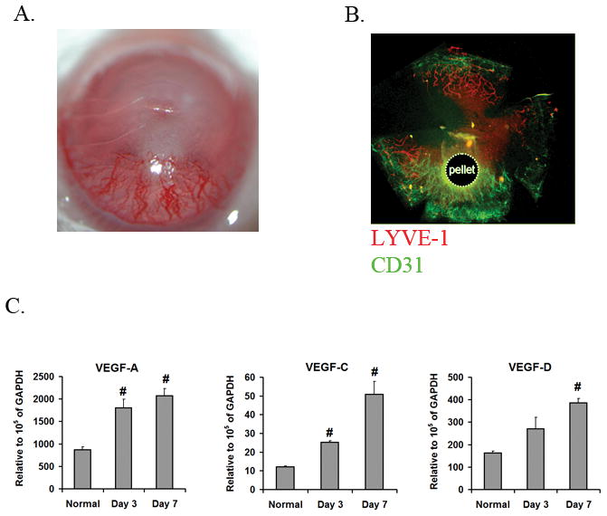
Figure 1.

b-FGF induces both blood and lymphatic vessel growth. A. Representative photographs of a cornea at 7 days after implantation of b-FGF micropellet demonstrating extensive neovascularization. B. Immunostaining of a corneal flat mount at 7 days after b-FGF micropellet implantation with CD31 (green) and LYVE-1 (red) antibodies demonstrates the development of blood (CD31high/LYVE-1low) and lymphatic (CD31low/LYVE-1high) vessels. C. VEGF ligands mRNA expression levels were determined by qRT-PCR in corneas of normal and b-FGF micropellet implanted mice. Corneal expression of all VEGF ligands are increased 7 days after b-FGF micropellet implantation (#: P<0.01, compared to normal corneas).
