

Abstract
Recurrent Takotsubo cardiomyopathy is a relatively uncommon condition seen in patients with severe physical or emotional stress. We report a case of a 51-year-old woman who had recurrent Takotsubo cardiomyopathy with involvement of apical left ventricular (LV) segments, induced by intense emotional stress. On two occasions she presented with symptoms of acute coronary syndrome accompanied by LV regional wall motion abnormalities without a culprit coronary stenosis, and exhibited complete resolution of symptoms and restoration of normal LV wall motion.
Background
Takotsubo (stress-induced) cardiomyopathy, also known as broken heart syndrome or transient left ventricular (LV) apical ballooning syndrome, is a reversible cause of LV dysfunction. It manifests as chest pain syndrome mimicking acute coronary syndrome that is preceded by intense physical or psychosocial stress and typically recovers fully with conventional management. This condition accounts for 1–2% of acute myocardial infarction.1 Recurrence of the condition is relatively uncommon. Only a few cases of recurrent Takotsubo cardiomyopathy have been previously reported.2 It is crucial to recognise Takotsubo cardiomyopathy, as long-term therapy is not indicated in this condition as opposed to acute coronary syndrome. We describe key aspects of clinical presentation, diagnostic modalities and management of stress-induced cardiomyopathy.
Case presentation
A 51-year-old woman with a history of systemic lupus erythematous, hypothyroidism and Takotsubo cardiomyopathy 2 years earlier presented with severe chest pain and shortness of breath lasting 24 h. Preceding the symptoms, she had received sudden, unexpected bad news regarding the death of her close friend that upset her. Shortly after, she noticed her heart pounding, associated with chest pain described as ‘elephant sitting on my chest’, shortness of breath, nausea, vomiting and diarrhoea. She did not have the conventional risk factors for coronary artery disease such as smoking, hypertension, hyperlipidaemia, diabetes or family history of premature coronary artery disease.
On physical examination, she appeared to be in distress. Her vital signs included blood pressure of 127/80 mm Hg and heart rate of 104 bpm. Systemic examination was within normal limits.
Investigations
Presenting ECG revealed sinus tachycardia with anterior and inferolateral T wave inversions, which were new compared with her baseline ECG. Routine haematological and biochemical investigations were within normal values. Presenting troponin I was elevated at 1.49 ng/ml and brain natriuretic peptide was normal at 38 pg/ml. Since she had acute onset of shortness of breath with sinus tachycardia, contrast-enhanced CT scan of the chest was performed which failed to demonstrate any evidence of pulmonary embolism. Transthoracic echocardiogram (TTE) showed anteroapical hypokinesis and hyperdynamic contractility at the base with an ejection fraction (EF) of 43%. In comparison, her previous episode of Takotsubo cardiomyopathy 2 years ago had revealed troponin I of 1.69 ng/ml, LVEF of 25% with complete akinesis of LV walls. On follow-up after 3 weeks, her EF had normalised to 61% with normal LV wall motion.
Differential diagnosis
Differential diagnoses of acute coronary syndrome and stress-induced cardiomyopathy were considered. A left heart catheterisation with coronary angiography was performed for concerns of acute coronary syndrome. Left ventriculography revealed anteroapical and inferoapical akinesis, with hyperdynamic contractility at the base (figure 1). Overall EF was reduced at 41%. Coronary angiography revealed normal coronary arteries.
Figure 1.
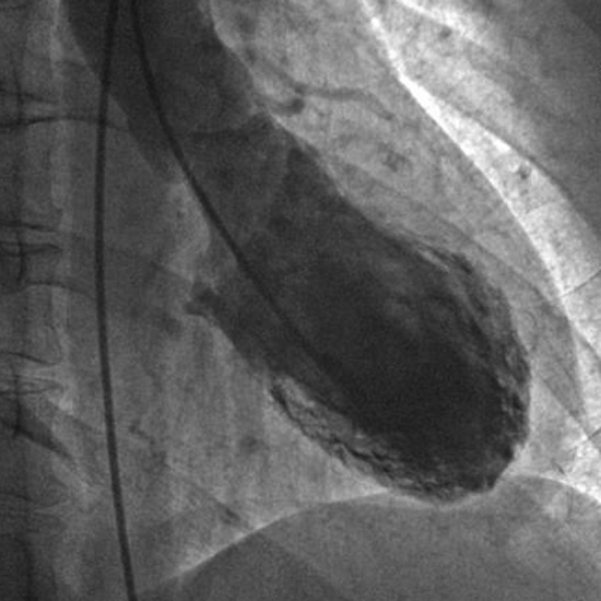
Left ventriculogram with basal hyperkinesis and apical hypokinesis
Treatment
She was kept on aspirin, ACE inhibitor and beta blocker. A TTE obtained 4 weeks after discharge revealed normal LV function with normalisation of EF, which was 61%. Based on the typical clinical presentation, LV wall motion abnormalities, normal coronary arteries and reversibility of the LV dysfunction, the diagnosis of Takotsubo cardiomyopathy was made.
Outcome and follow-up
At 6 month follow-up visit, she was asymptomatic and her repeat echocardiogram revealed normal LV function with an EF of 64%.
Discussion
Takotsubo cardiomyopathy is a syndrome characterised by the sudden onset of acute chest symptoms following intense physical or psychosocial stressors, ECG changes consistent with myocardial ischaemia, transient LV wall motion abnormalities and normal coronary arteries on coronary angiography with minimally elevated myocardial enzymes.3 It is more prevalent in postmenopausal women compared with men, with the mean age of 62–75 years.4–7 The recurrence rate for Takotsubo cardiomyopathy is 11.4% over 4 years after the first presentation.8 Bridgman et al9 described a case of a female who had two separate occasions of stress-induced cardiomyopathy with a different pattern of regional wall motion abnormality; both induced by major earthquakes. Individual variation in the location and density of cardiac adrenoreceptors has been proposed as the cause of different LV wall motion involvement in atypical variants.7 Though rare, recurrence and occurrence in the first-degree relatives may suggest that there is a genetic predisposition to this syndrome.10–12
A stress-induced surge in catecholamines leading to myocardial stunning is the proposed mechanism.13 14 Physicians should consider Takotsubo cardiomyopathy as a differential diagnosis of chest pain especially in postmenopausal women with recent unexpected physical or emotional stress.15
ECG findings on admission usually include ST segment elevation with diffuse precordial T wave inversion causing difficulty distinguishing acute myocardial infarction from Takotsubo cardiomyopathy.2 Echocardiography usually shows a left ventricle with well-preserved function in the base, moderate to severe dysfunction in the mid portion of the left ventricle and akinesis or dyskinesis in the apex of the heart.16 Confirmation of the diagnosis of Takotsubo cardiomyopathy is only possible with coronary angiography which can rule out obstructive coronary artery disease as a cause of LV dysfunction.2
LV dysfunction usually recovers within a few weeks with conservative management with beta blockers, ACE inhibitors and diuretics, if associated with pulmonary oedema.17–19 Long-term treatment with aspirin, beta blocker, ACE inhibitor and calcium channel blockers were not found to be beneficial in Takotsubo cardiomyopathy.20 Elesber et al8 observed no prognostic value of troponin or B-type natriuretic peptide levels in their 100 case series of Takotsubo cardiomyopathy and found that ECG findings at index hospitalisation did not predict short-term or long-term survival, unlike in acute coronary syndromes. Although fatal cases have been reported,16 prognosis of stress-induced cardiomyopathy is usually favourable.2 21 Involvement of the right ventricle has been described and is found to be associated with lower LVEF, longer hospitalisation, more frequent complications like congestive heart failure, need for intra-aortic balloon pump support or cardiopulmonary resuscitation.18 As in this case, even the recurrent cases have favourable prognosis with the conservative management.9 10
Learning points.
Takotsubo cardiomyopathy mimics acute coronary syndrome and is usually precipitated by intense physical or emotional stress.
The diagnosis requires coronary angiography to exclude coronary artery disease.
Prognosis is generally favourable even in recurrent cases.
Footnotes
Correction notice: This article has been corrected since it was published online on 23 May 2013. Figure 1 which was previously omitted has now been included.
Contributors: NRM wrote the primary draft of the manuscript. MRA and RP edited the primary draft. EJH reviewed and edited the final manuscript.
Competing interests: None.
Patient consent: Obtained.
Provenance and peer review: Not commissioned; externally peer reviewed.
References
- 1.Kurowski V, Kaiser A, Von Hof K, et al. Apical and midventricular transient left ventricular dysfunction syndrome (Takotsubo cardiomyopathy): frequency, mechanisms, and prognosis. Chest 2007;2013:809–16 [DOI] [PubMed] [Google Scholar]
- 2.Akashi YJ, Goldstein DS, Barbaro G, et al. Takotsubo cardiomyopathy: a new form of acute, reversible heart failure. Circulation 2008;2013:2754–62 [DOI] [PMC free article] [PubMed] [Google Scholar]
- 3.Seth PS, Aurigemma GP, Krasnow JM, et al. A syndrome of transient left ventricular apical wall motion abnormality in the absence of coronary disease: a perspective from the United States. Cardiology 2003;2013:61–6 [DOI] [PubMed] [Google Scholar]
- 4.Wittstein IS, Thiemann DR, Lima JAC, et al. Neurohumoral features of myocardial stunning due to sudden emotional stress. N Engl J Med 2005;2013:539–48 [DOI] [PubMed] [Google Scholar]
- 5.Sharkey SW, Lesser JR, Zenovich AG, et al. Acute and reversible cardiomyopathy provoked by stress in women from the United States. Circulation 2005;2013:472–9 [DOI] [PubMed] [Google Scholar]
- 6.Bybee KA, Kara T, Prasad A, et al. Systematic review: transient left ventricular apical ballooning: a syndrome that mimics ST-segment elevation myocardial infarction. Ann Intern Med 2004;2013:858–65 [DOI] [PubMed] [Google Scholar]
- 7.Wever-Pinzon O, Wever-Pinzon J, Tami L. Recurrent Takotsubo cardiomyopathy presenting with different morphologic patterns. Int J Cardiol 2011;2013:379–81 [DOI] [PubMed] [Google Scholar]
- 8.Elesber AA, Prasad A, Lennon RJ, et al. Four-year recurrence rate and prognosis of the apical ballooning syndrome. J Am Coll Cardiol 2007;2013:448–52 [DOI] [PubMed] [Google Scholar]
- 9.Bridgman PG, Chan CW, Elliott JM. A case of recurrent earthquake stress cardiomyopathy with a differing wall motion abnormality. Echocardiography 2012;2013:E26–7 [DOI] [PubMed] [Google Scholar]
- 10.Pathak H, Esses J, Pathak S, et al. A unique case of recurrent Takotsubo cardiomyopathy. South Med. J 2010;2013:805–6 [DOI] [PubMed] [Google Scholar]
- 11.Pison L, De Vusser P, Mullens W. Apical ballooning in relatives. Heart 2004;2013:e67. [DOI] [PMC free article] [PubMed] [Google Scholar]
- 12.Kushiro T, Saito F, Kusama J, et al. Takotsubo-shaped cardiomyopathy with type I CD36 deficiency. Heart Vessels 2005;2013:123–5 [DOI] [PubMed] [Google Scholar]
- 13.Burgdorf C, Von Hof K, Schunkert H, et al. Regional alterations in myocardial sympathetic innervation in patients with transient left-ventricular apical ballooning (Tako-Tsubo cardiomyopathy). J Nucl Cardiol 2008;2013:65–72 [DOI] [PubMed] [Google Scholar]
- 14.Wong CP, Chia PL. Recurrent Takotsubo cardiomyopathy precipitated by myasthenic crisis. Int J Cardiol 2012;2013:e11–12 [DOI] [PubMed] [Google Scholar]
- 15.Gianni M, Dentali F, Grandi AM, et al. Apical ballooning syndrome or Takotsubo cardiomyopathy: a systematic review. Eur Heart J 2006;2013:1523–9 [DOI] [PubMed] [Google Scholar]
- 16.Nykamp D, Titak JA. Takotsubo cardiomyopathy, or broken-heart syndrome. Ann Pharmacother 2010;2013:590–3 [DOI] [PubMed] [Google Scholar]
- 17.Kurisu S, Sato H, Kawagoe T, et al. Tako-tsubo-like left ventricular dysfunction with ST-segment elevation: a novel cardiac syndrome mimicking acute myocardial infarction. Am Heart J 2002;2013:448–55 [DOI] [PubMed] [Google Scholar]
- 18.Sato M, Fujita S, Saito A, et al. Increased incidence of transient left ventricular apical ballooning (so-called ‘Takotsubo’ cardiomyopathy) after the mid-Niigata Prefecture earthquake. Circ J 2006;2013:947–53 [DOI] [PubMed] [Google Scholar]
- 19.Sharma AK, Singh JP, Heist EK. Stress cardiomyopathy. Crit Pathw Cardiol 2011;2013:142–7 [DOI] [PubMed] [Google Scholar]
- 20.Fazio G, Pizzuto C, Barbaro G, et al. Chronic pharmacological treatment in Takotsubo cardiomyopathy. Int J Cardiol 2008;2013:121–3 [DOI] [PubMed] [Google Scholar]
- 21.Akashi YJ, Tejima T, Sakurada H, et al. Left ventricular rupture associated with Takotsubo cardiomyopathy. Mayo Clin Proc 2004;2013:821–4 [DOI] [PubMed] [Google Scholar]