Abstract
We showed that the shape of the thigh CT value histogram, which was reflecting muscle and fat, changed with the disease progression in a patient with Duchenne muscular dystrophy, and this shape of the histogram will employ a new analytical method. CT images of the middle part of the thigh were acquired in a patient with Duchenne muscular dystrophy once a year from 6 to 11 years of age. Regions apparently corresponding to subcutaneous fat, bone and bone marrow were manually excluded, and the CT values were calculated to prepare histograms. His motor disability was also evaluated employing Vignos functional rating scale. A single peak was noted in the muscle CT value range in the histogram at the youngest age. The muscle-to-fat ratio in muscle decreased with the worsening of his disease disability level and the peak of the histogram shifted from the muscle to the fat CT value.
Background
There has been no report in which fat in muscles on muscle CT was evaluated in patients with neuromuscular diseases. Investigation of the cross-sectional area analysis (CSA)1 2 employing muscle CT has been reported, and the correlation of CSA of the central region of the leg with the leg muscle strength and mass has been shown, but the muscle-to-fat ratio in muscle bundles has not been quantified. We introduce a new, simple method to investigate fat in muscle bundles.
Case presentation
We investigated the patient who was diagnosed as Duchhenne muscular dystrophy (DMD) with duplication of exon 8–29 when he was 6 years old. His serum creatine kinase (CK) was elevated at birth, and he had family history of DMD of his elder brother . He began to hold up his head steadily at 6 months of age, he began to keep sitting position at 10 months old, and he was able to walk at 1 year and 7 months old. He came to NHO Suzuka hospital suffering from his walking disability at 5 years old. His CK elevated to 7883 IU/l, and he began to use wheel chair from his waddling gait. He became non-ambulatory at 11 years.
Muscle images of the middle part of the thigh were acquired once a year for 6 years from 6 to 11 years old, clinically. The images were investigated following the NHO Suzuka hospital clinical ethics guidelines. In addition, the clinical stage was evaluated employing the leg grading scale of Vignos functional rating scale (table 1).3
Table 1.
Leg grading scale of Vignos functional rating scale
| Leg grading scale of Vignos functional rating scale | |
|---|---|
| Grade 1 | Walks and climbs stairs without assistance |
| Grade 2 | Walks and climbs stairs with aid of railing |
| Grade 3 | Walks and climbs stairs slowly with aid of railing (over 25 s for eight standard steps) |
| Grade 4 | Walks unassisted but cannot climb stairs or get out of chair |
| Grade 5 | Walks unassisted but cannot rise from chair or climb stairs |
| Grade 6 | Walks only with assistance or walks independently with long leg braces |
| Grade 7 | Walks in long leg braces but requires assistance for balance |
| Grade 8 | Stands in long leg braces but unable to walk even with assistance |
| Grade 9 | Wheelchair or bed bound; can only perform limited activities involving lower arm and hand muscles |
Investigations
CT images were acquired using a multidetector CT of HITACHI. The CT scan was applied to the central region of the thigh at a 1 cm slice thickness, with 512×512 pixels, and a 120 kV tube voltage. The DICOM files were obtained from the image server. After the regions apparently corresponding to subcutaneous fat, bone and bone marrow were manually excluded, histograms of the CT values of the DICOM images were drawn using MATLAB. CT values from −150 to 100 HU were plotted at 5-HU intervals on the X-axis of the histogram, and the frequencies (0–0.3) of the CT values were plotted on the Y axis. The CT values from 30 to 120 and from −200 to −60 were regarded as the standards for muscle and fat, respectively.1
Outcome and follow-up
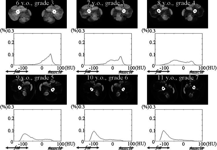
The muscle image and the CT value histogram of the thigh at each age are shown in (figure 1). The motor disability stage based on the leg grading scale of Vignos functional rating scale of the patient is also presented.
Figure 1.
CT value histograms and images of the thigh acquired over 6 years. At 6 years of age, a single peak was present within the range corresponding to muscle in the histogram, and a slightly low-intensity region reflecting fat substitution was noted in the great adductor and vastus lateralis muscle in the CT image of the thigh. However, no low-intensity region was noted in any other muscle. The muscle peak on the histogram declined while the peak in the intermediate range between the muscle and fat CT values rose with ageing, and the low-intensity region increased in the muscle bundles. Finally, the peak disappeared from the muscle CT value range, and the peak was present only in the fat CT value range. Most muscles other than the sartorius, gracilis, and rectus femoris muscles showed a low intensity. The leg grading scale of Vignos functional rating scale are also presented.
At 6 years, the histogram showed a single peak in the muscle CT value range. On muscle CT of the thigh, a region showing a slightly low intensity was present in the great adductor and vastus lateralis muscle, but no low-intensity region was noted in any other muscle. The peak muscle CT value declined with ageing, and the peak between the muscle and fat CT value ranges rose, increasing low-intensity regions in the muscle bundles. Finally, the muscle CT value peak disappeared, and only a single fat CT value peak was noted. Most muscles other than the sartorius, gracilis and rectus femoris muscles showed a low intensity.4 At the same time, motor disability aggravated with ageing. On summarising the above, the peak on the histogram shifted from the muscle to the fat CT value with the aggravation of motor disability.
Discussion
CT devices are widely used imaging systems capable of acquiring muscle images within a short time. Although there is a problem of x-ray exposure, they are applicable for children and mentally retarded patients who cannot keep still in an MR device.
The peak shifted from the muscle to the fat CT value on the histogram with the aggravation of motor disability, with which fat substitution lowering the intensity occurred on CT. Pathological fatty degeneration in the patient with Duchenne muscular dystrophy was reflected in this peak shift phenomenon.
The peak present in the intermediate range must reflect the CT value of muscle-fat mixed tissue. CT value of the tissue comprised of muscle, fat and connective tissue reflects the CT values of these three types of tissue. The fat ratio increases and the CT value decreases with disease progression, resulting in the substitution of almost all muscles for fat.
This procedure may be used as a simple method to evaluate fat substitution and motor disability of patients with Duchenne muscular dystrophy using CT.
Learning points.
A CT is capable of acquiring muscle image within a short time.
The peak on the histogram of CT value of thigh shifted from the muscle to the fat CT value with an aggravation of motor disability.
This histogram analysis may be used as a simple method to evaluate fat substitution and motor disability of patients with Duchenne muscular dystrophy using CT.
Footnotes
Contributors: All authors contributed to the study, including methods and discussion.
Competing interests: None.
Patient consent: Obtained.
Provenance and peer review: Not commissioned; externally peer reviewed.
References
- 1.Maughan RJ, Watson JS, Weir J. Strength and cross-sectional area of human skeletal muscle. J Physiol 1983;2013:37–49 [DOI] [PMC free article] [PubMed] [Google Scholar]
- 2.Liu M, Chino N, Ishihara T. Muscle damage progression in Duchenne muscular dystrophy evaluated by a new quantitative computed tomography method. Arch Phys Med Rehabil 1993;2013:507–14 [DOI] [PubMed] [Google Scholar]
- 3.Vignos PJ, Archibald KC. Maintenance of ambulation in childhood muscular dystrophy. Chronic Dis 1960;2013:273–90 [DOI] [PubMed] [Google Scholar]
- 4.Shimizu J, Matsumura K, Kawai M, et al. X-ray CT of Duchenne muscular dystrophy skeletal muscles—chronological study for five years. Rinsho Shinkeigaku 1991;2013:953–9 [PubMed] [Google Scholar]


