Summary
Hyperdontia is the condition of having supernumerary teeth, or teeth which appear in addition to the regular number of teeth. It is a developmental anomaly and has been argued to arise from multiple aetiologies. The most common site is the maxillary incisor region; but the prevalence of more than three teeth supernumerary tooth is less than 1%. A case of 13 year male patient is reported with a multiple impacted supernumerary tooth in maxillary anterior region hindering the eruption of right permanent central incisor. The supernumerary tooth was treated via surgical approach followed by an interim prosthesis for permanent central incisor which later on erupted in due course of time. Background Supernumerary teeth may be defined as any teeth or tooth substance in excess of the usual configuration of 20 deciduous and 32 permanent teeth.1 The presence of supernumerary teeth in the premaxillary region often poses unique diagnostic and managerial concerns for the practitioner. Rarely is the surplus number compensated by an absence or deficiency of other teeth. Therefore, the dysfunctional nature of supernumerary teeth and their ability to create a variety of pathological disturbances in the normal eruption and position of adjacent teeth warrants their early detection and prudent management.2
Approximately 76–86% of cases represent single-tooth hyperdontia, with two supernumerary teeth noted in 12–23% and three or more extra teeth noted in less than 1% of cases.3
Multiple supernumerary teeth are also associated with many syndromes like cleidocranial dysplasia and Gardner's syndrome etc. However, it is rare to find multiple supernumeraries in individuals with no other associated disease or syndrome. In such cases, the maxillary anterior region is the common site of occurrence.4
The exact aetiology is not clearly understood.2 The supernumerary teeth result from any disturbance in the initiation and proliferation stages of odontogenesis.5 6 There are several theories regarding the development of a supernumerary tooth—phylogenetic reversion (atavism) theory, dichotomy of tooth germ theory and hyperactivity of the dental lamina. The latter being the most accepted theory,2 states that the remnants of dental lamina or palatal offshoots of active dental lamina are induced to develop into an extra tooth bud, which results in the formation of a supernumerary tooth. Genetics is also considered to contribute to the development of supernumerary teeth, as these have been diagnosed in twins, siblings and sequential generations of a family.7
Classification of supernumerary teeth may be on the basis of position or form. Positional variations include mesiodens, paramolars, distomolars and parapremolars. Variations in form consist of conical types, tuberculate types, supplemental teeth and odontomes. Supernumerary teeth may, therefore, vary from a simple odontome, through a conical or tuberculate tooth to a supplemental tooth which closely resembles a normal tooth. Also, the site and number of supernumeraries can vary greatly.1
This report presents a case of a non-syndromic male patient with multiple supernumerary teeth and a permanent impacted tooth in the maxillary anterior region.
Case presentation
A 13-year-old man presented to the Department of Pedodontics and Preventive Dentistry with a rotated upper right front tooth. The patient had no significant medical and family history. Extra oral examination did not reveal any abnormality.
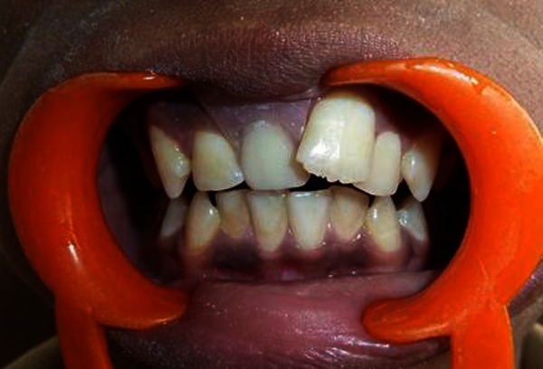
Clinical examination revealed permanent dentition, with normal alignment and occlusion except in the maxillary right anterior region, where a partially erupted, rotated tooth was present and resembled the central incisor (figure 1). All other permanent maxillary incisors were fully erupted.
Figure 1.
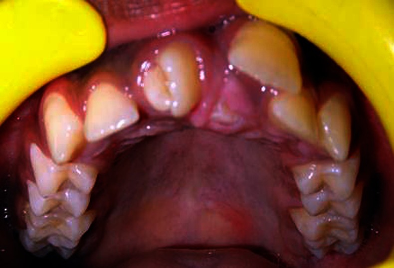
Photograph showing rotated supplemental tooth.
Investigations
Diagnostic maxillary occlusal radiograph (figure 2A) and an orthopantomograph (figure 2B), showed the presence of four supernumerary teeth which were present in the maxillary anterior region, and an impacted right permanent central incisor. Several clinical examination procedures were performed to discard the presence of any systemic pathology, and they all showed normal results.
Figure 2.
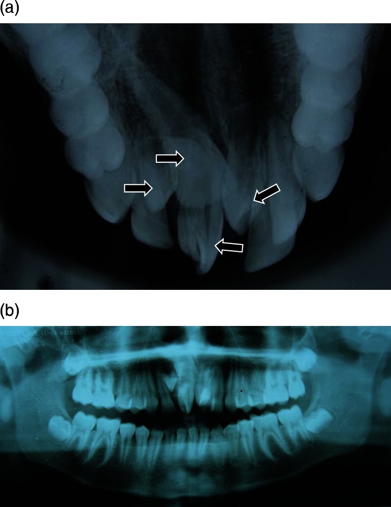
(A) Maxillary occlusal view is showing supernumerary teeth. (B) Orthopantomograph is showing supernumerary teeth.
Treatment
The devised treatment plan consisted of surgical extraction of all the four supernumerary teeth to initiate the eruption of the central incisor as the root of the impacted central incisor tooth was not fully formed.
The treatment was carried out under local anaesthesia, which involved surgical extraction of partially erupted, rotated supplemental tooth and the other two impacted teeth. Both buccal and palatal flap was raised and surgical extraction of all the four teeth was performed (figure 3). 3-0 Sutures were placed and the patient was recalled after 1 week for suture removal (figure 4).
Figure 3.
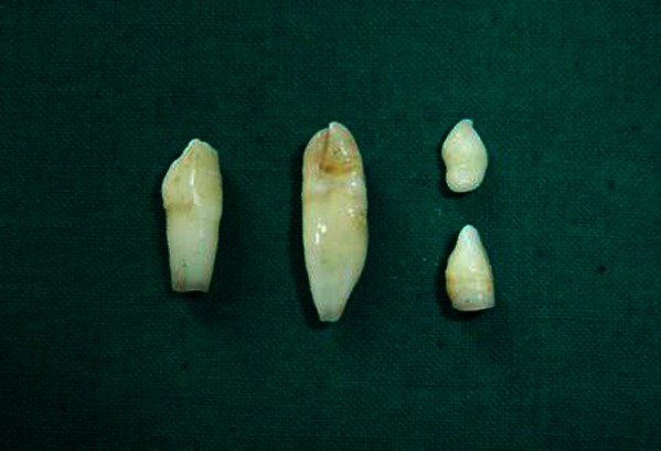
The extracted supernumerary teeth.
Figure 4.
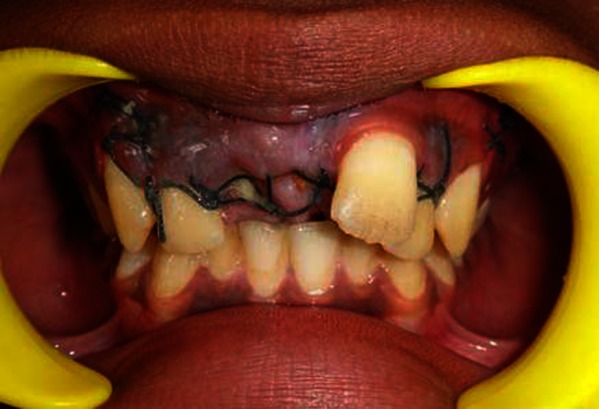
3-0 Suture placed.
Outcome and follow-up
The patient was being followed at regular intervals and the spontaneous eruption of impacted permanent maxillary right central incisor was observed and after 1 month of interval removable partial denture (figure 5) was given in respect to the maxillary right central incisor till the time central incisor erupted to the actual position.
Figure 5.
Removable partial denture given in respect to the maxillary right central incisor.
Discussion
Any delayed, ectopic or asymmetric eruption of maxillary permanent central incisors should alert the clinician to the possibility of an impacted mesiodens and requirement of careful monitoring of the case. The presence, position and relation of supernumerary teeth to the adjacent teeth, and the distance of the impacted permanent tooth from occlusal plane should be evaluated on the radiographic basis. An early recognition of the supernumerary teeth is essential for determining the appropriate treatment for each patient.7
In the present case, impaction of maxillary permanent right central incisor due to the presence of four supernumerary teeth placed right to the midline was observed in the male child who was in the permanent dentition stage with no associated craniofacial syndrome. Ersin et al8 reported mesiodens to be more in men (ie, in the ratio of 3 : 1), mean age for the diagnosis of mesiodens was 8.96 years and were more frequently located on the left side of the midline (and none at the midline). Majority of mesiodens were reported to be in a normal direction (91%), while 6% in an inverted direction and 3% in the horizontal direction.
The clinical complications of mesiodens include delayed eruption of permanent incisors, midline diastema, axial rotation or inclination of erupted permanent incisors, resorption of roots of adjacent teeth, root anomaly, cyst formation and intraoral infection.9–11 Gunduz et al9 reported the main complication as the delayed eruption of permanent incisors (38.8%), 55.2% of mesiodens were found to be in a vertical position, 37.6% in inverted position and 7% in horizontal position.
The common complications with the supernumerary tooth include impaction of adjacent teeth, crowding, diastema formation, rotation, displacement of teeth, occlusal interference, caries, periodontal problems, difficulty in mastication and compromised aesthetic. Impaction of permanent incisors due to supernumerary tooth is a rare entity encountered in the clinical practice. Similar alteration was noted in the present clinical case also, where a permanent right central incisor is found impacted due to the presence of multiple supernumerary teeth.
Two treatment options for delayed eruption of adjacent teeth due to the presence of supernumerary teeth include removal of only the supernumerary tooth if adequate space is available for the tooth to erupt or the removal of supernumerary tooth followed by a surgical-orthodontic treatment to re-establish space for the delayed tooth.12 The timing of surgical removal of supernumerary teeth is contentious and two alternatives exist. First, to remove the supernumerary tooth as soon as it has been diagnosed and second, to leave the supernumerary tooth as such till the root development of adjacent teeth is complete in order to prevent damage to their root apices. However, no evidence of root resorption, loss of vitality or any disturbance to root development has been reported by Hogstrom and Andersson.13
The exact aetiology of supernumerary teeth is still obscure although many theories have been proposed. Two popularly accepted theories are:
The dichotomy theory of tooth germs states that the tooth bud splits into two equal or different sized parts, resulting in two teeth of equal size or one normal and one dismorphic tooth, respectively. This hypothesis is supported by animal experiments in which split germs have been cultivated in vitro.
Localised and independent hyperactivity of dental lamina is the other accepted theory, which suggests supernumerary teeth are formed as a result of local, independent, conditioned hyperactivity of dental lamina.
Several researchers have also proposed that multiple supernumerary teeth are a part of postpermanent dentition. The exact mode of inheritance has not been established; however, a familial tendency has been noted.7
In non-syndromic patients, the multiple supernumerary teeth are more frequently found in the premolar region. However, that was not in accordance with the mandibular or maxillary predilection. In our findings, all the four supernumerary teeth were present in the incisal area.
In the present case, surgical extraction of supernumerary teeth was made as soon as it was diagnosed, without any damage to adjacent teeth. Removable partial denture was given with respect to the maxillary right central incisor and the patient was monitored at regular intervals for the spontaneous eruption of permanent maxillary right central incisor.
Spontaneous eruption of the delayed tooth following the removal of supernumerary tooth is reported to be in 54–75% and within 16–18 months of the removal of supernumerary tooth.14 15 Smailiene et al15 have suggested that spontaneous eruption of impacted maxillary incisor has an advantage over its surgical-orthodontic treatment.7
Research data indicate that the spontaneous eruption of impacted maxillary incisor may take up to 3 years and sometimes orthodontic treatment is necessary to achieve adequate alignment of the erupted tooth in the dental arch. If the root of the impacted tooth is still developing, the tooth may erupt normally; but, once the root apex has closed, the tooth has lost its potential to erupt. In the present case since the root was not completely formed it was desirable to wait for spontaneous eruption.16
Learning points.
The absence of any syndrome does not rule out the presence of multiple supernumeraries and this warrants the usage of more than one radiograph for a comprehensive evaluation.
Early diagnosis and proper treatment planning for such uncommon cases are necessary to avoid further complication.
Many supernumerary teeth never erupt, but they may delay eruption of nearby teeth or cause other dental problems.
Footnotes
Competing interests: None.
Patient consent: Obtained.
Provenance and peer review: Not commissioned; externally peer reviewed.
References
- 1.Scheiner MA, Sampson WJ. Supernumerary teeth: a review of the literature and four case reports. Aust Dent J 1997;2013:160–5 [DOI] [PubMed] [Google Scholar]
- 2.Primosch RE. Anterior supernumerary teeth—assessment and surgical intervention in children. Pediatr Dent 1981;2013:204–15 [PubMed] [Google Scholar]
- 3.Neville BW, Damm DD, Allen CM, et al. Oral and maxillofacial pathology. 2nd edn W.B. Saunders Company, 2002:70–71 [Google Scholar]
- 4.Gunduz K, Muglali M. Non-syndrome multiple supernumerary teeth: a case report. J Contemp Dent Pract 2007;2013:81–7 [PubMed] [Google Scholar]
- 5.Luten JR. The prevalence of supernumerary teeth in primary and mixed dentition. J Dent Child 1967;2013:346–53 [PubMed] [Google Scholar]
- 6.Bergstrom K. An orthopantomographic study of hypodontia, supernumeraries, and other anomalies in school children between the ages of 8–9 years—an epidemiological study. Swed Dent J 1977;2013:145–57 [PubMed] [Google Scholar]
- 7.Jafri SAH, Pannu PK, Galhotra V, et al. Management of an inverted impacted mesiodens, associated with a partially erupted supplemental tooth—a case report. Ind J Dent 2011;2013:40–3 [Google Scholar]
- 8.Ersin NK, Candan U, Alpoz AR, et al. Mesiodens in primary, mixed and permanent dentitions—a clinical and radiographic study. J Clin Pediatr Dent 2004;2013:295–8 [DOI] [PubMed] [Google Scholar]
- 9.Gunduz K, Celenk P, Zengin Z, et al. Mesiodens—a radiographic study in children. J Oral Sci 2008;2013:287–91 [DOI] [PubMed] [Google Scholar]
- 10.Goaz SW. Radiology principles and interpretation. St. Louis: Mosby Company, 1987 [Google Scholar]
- 11.Garvey MT, Barry HJ, Blake M. Supernumerary teeth—an overview of classification, diagnosis and management. J Can Dent Assoc 1999;2013:612–16 [PubMed] [Google Scholar]
- 12.Scheiner MA, Sampson WJ. Supernumerary teeth—a review of the literature and four case reports. Aust Dent J 1997;2013:160–5 [DOI] [PubMed] [Google Scholar]
- 13.Hogstrom A, Andersson L. Complications related to surgical removal of anterior supernumerary teeth in children. J Dent Child 1987;2013:341–3 [PubMed] [Google Scholar]
- 14.Di Biase DD. The effects of variations in tooth morphology and position on eruption. Dent Pract Dent Rec 1971;2013:95–108 [PubMed] [Google Scholar]
- 15.Smailiene D, Sidlauskas A, Bucinskiene J. Impaction of the central maxillary incisor associated with supernumerary teeth—initial position and spontaneous eruption timing. Stomatologija 2006;2013:103–7 [PubMed] [Google Scholar]
- 16.Shetty RM, Dixit U, Reddy H, et al. Impaction of the maxillary central incisor associated with supernumerary tooth: surgical and orthodontic treatment. People's J Sci Res 2011;2013:51–6 [Google Scholar]


