

Description
A 22-year-old man was presented to our orthopaedic clinic with right ankle pain after an inversion injury. On physical examination, there was no swelling and range of motion of the ankle joint was normal. The only symptom was pain on the dorsal part of calcaneocuboid joint. On anteroposterior and lateral x-rayof the right foot, a fracture at the anterior process of calcaneus was suspected. On medial oblique foot x-ray, bone fragment was seen between cuboid and calcaneus (figure 1). X-ray of the contralateral side did not show any similar finding (figure 2). For further radiological evaluation, CT of right foot was performed. On CT and three-dimensional volume rendering images, the bone fragment was found to have ovoid well corticated and blunt edges with regular shape (figures 3A, B and 4). These findings were compatible with OS calcaneus secundarius. He was treated with non-steroidal anti-inflammatory drugs and symptoms were relieved after 1 week. He was free of pain during mobilisation after 1 month.
Figure 1.
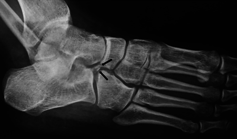
On medial oblique right foot x-ray, bone fragment (arrows) is shown between cuboid and calcaneus.
Figure 2.

On medial oblique left foot x-ray, any bone fragment (arrow) is not shown between cuboid and calcaneus.
Figure 3.

Coronal oblique (A) and sagittal (B) CT images show the os calcaneus secundarius (arrows).
Figure 4.

Three-dimensional volume rendering image shows the os calcaneus secundarius (arrows).
Os calcaneus secundarius is an accessory ossicle of the anterior facet of the calcaneus. It can easily confuse with anterior calcaneal process fracture.1 The os calcaneus secondarius can sometimes cause symptoms, like reduced motion at the subtalar joint and even chronic pain. The differential diagnosis is quite difficult with physical examination or classical x-rays.2 CT images show well corticated bony fragment with blunt edges and regular shape facilitates the diagnosis. If there is still doubt on CT images, the absence of bone marrow oedema and fracture on MRI could strengthen the diagnosis of os calcaneus secundarius.3
Learning points.
Os calcaneus secundarius is an accessory ossicle of the anterior facet of the calcaneus.
Os calcaneus secundarius can easily be confused with anterior calcaneal process fracture.
If there is doubt on CT images, the absence of bone marrow oedema and fracture on MRI could strengthen the diagnosis of os calcaneus secundarius.
Footnotes
Contributors: All authors have contributed equally in preparing the manuscript research, review, writing and all of them approved the final draft of the article.
Competing interests: None.
Patient consent: Obtained.
Provenance and peer review: Not commissioned; externally peer reviewed.
References
- 1.Mellado JM, Ramos A, Salvadó E, et al. Accessory ossicles and sesamoid bones of the ankle and foot: imaging findings, clinical significance and differential diagnosis. Eur Radiol 2003;2013:L164–77 [DOI] [PubMed] [Google Scholar]
- 2.Trnka HJ, Zettl R, Ritschl P. Fracture of the anterior superior process of the calcaneus: an often misdiagnosed fracture. Arch Orthop Trauma Surg 1998;2013:300–2 [DOI] [PubMed] [Google Scholar]
- 3.Kürklü M, Köse O, Yurttas Y, et al. Anterosuperior calcaneal process fracture or OS calcaneus secundarius? Am J Phys Med Rehabil 2010;2013:522. [DOI] [PubMed] [Google Scholar]