

Abstract
Significant numbers of patients visiting the paediatric dental clinics have aesthetically objectionable brown stains and desire treatment for them. Intrinsic tooth discolouration can be a significant aesthetic, and in some instances, functional, problem. Dental fluorosis, tetracycline staining, localised and chronological hypoplasia, and both amelogenesis and dentinogenesis imperfecta can all produce a cosmetically unsatisfactory dentition. The aetiology of intrinsic discolouration of enamel may sometimes be deduced from the patient's history, and one factor long associated with the problem has been a high level of fluoride intake. Optimal use of topical fluorides leads to a decrease in the caries prevalence but may show an increase in the prevalence of fluorosis staining because of metabolic alterations in the ameloblasts, causing a defective matrix formation and improper calcification. A 12-year-old male patient was screened at the dental clinic for routine dental care. He wanted us to remove and/or minimise the noticeable brown/yellow staining of his teeth. He requested the least invasive and most cost-effective treatment to change his smile. Various treatment modalities are present for the treatment of fluorosis stains. This report discusses the microabrasion technique in the patient having dental fluorosis.
Background
While there is a range of restorative interventions that can be used to change the appearance of fluorosed teeth, the goal of minimally invasive treatment for mild-moderate fluorosis is the one that should be evaluated first. In this presented case, a minimally invasive treatment option of microabrasion was shown to be a satisfactory approach for the aesthetic treatment of moderate fluorosis. This procedure is simple, easy to perform with no patient discomfort and removes the stains permanently.
Case presentation
A 12-year-old male patient was screened at the dental clinic for routine dental care. His wanted us to remove and/or minimise the noticeable brown/yellow staining of his teeth. He wanted the least invasive and most cost-effective treatment to change his smile. A review of his medical and dental history revealed no contraindications to dental treatment. Considering his age, the patient was not interested in treatment options that involved significant removal of tooth structure, such as porcelain or composite resin veneers which had previously been suggested to him by his previous dentist.
The patient's desire to change the appearance of his teeth in the aesthetic zone was to improve his smile and thereby his confidence. From the appearance of his teeth, a diagnosis of mild to moderate fluorosis staining (determined by using Dean's Fluorosis Index) was present on the anterior and posterior teeth in the aesthetic zone (white mottled enamel hypomineralisation), with the most significant staining occurring on the maxillary anterior teeth; teeth nos. 11 and 21 had dark brown streaks in the middle third of the facial surfaces (figure 1).
Figure 1.
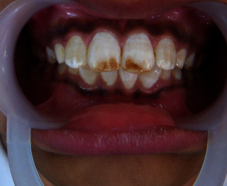
Teeth nos. 11 and 21 showing dark brown streaks in the middle third of the facial surfaces.
A review of his history and a complete dental examination revealed he was from Rajasthan, a state of India. He reported childhood friends as having the same discolouration of their teeth. Rajasthan is associated with endemic fluorosis. A treatment plan was presented to the patient that would fulfil his request for minimally invasive treatment which proposed microabrasion of the superficial enamel staining. Upon completion of treatment, the tooth shade would be evaluated.
Treatment
After a routine oral prophylaxis, the maxillary central incisors were isolated with a dental dam to protect the gingival tissues from the acidic microabrasion paste. A mixture of sodium bicarbonate and water was placed on the rubber dam behind the teeth for protection in case of spillage(figure 2).
Figure 2.
Rubber dam application to prevent spillage.
18% hydrochloric acid was mixed with pumice into slurry and a small amount was applied to the labial surface with either a slowly rotating rubber cup or rubbed over the surface for 5 s.1 It was then washed for 5 s directly by the aspirator. The procedure was repeated up to a maximum of 10×5 s applications per tooth until the stain is reduced.2 3 Any possible improvement would have occurred by this time. Topical fluoride varnish (bifluorid 12 VOCO, GmbH, Germany) was applied on the teeth for 3 min.
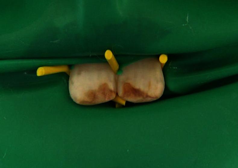
The procedure was repeated for three sittings, until a satisfactory cosmetic outcome was achieved (figure 3). In the final sitting the teeth were polished with graded Soflex discs and prophylactic polishing pastes.
Figure 3.

Satisfactory cosmetic outcome.
Outcome and follow-up
The patient was satisfied with the final aesthetic result. The patient was reviewed after 1 month for sensitivity testing.
Discussion
There is an ever-growing demand for aesthetic dental treatment. Treatment modalities include crowns, bleaching, and ceramic or composite veneers. A more conservative treatment option, enamel microabrasion, may be indicated in certain clinical scenarios.4 Enamel microabrasion was developed in the mid-1980s as a method of eliminating enamel discolouration defects and improving the appearance of teeth.5
Such kind of treatment is appropriate since it aims at the maximum preservation of the dental structures and avoids the damages that are inflicted during the operative recovery of the dental surfaces affected with fluorosis.
When hydrochloric acid is used as slurry with pumice, the abrasive property hastens the removal of the tooth structure by exposing a greater surface area of enamel. An interesting effect of enamel microabrasion has been that the appearance of some deeper enamel lesions can be improved, even if the discoloured defect is not completely removed. The reflective and refractive indices of the microabraded enamel are altered after treatment as the surfaces re-mineralise. After enamel microabrasion treatment, a superficial enamel structure may occur that has reflective and refractive properties that mask residual subjacent discolouration. Donly et al6 showed that a dense prismless layer is formed on the abraded enamel surface giving the tooth a glass-like lustre appearance.
According to data from the relevant literature, microabrasion removes an enamel layer between 100 and 200 μm.7 8
The active remineralisation using fluoride varnish ensures the resistance of the enamel layer only to a minimum degree, reduced by microabrasion. The method is not time-consuming and provides a solution for the complex situation created by dental fluorosis.
While the exact reason for the colour change that occurs after microabrasion is not known, the microabraded surface reflects and refracts light from the tooth surface in such a way that mild imperfections in the underlying enamel are camouflaged. The acid may also penetrate and bleach the organic compounds within the enamel, which explains the improvement in tooth colour. Mild surface abrasion of the enamel prisms with simultaneous acid erosion leads to compaction of the mineralised tissue within the organic region of the enamel, replacing the outer prism-free region. Light reflected off and refracted through this new surface is thought to act differently than light from an untreated enamel surface. In addition, subsurface stains may be camouflaged by the optical properties of the newly microabraded surface. Croll has named this phenomenon the ‘abrasion effect’. However, in this case it reduced the stains but did not remove them completely.
Use of microabrasion in the treatment of dental fluorosis should be performed cautiously. If after one cycle of 20 min no improvement is noted, a labial veneer restoration should be considered as an alternative treatment. Further treatment with acid can result in an unaesthetic, dished out effect or unaesthetic reduction in the mesial distal curvature of the labial surface and may also cause postoperative sensitivity.
Enamel microabrasion, however, cannot solve all tooth discolouration problems. Dentinal discolouration such as that seen with dentinogenesis imperfecta, tetracycline staining or tooth darkening associated with devitalisation or endodontic therapy cannot be affected by microabrasion; other colour correction methods are necessary in such cases. Likewise, deep enamel hypoplastic defects, once removed, leave a tooth form defect that requires replacement of lost anatomic structure with tooth-coloured restorative material. Sometimes you cannot be certain as to the depth of a lesion; therefore, it is unknown whether enamel microabrasion by itself will be the best treatment. In such cases, there is no risk to attempting microabrasion when composite resin bonding can subsequently be performed, as necessary.
Learning points.
A minimally invasive treatment option of microabrasion was shown to be a satisfactory approach for the aesthetic treatment of moderate fluorosis.
This procedure is simple, easy to perform.
No patient discomfort and removes the stains permanently.
The procedure does not require anaesthesia.
Multiple teeth can be treated at one time.
Footnotes
Competing interests: None.
Patient consent: Obtained.
Provenance and peer review: Not commissioned; externally peer reviewed.
References
- 1.Ismail AI, Hasson H. Fluoride supplements, dental caries and fluorosis: a systematic review. J Am Dent Assoc 2008;2013:1457–68 [DOI] [PubMed] [Google Scholar]
- 2.Wray A, Welbury R. Treatment of intrinsic discoloration in permanent anterior teeth in children and adolescents. http://www.rcseng.ac.uk/fds/publicationsclinicalguidelines/clinical_guidelines/documents/discolor.pdf [DOI] [PubMed]
- 3.Croll TP. Enamel microabrasion for removal of superficial dysmineralization and decalcification defects. J Am Dent Assoc 1990;2013:411–15 [DOI] [PubMed] [Google Scholar]
- 4.Lynch CD, McConnell RJ. The use of microabrasion to remove discoloured enamel: a clinical report. J Prosthet Dent 2003;2013:417–19 [DOI] [PubMed] [Google Scholar]
- 5.Croll TP. Enamel microabrasion: 10 year s experience. Asian J Aesthet Dent 1995;2013:9–15 [PubMed] [Google Scholar]
- 6.Donly KJ, O'Neill M, Croll T. Enamel microabrasion: a microscopic evaluation of the ‘abrosion effect’. Quintessence Int 1992;2013:175–9 [PubMed] [Google Scholar]
- 7.Tong LS, Pang MK, Mok NY, et al. The effects of etching, micro-abrasion, and bleaching on surface enamel. J Dent Res 1993;2013:67–71 [DOI] [PubMed] [Google Scholar]
- 8.Dalzell DP, Howes RI, Hubler PM. Microabrasion: effect of time, number of applications, and pressure on enamel loss. Pediatr Dent 1995;2013:207–11 [PubMed] [Google Scholar]