

Abstract
Sellar tumours in adults are most commonly pituitary adenomas. Primary spindle cell sarcoma of the sella turcica without a prior history of cranial radiation is extremely rare. We report a case of a large sellar mass with suprasellar and cavernous sinus extension in a geriatric male patient who presented with complete left oculomotor nerve palsy and panhypopituitarism. The patient underwent partial resection of the sellar mass through transcranial route. The pathology of the mass revealed a poorly differentiated spindle cell neoplasm most consistent with a sarcoma. Postoperatively, the size of the residual sellar mass decreased significantly following six cycles of external beam radiation in conjunction with temozolomide.
Background
Most common adult sellar tumours are pituitary adenomas; however, about 10% of adult sellar tumours are non-adenohypophysial in nature. Occurrence of primary sellar spindle cell sarcoma is extremely rare, especially in the absence of any history of radiation exposure. We report a case of undifferentiated primary sellar spindle cell sarcoma in an elderly man, which occurred without any history of radiation exposure and was successfully treated with partial surgical resection followed by external radiotherapy and adjuvant chemotherapy with temozolamide.
To the best of our knowledge, our case represents the second case in medical literature of primary undifferentiated sellar spindle cell sarcoma in an adult, which occurred without prior radiation therapy. Also, it represents the first report of successful use of adjuvant chemotherapy (temozolamide) in primary undifferentiated sellar spindle cell sarcoma.
Case presentation
An 85-year-old man presented to our hospital with gradually progressing left-sided ptosis and fatigue. He reported that his initial symptoms started 4 months ago with diplopia and drooping eyelid. The diplopia resolved 2 months ago after the development of complete ptosis. Other associated symptoms were generalised weakness and fatigue. Prior to this hospitalisation, he had seen his primary care physician for increasing fatigue and weakness. He was found to be hypotensive and bradycardic, which prompted his referral to our facility. The patient’s extensive outpatient work-up, including serological markers for myasthenia gravis, diabetes mellitus and vasculitis, in the last 4 weeks before his current admission was negative. His medical history was significant for hypertension and coronary artery disease. Family, childhood and social histories were unremarkable. Physical examination revealed hypotension, complete left-sided ptosis, mydriasis and absent medial left eye movement, consistent with left oculomotor nerve palsy (figures 1A–1B).
Figure 1.

(A) Figure reveals complete ptosis of the left eye. (B) The left eye has mydriasis and fixed ‘downward and outward gaze’, consistent with dense left oculomotor palsy. (C). Contrast-enhanced T1 coronal section of MRI of the brain showing a large bilobed left sided, sellar mass with cavernous sinus extension. (D and E) Haematoxylin and eosin stained sellar tissue sections on high and low magnifications respectively. Spindle cells with fusiform nuclei with finely granular chromatin and insignificant nucleoli reveal characteristics of spindle cell sarcoma.
Investigations
The routine laboratory work-up including the haemogram and the basic metabolic panel were normal. The results of hormonal work-up were consistent with central hypothyroidism, central adrenal insufficiency with modestly elevated prolactin (table 1).
Table 1.
Results of the hormonal laboratory work-up
| Test | Patient's value | Reference range for our hospital |
|---|---|---|
| Free T4 | 0.4 ng/dl | 0.7–1.7 ng/dl |
| Thyroid stimulating hormone | 1.04 U/l | 0.45–4.5 U/l |
| Cortisol morning | <0.5 µg/dl | 2.3–19.4 µg/dl |
| Luteinizing hormone | 0.7 mIU/ml | 3.0–34.6 mIU/ml |
| Follicular stimulating hormone | 2.2 mIU/ml | 0.2–18.1 mIU/ml |
| Prolactin | 60 ng/ml | 5–20 ng/ml |
| Adrenocorticotropic hormone | 3.3 pg/ml | 7.2–63.3 pg/ml |
MRI of the brain revealed a large bilobed left sided, sellar mass (measuring 2.1×2.8 ×1.8 cm) with suprasellar and cavernous sinus extension (figure 1C).
Differential diagnosis
Pituitary adenoma
Sarcoma (fibrosarcoma, osteosarcoma, rhabdomyosarcoma, leiomyosarcoma or undifferentiated sarcoma)
Meningioma
Germ cell tumor
Metastases
Craniopharyngioma
Treatment
Treatment with levothyroxine and hydrocortisone resulted in a remarkable improvement in clinical symptoms of fatigue and weakness. He was subsequently referred to an outside facility for surgical resection of sellar tumour in the light of hypopituitarism and cranial nerve palsy. The patient underwent uncomplicated partial resection of the tumour through the transcranial route after initial attempt through the transsphenoidal route was unsuccessful.
Outcome and follow-up
Histopathology was consistent with a poorly differentiated malignant spindle cell neoplasm, likely consistent with a sarcoma of unknown origin (figures 1D–1E). Immunohistochemistry was positive for BCL-2 but demonstrated a negative staining profile with glial fibrillar acidic protein, oligodendrocyte transcription factor 2, desmin, chromogranin, smooth muscle actin, CD34, pan-keratin, S100, cytokeratin CAM5.2, leucocyte common antigen and epithelial membrane protein. Karyotyping performed at a level of 400 bands or greater, did not demonstrate any cytogenetic aberrations (46 XY on 13 metaphases). Positron emission tomography (PET) scan revealed no evidence of metastases or any other primary tumour. The patient subsequently underwent six cycles of external beam radiation treatment and chemotherapy using timozolomide. There was a significant decrease in the size of the residual tumour after 2 months, as seen on the follow-up MRI (from 2.3 cm in its largest linear dimension to 1.6 cm in the largest linear dimension). After 6 months following his surgery and chemo-radiotherapy, he is doing really well clinically and biochemically on maintenance doses of hydrocortisone and levothyroxine. His vision and ptosis also have shown slow improvement during the follow-up period (figure 2).
Figure 2.
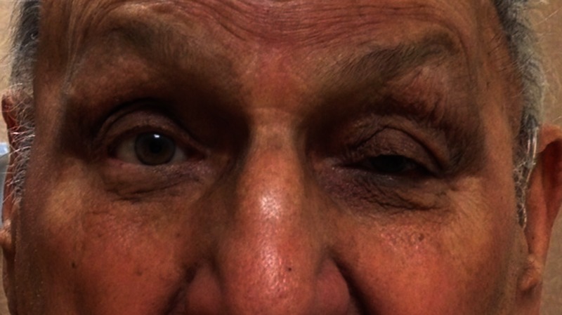
Figure represents a patient follow-up image 6 months after postoperative surgical resection and radiotherapy. The patient is currently on temozolamide. There is significant improvement in his visual symptoms. Left eye ptosis has partially resolved.
Discussion
Our patient presented with a large sellar mass with compressive symptoms and panhypopituitarism. The pathology revealed primary spindle cell neoplasm of the sella, most likely poorly differentiated sarcoma. Although, pituitary adenomas are the most common tumours of the sellar–suprasellar region, nearly 10% are non-adenohypophysial in nature.1 Most well-recognised primary sarcomas of this region include fibrosarcoma, which almost always occur after radiotherapy for pituitary adenoma.1 2 Benign spindle cell lesions of the sella previously well described in literature include meningioma, schwannoma, solitary fibrous tumour, myofibroblastoma, pilocytic astrocytoma, pituitary adenoma with spindle cell features, and pituicytoma.3–5 These are readily distinguished from the obviously anaplastic tumours such as the one in our patient. Malignant mesenchymal lesions include a typically post-irradiation lesion: haemangiopericytoma, fibrosarcoma, osteosarcoma, chondrosarcoma, rare types of synovial sarcomas, leiomyosarcoma and rhabdomyosarcoma.2 6–11 Rhabdomyosarcomas comprise 19% of all sarcomas, and in fact among children, they represent 60% of soft tissue sarcomas, making them the most common sarcoma in this age group.12 13 Only two cases of primary intrasellar rhabdomyosarcoma have been reported so far.11 14
Occurrence of primary spindle cell sarcoma of the sella turcica in the absence of history of radiation exposure is a rarity.2 Our extensive review of the Pubmed and Medline literatures have revealed only three case reports to date of primary intrasellar spindle cell sarcomas without prior radiation exposure.2 15 16 Two of these reports identified the primary sellar tumour as fibrosarcoma,2 15 and one report was of an undifferentiated sarcoma.16 That one case represented the first sellar example of an undifferentiated spindle cell sarcoma in medical literature, which showed desmin reactivity alone, without other immunohistochemical or ultrastructural features of rhabdomyosarcoma.16 Our case exhibited histopathological features of a poorly differentiated spindle cell neoplasm which showed largely non-reactive immunohistochemistry profile except BCL-2 positivity, suggesting an undifferentiated sarcoma possibly of mesenchymal origin. To the best of our knowledge, our case represents the second reporting of a sellar undifferentiated spindle cell sarcoma. Interestingly, in the previous report of undifferentiated sellar spindle cell sarcoma, a consideration was that the tumour in some way may have been related to the patient’s previous testicular seminoma, either as a metastasis or as a separate de novo lesion.16 Our case represented a true form of primary intrasellar undifferentiated tumour, as the patient had no history of malignancy and his PET scan did not show any evidence of other tumours. Neoplasms lacking cellular differentiation not only pose a diagnostic challenge to clinicians and histopathologists, but also a management dilemma. Their pathological evaluation focuses upon identifying the basic tumoral category to which they belong. Indeed, for therapeutic purposes, it is essential to further define these tumours as being epithelial, mesenchymal, melanocytic or haematopoietic. Given the lack of significant literature regarding undifferentiated sellar sarcoma, the treatment opportunities may pose a dilemma. Previously, one report of primary sellar fibrosarcoma has reported the use of subtotal surgical resection followed by gamma knife surgery (GKS) for residual disease2 and the second report described the use of GKS followed by postoperative external beam radiotherapy.15 The first described patient developed a disease recurrence after 4 months2; however, the second patient did well after 2 years of follow-up without recurrence.15
In general, management of sarcomas consists of resection followed by adjuvant therapy in high-risk cases. Residual disease and local tumour recurrence is common in patients with sellar tumours, and postoperative external beam radiotherapy is often administered; however, the dose required to control sarcomas is higher than tolerated by neighbouring normal structures. The combination of GKS and postoperative external beam radiotherapy can permit an increase in the dose prescribed for residual tumours while minimising the dose received by the optic chiasm. In both the cases of primary sellar fibrosarcoma, chemotherapy was not used as an adjuvant, as its role for these tumours was not well-defined.2 15 In the case of undifferentiated sellar sarcoma previously described, the patient underwent subtotal tumour resection and radiotherapy and was planned for chemotherapy (mesna, adriamycin, ifosfamide); however, post-therapy follow-up was not provided16 The patient in our case, who had undifferentiated sellar sarcoma, was treated with subtotal resection, postoperative radiotherapy and chemotherapy with temozolomide. To the best of our knowledge, this is the second report of primary sellar undifferentiated spindle cell sarcoma and the first report of the use of temozolomide in this setting. Our patient showed remarkable improvement with the use of this treatment regimen and residual tumour regressed significantly over a few weeks. This may suggest the need for exploration of the potential role of chemotherapy in these rare sellar tumours, which is currently not well-defined. As an example, currently, all patients with rhabdomyosarcoma are presumed to have micrometastatic disease at presentation, thus the rationale for multimodality therapy, that is, attempted complete resection and/or local irradiation coupled with chemotherapy; while about five decades ago, only surgery was considered to be the primary treatment for such tumours and chemotherapy used to be reserved for distant metastases.17 18
Learning points.
Our case represents one of rarest examples of a primary sellar undifferentiated sarcoma which lacked response to most immunomarkers. The case also demonstrates the difficulties inherent in the differential diagnosis of undifferentiated sellar neoplasms.
The role of adjuvant chemotherapy is not clearly defined in these tumour types because of their rare occurrence and lack of prior significant experimental evidence, but successful treatment in our case was achieved with partial surgical resection and adjuvant radiotherapy and chemotherapy (temozolomide).
This suggests a potential emerging role of adjuvant chemotherapeutic agents like temozolomide in the treatment of undifferentiated sellar tumours.
Acknowledgments
All authors are thankful to Dr Thomas Ukena (Head, Department of Pathology at Saint Vincent Hospital) for his help in the preparation of stained histopathology slides for this manuscript.
Footnotes
Competing interests: None.
Patient consent: Obtained.
Provenance and peer review: Not commissioned; externally peer reviewed.
References
- 1.Huang BY, Castillo M. Nonadenomatous tumors of the pituitary and sella turcica. Top Magn Reson Imaging 2005;2013:289–99 [DOI] [PubMed] [Google Scholar]
- 2.Lopes MB, Lanzino G, Cloft HJ, et al. Primary fibrosarcoma of the sella unrelated to previous radiation therapy. Mod Pathol 1998;2013:579–84 [PubMed] [Google Scholar]
- 3.Schultz AB, Brat DJ, Oyesiku NM, et al. Intrasellar pituicytoma in a patient with other endocrine neoplasms. Arch Pathol Lab Med 2001;2013:527–30 [DOI] [PubMed] [Google Scholar]
- 4.Carneiro SS, Scheithauer BW, Nascimento AG, et al. Solitary fibrous tumor of the meninges: a lesion distinct from fibrous meningioma. A clinicopathologic and immunohistochemical study. Am J Clin Pathol 1996;2013:217–24 [DOI] [PubMed] [Google Scholar]
- 5.Shinojima N, Ohta K, Yano S, et al. Myofibroblastoma in the suprasellar region. Case report. J Neurosurg 2002;2013:1203–7 [DOI] [PubMed] [Google Scholar]
- 6.Mena H, Ribas JL, Pezeshkpour GH, et al. Hemangiopericytoma of the central nervous system: a review of 94 cases. Hum Pathol 1991;2013:84–91 [DOI] [PubMed] [Google Scholar]
- 7.Allan CA, Kaltsas G, Evanson J, et al. Pituitary chondrosarcoma: an unusual cause of a sellar mass presenting as a pituitary adenoma. J Clin Endocrinol Metab 2001;2013:386–91 [DOI] [PubMed] [Google Scholar]
- 8.Ashkan K, Pollock J, D'Arrigo C, et al. Intracranial osteosarcomas: report of four cases and review of the literature. J Neurooncol 1998;2013:87–96 [DOI] [PubMed] [Google Scholar]
- 9.Scheithauer BW, Silva AI, Kattner K, et al. Synovial sarcoma of the sellar region. Neuro Oncol 2007;2013:454–9 [DOI] [PMC free article] [PubMed] [Google Scholar]
- 10.Anderson WR, Cameron JD, Tsai SH. Primary intracranial leiomyosarcoma. Case report with ultrastructural study. J Neurosurg 1980;2013:401–5 [DOI] [PubMed] [Google Scholar]
- 11.Arita K, Sugiyama K, Tominaga A, et al. Intrasellar rhabdomyosarcoma: case report. Neurosurgery 2001;2013:677–80 [DOI] [PubMed] [Google Scholar]
- 12.Stuart A, Radhakrishnan J. Rhabdomyosarcoma. Indian J Pediatr 2004;2013:331–7 [DOI] [PubMed] [Google Scholar]
- 13.Dagher R, Helman L. Rhabdomyosarcoma: an overview. Oncologist 1999;2013:34–44 [PubMed] [Google Scholar]
- 14.Zhong J, Li ST, Yao XH, et al. An intrasellar rhabdomyosarcoma misdiagnosed as pituitary adenoma. Surg Neurol 2007;2013(Suppl 2):S29–33; discussion S33 [DOI] [PubMed] [Google Scholar]
- 15.Alpert TE, Hahn SS, Chung CT, et al. Successful treatment of spindle cell sarcoma of the sella turcica. Case report. J Neurosurg 2002;2013(5 Suppl):438–40 [DOI] [PubMed] [Google Scholar]
- 16.Manoranjan B, Syro LV, Scheithauer BW, et al. Undifferentiated sarcoma of the sellar region. Endocr Pathol 2011;2013:159–64 [DOI] [PubMed] [Google Scholar]
- 17.Breitfeld PP, Meyer WH. Rhabdomyosarcoma: new windows of opportunity. Oncologist 2005;2013:518–27 [DOI] [PubMed] [Google Scholar]
- 18.Crist WM, Anderson JR, Meza JL, et al. Intergroup rhabdomyosarcoma study-IV: results for patients with nonmetastatic disease. J Clin Oncol 2001;2013:3091–102 [DOI] [PubMed] [Google Scholar]