

Abstract
Subcutaneous emphysema occurs when air is forced beneath the tissue, leading to swelling, crepitus on palpation and has the potential to spread along the fascial planes. This report describes the youngest case of subcutaneous emphysema related to dental treatment that has been documented to date. In addition to the patient's age, the case is of interest because periorbital subcutaneous emphysema is a rarest complication of stainless steel crown procedure.
Background
Subcutaneous emphysema is a rare occurrence in the dental setting. When it does occur, the entity may be mistaken for an anaphylactic reaction to a local anaesthetic agent or other medications used in dental surgery.1 Emphysema is a swelling due to the presence of air or gas in the interstices of the connective tissue. Various other terms have been used such as subcutaneous emphysema,1 surgical emphysema,2 interstitial emphysema3 and barotraumas4 for this condition.
Quite a few cases have been reported during routine dental examination. According to a review by Heyman and Babyof,5 a retrospective study published in 1995 reviewed 74 reports of subcutaneous emphysema between the years 1960 and 1993, whereas a comprehensive search from 1993 to 20086 reviewed yielded 32 case reports of subcutaneous emphysema. The most frequently reported cases are those occurring during or after oral surgery, and to a lesser degree during or after endodontics, periodontics and operative dentistry. This report describes the youngest case of subcutaneous emphysema related to stainless steel crown that has been documented to date. Also, there appears to be no evidence in the literature till date having periorbital oedema with dental treatment in a young patient, only one case of periorbital oedema complication is reported in an adult.7 Emphysemas originating from the oral cavity are most likely caused by a positive pressure produced by the patient himself or by compressed air from the dental unit.
Case presentation
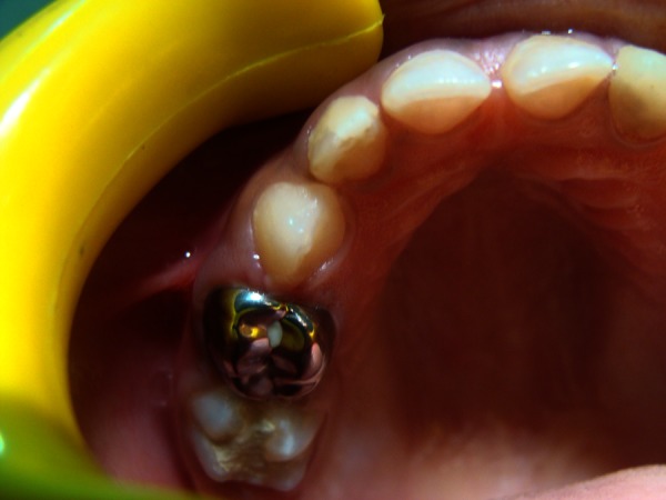
A healthy 4.5-year-old girl was seen for the placement of a stainless steel crown for a multisurface carious tooth of the right maxillary first molar. The patient was placed in a recumbent position, and crown preparation was initiated using a high speed hand-piece with an air-water spray. Intermittent spurts of air from the air syringe were also used during the procedure (figure 1). After completion of the procedure, the patient was discharged. She returned an hour later with an inability to open her left eye due to severe periorbital oedema and surgical emphysema of right side. A distinct swelling of the right periorbital area was evident (figures 2 and 3). Examination of the area revealed a painless swelling which, upon palpation, elicited a definite crepitus. The patient was assured that the swelling would resolve itself in several days. She was placed on prophylactic amoxicillin. After 48 h, the facial swelling had reduced slightly. Her recovery was uncomplicated and was complete after 5 days (figure 4).
Figure 1.

Photograph of intraoral region where stainless steel crown was delivered.
Figure 2.

Photograph showing a distinct swelling of the right periorbital area.
Figure 3.
Photograph showing a distinct swelling of the right periorbital area.
Figure 4.

Photograph after recovery.
Discussion
Emphysema is an infrequent complication in the dental practice; however, it is necessary to know and realise the clinical differential diagnosis with other diseases that produce increased volume in the affected areas8; among this includes haematoma, allergic reaction, angioedema, anaphylactic reaction or cellulitis.2 Emphysema is differentiated from other similar appearing swellings mainly by its fast onset, presence of crepitus or a ‘Velcro’ sensation, which provides an important element of differentiation9; radiography of an affected area can make an accurate diagnosis by observing the presence of air vacuoles.10 In our case, the swelling was sudden in onset, there was a presence of crepitus; though radiographs were not taken, emphysema was corroborated clinically.
The increased air pressures from high-speed hand-pieces and air syringes in use today would seem to augment the possibility of emphysema occurring during restorative procedures.11 The use of an air syringe, high speed hand-pieces or their combination was reported in 71% of emphysema cases.12 In this case, though stainless steel crown preparation was performed, the process did not need extensive preparation. It is speculated that much of the damage was done during the proximal preparation.
Emphysematic condition is rarely serious, it might be expected that bacteria would be forced into the tissues from the oral cavity. Antibiotic administration is controversial; however, it seems prudent to give these patients an anti-infective agent that covers Staphylococcus sp., Streptococcus sp. and anaerobes that may be transmitted from the oral cavity.13
The treatment is usually symptomatic only, and has good resolution; it requires clinical follow-up daily and its regression usually occurs without complications. But sometimes depending on the anatomical location and volume of air injected, the deep fascial spaces can be compromised to and progress to life-threatening infections or cardio-respiratory complications.14 Complications may rarely include cardiac tempanade, airway compromise or air embolism.15
Adequate knowledge of all aspects associated with subcutaneous emphysema is essential for the dentist to prevent its occurrence and adequate treatment.16 To visualise the area around the gingival margin, one should be careful to blow air gently towards the occlusal to drive fluid away and to dry the sulcus, use sideways blow and make use of cotton rolls and other absorbents. Avoid directing air towards the sulcus as far as possible. Care should be taken when using air-driven hand-pieces.
Subcutaneous emphysema is a potential complication of procedures that interrupt the epithelium of the oral cavity and introduces air, under pressure, along or into the fascial spaces of the head and neck. Although the frequency is rare, the iatrogenic subcutaneous emphysema can have serious and potentially life-threatening effects. When subcutaneous emphysema does arise, it must be quickly diagnosed, understood and effectively managed to reduce the incidence of further complications.
Learning points.
This paper presents a case of emphysema in a 4.5-year-old child. An uncommon complication that can occur during routine restorative procedure when compressed air is used to operative air turbine hand-pieces and discusses the management technique.
Simple palpation of the swollen facial areas is helpful in making the correct diagnosis.
Adequate knowledge of all aspects associated with subcutaneous emphysema is essential for the dentist to prevent its occurrence and adequate treatment.
Footnotes
Competing interests: None.
Patient consent: Obtained.
Provenance and peer review: Not commissioned; externally peer reviewed.
References
- 1.Steelman RJ, Johannes PW. Subcutaneous emphysema during restorative dentistry. Int J Paediatr Dent 2007;2013:228–9 [DOI] [PubMed] [Google Scholar]
- 2.Ali A, Cunliffe DR, Watt-Smith SR. Surgical emphysema and pneumomediastinum complicating dental extraction. Br Dent J 2000;2013:589–90 [DOI] [PubMed] [Google Scholar]
- 3.Childers JM, Traeger KA. Interstitial emphysema: an insidious complication of operative dentistry. Oper Dent 1982;2013:55–7 [PubMed] [Google Scholar]
- 4.Goorhuis H, Rothrock SG. Cervicofacial and thoracic barotrauma following a minor dental procedure. Pediatr Emerg Care 1993;2013:29–32 [DOI] [PubMed] [Google Scholar]
- 5.Heyman SN, Babyof I. Emphysematous complications in dentistry,1960–1993: an illustrative case and review of the literature. Quintessence Int 1995;2013:535. [PubMed] [Google Scholar]
- 6.Parkar A, Medhurst C, Irbash M, et al. Periorbital oedema and surgical emphysema, an unusual complication of a dental procedure: a case report. Cases J 2009;2013:8108. [DOI] [PMC free article] [PubMed] [Google Scholar]
- 7.McKenzie WS, Rosenberg M. Iatrogenic subcutaneous emphysema of dental and surgical origin: a literature review. J Oral Maxillofac Surg 2009;2013:1265–8 [DOI] [PubMed] [Google Scholar]
- 8.Bach CA, Derbez R, Baujat B, et al. Subcutaneous cervicofacial and mediastinal emphysema complicating tooth extraction: case report. Rev Stomatol Chir Maxillofac 2005:2013:130–2 [DOI] [PubMed] [Google Scholar]
- 9.Spaulding CR. Soft tissue emphysema. J Am Dent Assoc 1979;2013:587. [DOI] [PubMed] [Google Scholar]
- 10.Vargas V, Heras M, Torres D, et al. El enfisema como complicación en odontología. Revista SECIB On Line 2007;2013:1–4 http/www.secibonline.com/web/pdf/vol3_2007_articulo_actualizacion.pdf [Google Scholar]
- 11.CPT Stanley F, Koss DC. Subcutaneous emphysema of the infraorbital region after a dental restorative procedure . Med Bull US Army Europe 1970;2013:276–7 [Google Scholar]
- 12.Heyman SN, Babayof I. Emphysematous complications in dentistry 1960–1993: an illustrative case and review of the literature. Quintessence Int 1995;2013:535–43 [PubMed] [Google Scholar]
- 13.Torgay A, Aydin E, Celasun U, et al. Subcutaneous emphysema after dental treatment: a case report. Pediatr Anaesth 2006;2013:314–17 [DOI] [PubMed] [Google Scholar]
- 14.Guimarães BR, Moraes RB. Rubens Camino Júnior, João Gualberto Cerqueira Luz Subcutaneous emphysema during removal of third molars—aspects of interest to the dental surgeon. RFO Passo Fundo 2010;2013:165–70 [Google Scholar]
- 15.Sood T, Pullinger R. Pneumomediastinum secondary to dental extraction. Emerg Med J 2001;2013:517–18 [DOI] [PMC free article] [PubMed] [Google Scholar]
- 16.Wright KJ, Derkson GD, Riding KH. Tissue space emphysema, tissue necrosis, and infection following use of compressed air during pulp therapy: case report. Pediatr Dent 1991;2013:110–13 [PubMed] [Google Scholar]