Abstract
Over the last decade, several adult stem cell populations have been identified in human skin 1-4. The isolation of multipotent adult dermal precursors was first reported by Miller F. D laboratory 5, 6. These early studies described a multipotent precursor cell population from adult mammalian dermis 5. These cells--termed SKPs, for skin-derived precursors-- were isolated and expanded from rodent and human skin and differentiated into both neural and mesodermal progeny, including cell types never found in skin, such as neurons 5. Immunocytochemical studies on cultured SKPs revealed that cells expressed vimentin and nestin, an intermediate filament protein expressed in neural and skeletal muscle precursors, in addition to fibronectin and multipotent stem cell markers 6. Until now, the adult stem cells population SKPs have been isolated from freshly collected mammalian skin biopsies.
Recently, we have established and reported that a population of skin derived precursor cells could remain present in primary fibroblast cultures established from skin biopsies 7. The assumption that a few somatic stem cells might reside in primary fibroblast cultures at early population doublings was based upon the following observations: (1) SKPs and primary fibroblast cultures are derived from the dermis, and therefore a small number of SKP cells could remain present in primary dermal fibroblast cultures and (2) primary fibroblast cultures grown from frozen aliquots that have been subjected to unfavorable temperature during storage or transfer contained a small number of cells that remained viable 7. These rare cells were able to expand and could be passaged several times. This observation suggested that a small number of cells with high proliferation potency and resistance to stress were present in human fibroblast cultures 7.
We took advantage of these findings to establish a protocol for rapid isolation of adult stem cells from primary fibroblast cultures that are readily available from tissue banks around the world (Figure 1). This method has important significance as it allows the isolation of precursor cells when skin samples are not accessible while fibroblast cultures may be available from tissue banks, thus, opening new opportunities to dissect the molecular mechanisms underlying rare genetic diseases as well as modeling diseases in a dish.
Keywords: Stem Cell Biology, Issue 75, Cellular Biology, Molecular Biology, Anatomy, Physiology, Biomedical Engineering, Medicine, Dermatology, Cells, Cultured, Stem Cells, biology (general), Skin and Connective Tissue Diseases, Biological Phenomena, Adult stem cells, skin derived precursor cells, fibroblasts, sphere culture, skin-derived precursors, SKP, PCR, qPCR, immunocytochemistry, isolation, cell culture
Protocol
1. SKP Isolation from Primary Fibroblast Cultures
Fibroblast cultures either from cell banks or directly obtained from skin biopsies are maintained in culture in fibroblast growth medium DMEM containing 15% fetal calf serum, 2 mM glutamine, 10 mg/ml penicillin, and 10 mg/ml streptomycin.
Human fibroblasts GMO3349C and GMO8398A were obtained from the Coriell Institute for Medical Research (Camden, NJ) and were used in this study.
Cultures from population doublings (PPDs) 20 to 35 were used for SKP cultures at a confluency of 80%. One 10 cm tissue culture dish (BD Falcon) contains approximately 1.5x10⁶ cells.
Wash cells with PBS and incubate with 2 ml trypsin solution (0.25%, Invitrogen) for 1 hr at 37 °C.
Collect cells from the plate with 5 ml PBS and transfer the cell suspension to a 15 ml falcon tube.
Incubate the cells at 4 °C for 24 hr.
Prepare SKP growth medium consisting of DMEM-F12, 3:1 (V/V) and 40 ng/ml FGF2 (BD Biosciences, 20 ng/ml EGF (BD Biosciences), B27 serum free supplement 2% (Invitrogen), 1 μg/ml Fungizone (Invitrogen) and 25 μm/ml Gentamycin (Invitrogen).
Pellet the cells at 1,200 rpm for 5 min and resuspend the cell pellet directly in 4 ml SKP growth medium. Transfer the cell suspension to a 25 cm2 tissue culture flask (BD Falcon).
Incubate the flask at 37 °C and monitor the culture for sphere formation daily for 3 to 4 weeks.
2. Sphere Culture Conditions
Culture cells in a 25 cm2 flask (BD Falcon) at 37 °C, 5% CO2.
Shake the flask vigorously daily to avoid cells to adhere to the bottom of the flask. If necessary, pipette up and down with a 2 ml sterile pipette to detach adherent cells and transfer the culture to a new flask.
First spheres start to build within 3 to 4 days.
Let spheres sediment to the bottom of the flask and change half of the medium every 3 days. To keep the same final concentration of growth factors add 2X growth factors to the freshly prepared SKP growth medium.
Keep the volume constant maximum 4 ml.
When spheres reach a size of ~ 200 mm they should be passaged by breaking down the spheres to smaller size by vigorous pipetting up and down with a 2 ml pipette.
The culture is split into two 25 cm2 flasks (BD Falcon) and spheres cultures are feed as described in step 2.4).
Spheres are collected for stem cell markers screening by day 16 to 21.
The procedure from 2.6) and 2.7) describe the propagation of the spheres to (1) allows nutriments and growth factors included in SKP medium to access all cells within each sphere and (2) to expand the SKP sphere culture prior use.
Typically Sphere cultures under our culture conditions maintain the capacity to grow over a period of three months and are passaged between 3 to 4 times.
3. Immunocytochemistry for Stem Cell Markers
Sphere cultures are harvested in PBS by day 16 to 21. Cultures are pelleted at 1,000 rpm for 5 min.
Spheres are resuspended in a small volume of PBS.
Draw two circles with a DAKO pen of approximately 0.5 μm in diameter on microscope slides (Fisher brand).
Add 50 μl of sphere suspension into the circles.
Check by light microscopy for the presence of spheres in each drop.
Let the drops dry under the hood.
Either freeze the slides at -80 °C or proceed with the immunofluoresence staining protocol.
For immunofluorescence staining, fix the slides with pre-cooled Methanol (100%) at -20 °C for 10 min.
Wash the slides with PBS.
Block the slides with PBS/10% Fetal bovine serum / 0.02% Triton X100 for at least 1 hr.
Incubate with primary Antibody (e.g. anti-Nestin, anti-Oct4, anti-TG30, anti-Nanog antibodies, Table 3) diluted in blocking buffer: PBS/10% FBS for 2 hr at room temperature or overnight at 4 °C.
Wash 3 times with blocking buffer and incubate with secondary antibody for 1 hr at room temperature.
Wash 3 times with blocking buffer and 3 times with PBS.
Mount slides with Vectashield mounting medium (Vector Inc.).
Analyze samples for expression of stem cell markers by immunofluorescence microscopy.
4. Realtime PCR Analysis of Expression Levels of Stem Cell Markers
Take approximately 2-3 ml Sphere cultures from day 18 to 21.
Pellet the spheres at 1,200 rpm for 5 min and aspirate all medium.
Isolate RNA from the spheres using the RNeasy Minikit (Qiagen, Valencia, CA).
Assess RNA purity by spectrometry and agarose gel.
Synthesize cDNA (Omniscript Reverse Transcriptase (Qiagen)) using the total cellular RNA as template.
Validate the stem cell markers by Realtime-PCR using primers for stem cell markers (shown in Table 1) in a Power SYBR Green PCR Mastermix (Applied Biosystems) with a concentration of 375 nM of each primer and 50 ng of template in a 20 μl reaction volume. GAPDH is used as endogenous control.
For the amplification use an initial denaturation at 95 °C for 2.5 min followed by 40 cycles at 95 °C for 5 sec and 60 °C for 20 sec.
Analyze the run with the 2(ΔΔCt) method 8.
5. Directed Differentiation into Smooth Muscle Cells
Spheres from day 21 to 26 were plated into 6 well culture dishes (BD Falcon) in SKP medium.
Cells were allowed to adhere and outgrow from the spheres in SKP medium for 72 hr.
Smooth muscle cells (SMCs) differentiation started initiated by replaced the medium with SMC differentiation medium consisting of high-glucose Dulbecco's modified Eagle medium (Invitrogen) containing 5% FBS, 5 ng/ml PDGF-BB (Invitrogen), and 2.5 ng/ml TGF-b1 (Invitrogen).
Medium was changed every 3 days with freshly prepared SMC differentiation medium for a period of 3 to 4 weeks in cultures.
Screening for SMC markers was performed after 3 to 4 weeks by immunohistochemistry.
Cells were fixed with 4% paraformaldehyde solution in PBS for 10 min.
Cells were permeabilized with PBS containing 0.3% Triton X-100 for 30 min.
Cells were incubated with antibodies against αSMA (MO851, Dako, 1:100), or calponin (M3556, Dako, 1:100) for 1 hr at room temperature and further processed as described in 3.12) to 3.15).
Representative Results
We show that a population of cells that selectively expand to generate SKP spheres under controlled growth condition consisting of EGF and FGF2 are present in primary dermal fibroblast cultures (Figure 1) as we reported recently 7.
Fibroblast cultures from PPDs 15 to 25 that typically correspond to the primary fibroblasts strains available from cell banks were used in this study. Fibroblast cultures submitted to the double treatment consisting of cold temperature treatment together with nutriment depletion for 24 hr described in this method reproducibly generated cultures containing floating spheres (also referred as embryoid body EB) sharing similar growth characteristics as the one described previously for SKPs derived directly from skin samples 7, 9.
Here we show that using the method that we recently reported 7, we isolate a population of cells from primary fibroblast cultures that exhibit similar properties to the multipotent neural stem/precursor cells, isolated and identified in human and mouse dermis using neurosphere culture conditions 5, 10, 11. These SKP-derived from primary human fibroblast cultures show self-renewing potency as they can be expanded and passaged for at least a period of three months in vitro (please refer to method section). When spheres reached a size of 150 to 200 μM in diameter after 18 to 21 days in culture, they were mechanically broken down into an average of 50 μM in diameter and allowed to re-expand in suspension culture in SKP medium. These broken down spheres were allowed to grow until they reached 200 μM prior being passaged again as described in Method section. SKP spheres could be passaged at least three times. This observation indicates that cells within the spheres were capable to self renew and expand under neurosphere culture conditions as others and we reported 5, 7.
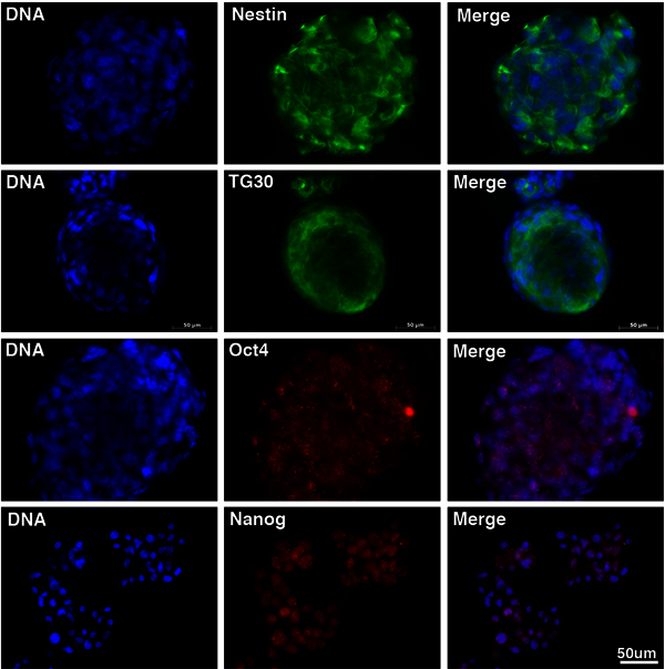
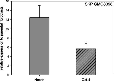
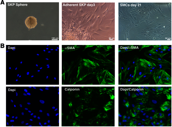
Moreover, these SKP cells derived from fibroblast cultures expressed neural crest and neuron stem cell marker nestin as well as the embryonic stem cell transcription factors Nanog and Oct4 and the pluripotent cell surface marker TG30 (Figure 2), similarly to the SKP derived directly from skin biopsies 5, 7, 10. Immunohistochemistry on SKP spheres indicated nestin positive signal in the cytoplasm, Oct 4 exhibited a punctuated nuclear signal and Nanog showed a more diffused nuclear signal (Figure 2). Additionally, the cell surface marker of pluripotent human embryonic stem cells TG30 gave a positive cytoplasmic membrane staining on SKP sphere preparations (Figure 2). Using real time PCR, we also confirmed the presence of mRNAs encoding nestin and Oct4 in RNA preparation isolated from SKP spheres collected by day 18 in culture (Figure 3). These findings indicate that cells within these SKP spheres express multipotent stem cell markers as previously reported for SKPs derived from mouse and human skin biopsies 5,10. To further test the multipotency of the SKP derived from fibroblast cultures, we induced these cells to differentiate into smooth muscle cells (Figure 4). After 3 weeks induction in medium containing the growth factors PDGF-BB and TGF-b1 12, immunocytochemistry confirmed the expression of the SMC markers, αSMA, calponin in the SMCs derived from these SKPs (Figure 4).
Collectively, this method and findings demonstrate that SKP spheres isolated from primary fibroblast cultures share similar properties to the ones derived from skin biopsies and must derive from an adult stem cell population residing within the human skin dermis.
| Name | Entrez ID | Primer Sequence | Amplicon |
| GAPDH | 2597 | F- CTC TGC TCC TCC TGT TCG AC | 144 bp |
| R- TTA AAA GCA GCC CTG GTG AC | |||
| Nestin | 10763 | F- GCCCTGACCACTCCAGTTTA | 200 bp |
| R- GGAGTCCTGGATTTCCTTCC | |||
| 4-Oct | 5460 | F- GAT GGC GTA CTG TGG GCC C | 195 bp |
| R- TGG GAC TCC TCC GGG TTT TG |
Table 1. List of primers used for real time PCR and qPCR.
| Antibody | Company | Catalogue number | Comments |
| anti-Nestin | Chemicon | MAB5326 | 1/100 |
| anti-Oct4 | abcam | ab19857 | 1/400 |
| anti-TG30 | Millipore | TG30 | 1/400 |
| anti-Nanog | abcam | ab21624 | 1/400 |
| anti-αSMA | Dako | MO851 | 1/100 |
| anti-Calponin 1 | Dako | M3556 | 1/100 |
| Alexa Fluor; 555 donkey anti-rabbit IgG (H+L) | Invitrogen | A31572 | 1/800 |
| Alexa Fluor; 555 donkey anti-mouse IgG (H+L) | Invitrogen | A31570 | 1/800 |
| Alexa Fluor; 488 donkey anti-mouse IgG (H+L) | Invitrogen | A21202 | 1/800 |
| Alexa Fluor; 488 donkey anti-rabbit IgG (H+L) | Invitrogen | A21206 | 1/800 |
Table 3. List of antibodies origin.
 Figure 1. Isolation of SKPs from primary fibroblast cultures. The method for SKP sphere culture starting from primary dermal fibroblast cultures GMO3349C at passage 12 is outlined. Day 0 shows the starting fibroblast suspension in SKP growth medium. Day 4 shows visible sphere formation. Day 17 shows a typical 3D SKP sphere. Scale bar: 50 mm and 100 μm as indicated. Click here to view larger figure.
 Figure 2. Immunofluorescence staining of SKP spheres derived from primary fibroblast cultures. Immunofluorescence analysis performed on SKPs derived from human primary fibroblast cultures (GMO3349C). Sphere from day 16 were deposited on microscope slides and stained with anti-Nestin, anti-TG30, anti-Oct4 and anti-Nanog antibodies as indicated. Nuclei were counterstained with a DNA stain dapi (blue). Scale bar: 20 mm. Click here to view larger figure.
 Figure 3. Realtime PCR Analysis of stem cell markers Nestin and Oct-4. Analysis of total RNA isolated from SKP spheres derived from fibroblast GMO8398A at day 18 of culture. Relative expression levels of Nestin and Oct-4 by comparison to the parental fibroblast culture are indicated. All values are presented as mean +/- S.D. (P<0.05; n=3).
 Figure 4. Smooth muscle cells-derived from SKP spheres isolated from primary fibroblast cultures. SKP-spheres derived from primary fibroblasts were directed to differentiate into smooth muscle cells (SMCs). (A) Phase contrast imaging recapitulating the different culture steps from the SKP sphere culture to the SMCs differentiation (upper panel). (B) SMCs derived from SKP spheres were immunostained for indicated SMC markers, α-smooth muscle actin (αSMA) and calponin as indicated. Click here to view larger figure.
Discussion
Using the method described herein, naïve dermal stem cells can be isolated from primary dermal fibroblast cultures. Using this approach, we recently reported the isolation and characterization of adult stem cells from fibroblast cultures derived from patients with a rare genetic syndrome, Hutchinson-Gilford progeria syndrome 7. As show herein those precursor cells express stem cell markers are capable of self-renewal and can be directed to differentiate into different cellular lineages including fibroblasts and smooth muscle cells (please refer to Figure 4 by Wenzel et al., 2012) 7. This method offers several advantages to the method previously reported for the isolation of SKPs from mammalian skin samples 10. SKP can be isolated from primary fibroblast cultures that are either freshly established from skin biopsies or from primary fibroblast cultures that already exist and can be obtained from cell banks around the world. This new approach offers the possibility to study the implication of adult stem cells in the pathogenesis of various genetic and rare diseases for which skin samples are not readily available. Finally this method provides a window of opportunity to explore adult stem cell biology and dissect their impact during physiological and disease state. In conclusion this approach offers a novel and rapid way to isolate skin derived precursor cells.
Disclosures
We have nothing to disclose.
Acknowledgments
This work was supported by the Alexander von Humboldt Foundation (5090371), the Christine Kühne Center for Allergy Research and Education (CK-CARE), and the Bayerischen Staatsministerium (to K.D.).
References
- Jahoda CA, Whitehouse J, Reynolds AJ, Hole N. Hair follicle dermal cells differentiate into adipogenic and osteogenic lineages. Exp. Dermatol. 2003;12:849. doi: 10.1111/j.0906-6705.2003.00161.x. [DOI] [PubMed] [Google Scholar]
- Watt FM, Celso Lo, C , Silva-Vargas V. Epidermal stem cells: an update. Curr. Opin. Genet. Dev. 2006;16:518. doi: 10.1016/j.gde.2006.08.006. [DOI] [PubMed] [Google Scholar]
- Blanpain C, Horsley V, Fuchs E. Epithelial stem cells: turning over new leaves. Cell. 2007;128:445. doi: 10.1016/j.cell.2007.01.014. [DOI] [PMC free article] [PubMed] [Google Scholar]
- Hunt DP, Jahoda C, Chandran S. Multipotent skin-derived precursors: from biology to clinical translation. Vurr. Opin. Biotechnol. 2009;20:522. doi: 10.1016/j.copbio.2009.10.004. [DOI] [PubMed] [Google Scholar]
- Toma JG, et al. Isolation of multipotent adult stem cells from the dermis of mammalian skin. Nat. Cell Biol. 2001;3:778. doi: 10.1038/ncb0901-778. [DOI] [PubMed] [Google Scholar]
- Fernandes KJL, et al. A dermal niche for multipotent adult skin-derived precursor cells. Nat. Cell Biol. 2004;6:1082. doi: 10.1038/ncb1181. [DOI] [PubMed] [Google Scholar]
- Wenzel V, et al. Naïve adult stem cells from patients with Hutchinson-Gilford progeria syndrome express low levels of progerin in vivo. Bio. Open. 2012;1 doi: 10.1242/bio.20121149. [DOI] [PMC free article] [PubMed] [Google Scholar]
- Livak KJ, Schmittgen TD. Analysis of relative gene expression data using real-time quantitative PCR and the 2(-Delta Delta C(T)) Method. Methods. 2001;25:402. doi: 10.1006/meth.2001.1262. [DOI] [PubMed] [Google Scholar]
- Biernaskie JA, McKenzie IA, Toma JT, Miller FD. Isolation of skin-derived precursors (SKPs) and differentiation and enrichment of their Schwann cell progeny. Nature Protocol. 2006;1:2803. doi: 10.1038/nprot.2006.422. [DOI] [PubMed] [Google Scholar]
- Toma JG, McKenzie I, Bagli D, Miller FD. Isolation and characterization of multipotent skin-derived precursors from human skin. Stem Cells. 2005;23:727. doi: 10.1634/stemcells.2004-0134. [DOI] [PubMed] [Google Scholar]
- Fernandes KJ, et al. Analysis of the neurogenic potential of multipotent skin-derived precursors. Electrophoresis. 2006;201:32. doi: 10.1016/j.expneurol.2006.03.018. [DOI] [PubMed] [Google Scholar]
- Hill lKL, et al. Human embryonic stem cell-derived vascular progenitor cells capable of endothelial and smooth muscle cell function. Exp. Hematol. 2010;38:246. doi: 10.1016/j.exphem.2010.01.001. [DOI] [PMC free article] [PubMed] [Google Scholar]


