Abstract
Inflammatory myofibroblastic tumor (IMT), which usually affects young adults and children, is a solid neoplastic mesenchymal proliferation composed of myofibroblastic spindle cells admixed with inflammatory infiltrates. Numerous extrapulmonary sites of these tumors have been found, but intestinal IMT is rare, especially in elderly patient. Its diagnosis is recognized as difficult because the patients usually do not have a specific symptom. Here, we present the case of a 79-year-old man with an IMT that caused small intestinal intussusception, which was diagnosed by abdominal ultrasonography. We review the literature on IMT and specially focus on the diagnostic modalities for this disease.
Key Words: Adult intussusception, Inflammatory myofibroblastic tumor, Ultrasonography
Introduction
Inflammatory myofibroblastic tumor (IMT) is a distinctive pseudosarcomatous lesion that occurs in the viscera and soft tissue of children and young adults. It was first described in the lung, however, extrapulmonary IMTs have also been described in the mesentery, liver, head and neck, and heart [1]. The pathogenesis of IMT remains unclear, although various allergic, immunologic and infectious mechanisms have been postulated [2].
The diagnosis of intestinal tumor is difficult because the symptoms are non-specific. Recently, new imaging modalities for diagnosis have been improved and intestinal tumor is becoming detectable. Especially, abdominal ultrasonography was described as a useful and non-invasive technique to demonstrate intestinal tumors.
The previously described cases of primary intestinal IMTs were in the form of case reports or small series [1, 3]. Here we present the case of a primary intestinal IMT in an elderly man and specially focus on the diagnostic modalities for this disease.
Case Report
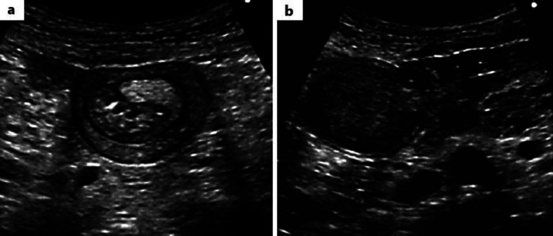
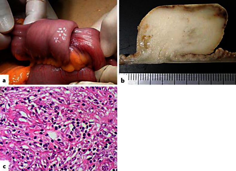
A 79-year-old man presented to our hospital with a 3-month history of nausea, constipation and abdominal distention. Laboratory tests were within normal range. Abdominal ultrasonography revealed a 5-cm mobile, well-defined, elastic hard mass in the left upper quadrant of the abdomen. Ultrasonographic images in the transverse plane showed multiple concentric ring sign (fig. 1a). In the longitudinal section, multiple parallel lines provided the so-called ‘sandwich appearance’, and a 3.5-cm solid mass was observed in the presenting part (fig. 1b). In addition, color Doppler ultrasonography showed abundant blood flow surrounding the mass. Computed tomography (CT) revealed an enhanced homogeneous mass and slight dilatation of the oral side of the intestine (fig. 2). Based on these findings, we diagnosed the patient with intussusception caused by an intestinal tumor. We performed elective laparotomy that revealed an ileal intussusception located 80 cm proximal to the end of the ileum. The intussusception was found to be caused by an ileal tumor located 70 cm from Bauhin's valve. We partially resected the ileum maintaining a 10-cm margin from the tumor, and ileo-ileal anastomosis was performed in an end-to-end manner (fig. 3a). The tumor was a homogeneous solid mass, 3.5 × 2.5 cm in diameter, and was well-circumscribed and grayish-white in color (fig. 3b). Histopathological examination of the tumor revealed proliferation of spindle-shaped cells with infiltration of plasma cells and lymphocytes (fig. 3c). Pathologically, the tumor was diagnosed as an IMT of the small intestine. The patient had no tumor recurrence 12 months after surgery.
Fig. 1.
Abdominal ultrasonography findings. a Transverse ultrasonographic image shows the hypoechoic outer layer of edematous bowel wall with echogenic layers, known as the ‘target sign’. b Longitudinal ultrasonographic image shows a tumor and ‘sandwich appearance’.
Fig. 2.
Enhanced CT scan shows the edematous wall of the intussusception and an enhanced homogeneous mass.
Fig. 3.
Operative findings and specimen. a Laparotomy revealed an ileal intussusception. b Macroscopic findings of the resected specimen. c Histopathological examination with hematoxylin and eosin staining (×400).
Discussion
IMT is a ‘tumor composed of differentiated myofibroblastic spindle cells usually accompanied by numerous plasma cells and/or lymphocytes’ [4]. It was originally described in the lungs; however, extrapulmonary IMT is known to occur in any anatomic location regardless of age. The most common sites of extrapulmonary IMT occurrence are the mesentery and the omentum. Coffin et al. [1] showed that IMT developed at a mean age of 9.7 years, and in 36 of 84 cases (43%), IMTs arose from the mesentery and omentum. On the other hand, only 1 case (1.2%) exhibited an IMT of ileal origin. To our knowledge, our patient is the first reported case aged >70 years with IMT.
Intussusception is more common in the pediatric population than in adults. An estimated 5% of all intussusceptions occur in adults. Recent studies showed that approximately 30% of all small bowel intussusceptions are caused by malignancy, whereas the remainder are caused by benign lesions (60%) or are idiopathic (10%) [5, 6]. Intussusception is not easily diagnosed in adults because patients usually present with non-specific, vague symptoms such as abdominal pain, nausea and vomiting. Thus, diagnostic imaging plays an important role in the diagnosis of this condition. CT is the most commonly used imaging technique, but sonography is also appropriate and useful in the diagnosis of bowel intussusception [7].
Ultrasonography facilitates examination in all planes in a non-invasive manner and in real time. These advantages of ultrasonography prove to be beneficial in emergencies. In our case, ultrasonography enabled an accurate and rapid diagnosis at the first examination. The classic appearance of an intussuscepted bowel on an ultrasonographic image in a transverse plane is called the ‘target sign’ and presents as multiple concentric rings in the bowel. The longitudinal appearance of intussusception is usually viewed as multiple parallel lines, which is termed as the ‘sandwich appearance’ or ‘pseudo-kidney sign’. Target or sausage shapes are observed both on CT as well as on ultrasonographic images. Diagnosis of intussusceptions by CT enables the differentiation between non-pathologic and pathologic intussusceptions. The presence of a mass imaged as a lead on CT is indicative of a neoplasm and the need for operative treatment [8].
In conclusion, the possibility of IMT should be considered during differential diagnosis in elderly patients with a small intestinal tumor. Furthermore, abdominal ultrasonography is a useful technique to demonstrate intestinal intussusception in adults.
Disclosure Statement
The authors have no conflicts of interest to declare.
References
- 1.Coffin CM, Watterson J, Priest JR, Dehner LP. Extrapulmonary inflammatory myofibroblastic tumor (inflammatory pseudotumor). A clinicopathologic and immunohistochemical study of 84 cases. Am J Surg Pathol. 1995;19:859–872. doi: 10.1097/00000478-199508000-00001. [DOI] [PubMed] [Google Scholar]
- 2.Horiuchi R, Uchida T, Kojima T, Shikata T. Inflammatory pseudotumor of the liver. Clinicopathologic study and review of the literature. Cancer. 1990;65:1583–1590. doi: 10.1002/1097-0142(19900401)65:7<1583::aid-cncr2820650722>3.0.co;2-l. [DOI] [PubMed] [Google Scholar]
- 3.Difiore JW, Goldblum JR. Inflammatory myofibroblastic tumor of the small intestine. J Am Coll Surg. 2002;194:502–506. doi: 10.1016/s1072-7515(02)01118-3. [DOI] [PubMed] [Google Scholar]
- 4.Coffin CM, Hornick JL, Fletcher CD. Inflammatory myofibroblastic tumor: comparison of clinicopathologic, histologic, and immunohistochemical features including ALK expression in atypical and aggressive cases. Am J Surg Pathol. 2007;31:509–520. doi: 10.1097/01.pas.0000213393.57322.c7. [DOI] [PubMed] [Google Scholar]
- 5.Azar T, Berger DL. Adult intussusception. Ann Surg. 1997;226:134–138. doi: 10.1097/00000658-199708000-00003. [DOI] [PMC free article] [PubMed] [Google Scholar]
- 6.Zubaidi A, Al-Saif F, Silverman R. Adult intussusception: a retrospective review. Dis Colon Rectum. 2006;49:1546–1551. doi: 10.1007/s10350-006-0664-5. [DOI] [PubMed] [Google Scholar]
- 7.Cerro P, Magrini L, Porcari P, De Angelis O. Sonographic diagnosis of intussusceptions in adults. Abdom Imaging. 2000;25:45–47. doi: 10.1007/s002619910008. [DOI] [PubMed] [Google Scholar]
- 8.Onkendi EO, Grotz TE, Murray JA, Donohue JH. Adult intussusception in the last 25 years of modern imaging: is surgery still indicated? J Gastrointest Surg. 2011;15:1699–1705. doi: 10.1007/s11605-011-1609-4. [DOI] [PubMed] [Google Scholar]


