Abstract
Purpose:
To review malpractice claims associated with retained lens fragments during cataract surgery to identify ways to improve patient outcomes.
Methods:
Retrospective, noncomparative, consecutive case series. Closed claims data related to cataract surgeries complicated by retained lens fragments (1989 through 2009) from an ophthalmic insurance carrier were reviewed. Factors associated with these claims and claims outcomes were analyzed.
Results:
During the 21-year period, 117 (12.5%) of 937 closed claims associated with cataract surgery were related to retained lens fragments with 108 unique cataract surgeries, 97% against cataract surgeon and 3% against retinal surgeon. Twelve (11%) of 108 claims were resolved by a trial, 30 (28%) were settled, and 66 (61%) were dismissed. The defendant prevailed in 83% of trials. Indemnity payments totaling more than $3,586,000 were made in 32 (30%) of the claims (median payment, $90,000). The difference between the preoperative visual acuity and the final visual acuity was predictive of an indemnity payment (odds ratio [OR], 2.28; P=.001) and going to a trial (OR, 2.93; P=.000). Development of corneal edema was associated with an indemnity payment (OR, 3.50; P=.037). Timing of referral and elevated intraocular pressure (IOP) were statistically significant in univariate analyses but not in multivariate analyses for a trial.
Conclusions:
Whereas the majority of claims were dismissed, claims associated with greater visual acuity decline, corneal edema, or elevated IOP were more likely to result in a trial or payment. Ways to reduce significant vision loss, including improved management of corneal edema and IOP, and timely referral to a subspecialist should be considered.
INTRODUCTION
In the practice of medicine, some adverse outcomes are unavoidable because of the nature of the underlying disease, variation in response to treatment, and diagnostic uncertainty. In addition, there are potential complications associated with any surgical procedure due to unavoidable risks despite appropriate care, complications that are unexpected or unpredictable, or decisions that were made carefully by the patient and physician with informed consent but, in retrospect, were less than optimal owing to the uncertainties inherent to the practice of medicine. Malpractice, in contrast, requires demonstration of negligence, defined as substandard care that resulted in harm.1 Malpractice suits are usually based on the legal theory of negligence, requiring the presence of the following four elements: (1) duty to treat, (2) breach of duty, (3) cause, and (4) damages. Duty to treat means that a doctor-patient relationship must be established prior to the alleged negligent act. Breach of duty occurs when the physician fails to follow the standard of care for the patient’s condition. Standard of care is what a “reasonable” physician would do in similar circumstances. The negligent act must be a “proximate cause” of the plaintiff’s injuries, which means the act was necessary for the injury when and in the manner it occurred, and the injury must be a foreseeable consequence of the negligent act. Finally, the patient must have suffered actual damage or injury as a result of negligence.
A number of studies have found that there is substantial variation in the likelihood of malpractice suits across specialties and the cumulative risk of facing a malpractice claim is high in all specialties.2–6 The Physician Practice Information Survey by the American Medical Association of 5,825 physicians across 42 medical specialties, fielded in 2007 and 2008, found that an average of 95 claims were filed for every 100 physicians, almost 1 per physician, as a group.2 However, the chance of being sued each year for a physician was about 5%. According to this report, 42% of physicians have been sued for medical malpractice at some point in their careers and 20% were sued at least twice during their careers.2 This survey found a wide variation in the incidence of liability claims between specialties. The number of claims per 100 physicians was more than 5 times greater for general surgeons and obstetricians and gynecologists than it was for pediatricians and psychiatrists. More than 50% of obstetricians and gynecologists have already been sued before they reached the age of 40 years, and 90% of general surgeons aged 55 years and older have been sued.
Another study found that 7.4% of all physicians had a malpractice claim each year, with 1.6% having a claim leading to a payment.5 The proportion of physicians facing a claim each year ranged from 2.6% in psychiatry to 19.1% in neurosurgery. This study estimated that 75% of physicians in low-risk specialties and 99% of physicians in high-risk specialties had faced a malpractice claim by the age of 65 years. Furthermore, there was a wide variation in the size of indemnity payment (payment to a plaintiff) across specialties, and the specialties that were most likely to face indemnity claims were often not those with the highest average payments.5 For example, pediatrics was 24th among 25 specialties with regard to proportion of physicians facing a malpractice claim annually, but it had the highest mean amount of indemnity payment.
Although these findings may cause fear and increased practice of defensive medicine by physicians, better understanding of the incidence, associated factors, and outcomes of medical malpractice claims may result in increased knowledge to the physicians and more effective and improved care to the patients. Previous studies have shown that useful information can be gained from evaluation of malpractice claims data.3,5–15 However, most of the previous studies that estimated specialty-specific malpractice risk from actual claims data are not recent, and only a handful of studies specifically address the specialty of ophthalmology.5–16 In the most recently published study, Jena and colleagues5 analyzed closed malpractice claims for 40,916 physicians who were covered for at least one policy year from 1991 through 2005, including 807 ophthalmologists insured during the study period. They found that the claims frequency for ophthalmology was slightly lower than the average for all specialties and was in between nephrology and diagnostic radiology.
Given the differences in the frequency of claims for various medical specialties and the limited number of studies in the literature related to malpractice claims in ophthalmology, this current study used the available data from a large ophthalmology-specific insurance company in an effort to gather specialty-specific data. Claims data from the Ophthalmic Mutual Insurance Company (OMIC) represent a unique opportunity to examine the medicolegal risks associated with ophthalmology. Sponsored by the American Academy of Ophthalmology, OMIC is the largest professional liability insurer for ophthalmologists in the United States, currently insuring over 4,300 ophthalmologists throughout the 49 states (all states except Wisconsin). With OMIC having 40% of the ophthalmology market share in 2010, OMIC policyholders compare favorably with current demographics of ophthalmologists.17 Because it is a single-specialty insurer with the ability to collect and analyze data on a large number of professional liability claims related to ophthalmology, gathering of information on malpractice claims related to a specific ophthalmic procedure is possible. Furthermore, these malpractice claims data can be used to identify ways to improve patient safety, develop risk management programs, and provide an excellent opportunity to enhance patient care related to an ophthalmic subspecialty or an ophthalmic procedure.
Over 3 million cataract surgeries are performed annually in the United States.18 Given the frequency of this procedure, perhaps it is not surprising that cataract surgery is the single most frequently named procedure in malpractice actions against ophthalmologists.13–15 An uncommon but potentially devastating complication of cataract surgery that can affect both the anterior segment and the posterior segment surgeons is posterior dislocation or retention of lens fragments during cataract surgery.
There has been a large interest over the years in clinical outcomes and management of retained lens fragments as evidenced by the substantial number of articles continuing to be written on this topic.19–78 The incidence of retained or dropped lens fragments during cataract surgery is estimated to be between 0.1% and 1.6% of cataract surgeries.18,19,23,29,45,54,64 There are numerous articles to indicate that a capsular tear with retained lens fragment is a well-known complication of cataract surgery.20–49 Studies show that reasonably favorable visual outcome can be obtained with intervention usually in the form of pars plana vitrectomy.20–49,74–77 Therefore, encountering this complication in itself would not be a malpractice. However, how this complication was managed intraoperatively and postoperatively, what degree of injury resulted, as well as how the informed consent was presented preoperatively, will determine whether or not malpractice occurred due to substandard care that resulted in harm to the patient.
Medical malpractice claims stemming from cataract surgery–related ophthalmic care present a unique opportunity to examine the risks associated with this frequently performed intraocular surgery and to improve the safety of patients. In the current study, closed claims from cataract surgeries complicated by retained lens fragments were evaluated to identify factors that are associated with indemnity payment or resulting in a trial. This study was carried out for a number of reasons: (1) the absence of published studies addressing the legal outcomes for this complication despite the number of cataract surgeries being performed in the United States; (2) tremendous interest in the management and outcomes of this potentially visually devastating complication based on the large number of published studies on this topic; (3) the relevance of study findings to both the anterior and posterior segment specialists; and (4) a potential to improve patient outcomes.
Legal outcomes were categorized as those claims resulting in a trial, settlement, or dismissal, and indemnity payment was evaluated for those claims ending in a settlement or in favor of the plaintiff after a trial. Associated factors were analyzed for (1) going on to a trial or settlement rather than being dismissed, and for (2) indemnity payment vs no payment. The first categorization was needed to evaluate legal costs incurred for each category of legal outcomes. Most previous studies on malpractice claims compared only the groups that went on to indemnity payment vs no payment. Whereas indemnity payment is usually associated with all settled claims, claims that go on to a trial may or may not result in an indemnity payment, depending on the verdict. Additional categorization and analyses were performed in this study to include claims outcomes of trial vs settlement vs dismissal in hopes of gaining additional information, such as legal expenses that may differ for these groupings, as well as to highlight factors associated with claims that result in a verdict for the plaintiff vs that for the defendant when there was a trial.
The aims of this study were to evaluate the medical malpractice claims resulting from the retained lens fragments during cataract surgery and to identify ways to improve patient outcomes. The hypothesis of the current study is that there may be differences among the groups of cases with different legal outcomes. Through highlighting circumstances of pertinent claims and identifying factors associated with malpractice claims resulting in an indemnity payment or going to a trial, this current study sought to ascertain steps that can be taken by ophthalmologists to improve patient care and safety as well as assist in risk management when cataract surgery is complicated by retained lens fragments.
METHODS
CLOSED CLAIMS DATA FROM OPHTHALMIC MUTUAL INSURANCE COMPANY
Closed claims data from OMIC were chosen to be the basis of this study because OMIC provides coverage to a large number of ophthalmologists and can provide data specific to an ophthalmic procedure. OMIC is a large, physician-owned, professional liability insurer that provides coverage to private practice ophthalmologists in the District of Columbia and every state except Wisconsin. To be insured by OMIC, an ophthalmologist must be a member of the American Academy of Ophthalmology. Currently OMIC is the largest insurer of ophthalmologists, with 40% of the market share, and has twice as many ophthalmologists as policyholders as the next largest insurer of ophthalmologists.17 Claims data from OMIC has been utilized in other previous studies related to ophthalmology.9–11 The OMIC Risk Management Committee gave approval for this study and granted access to the data under agreements protecting the identities of the patients, surgeons, and institutions. The data accumulation adhered to the Declaration of Helsinki and conformed with all federal and state laws and HIPAA guidelines.
A retrospective review was performed of all closed claims during the 21 years from 1989 through 2009 of those insured by OMIC to identify cases associated with cataract surgeries complicated by retained lens fragments (see “Inclusion and Exclusion Criteria” section that follows). OMIC underwriting applications and claims records were reviewed. The data collected were chosen based on the review of the literature to have a potential relevance to the outcome of litigations in ophthalmology9–16 or to the clinical outcomes20–65 and were obtainable from the available documents from OMIC. The items collected during the review of the claims are listed in Table 1. These items can be broadly separated into those pertaining to (1) the physician, (2) the patient, (3) preoperative, intraoperative, and postoperative clinical data, and (4) the litigation. Preoperative visual acuity was the visual acuity shortly prior to cataract surgery. Final visual acuity was the last recorded visual acuity. Glaucoma was defined as elevated intraocular pressure requiring pressure-lowering medication or documented visual field defect. In some categories of data, not all data points were available, and those are indicated in the appropriate tables.
TABLE 1.
ITEMS REVIEWED FOR POTENTIAL ASSOCIATED FACTORS FOR LITIGATION OUTCOMES FROM CLOSED CLAIMS RELATED TO CATARACT SURGERY COMPLICATE BY RETAINED LENS FRAGMENTS
| DEFENDANT (SURGEON) | PLAINTIFF (PATIENT) | RELATED TO SURGERY | LITIGATION |
|---|---|---|---|
| Age | Age | OD or OS | Date of cataract surgery |
| Gender | Gender | Preoperative VA | Date reported to OMIC |
| Location state | Location state | Final VA | Date opened |
| Prior surgery by the same surgeon | Preexisting ocular conditions | Date closed | |
| Fellow eye VA | Suit vs claim | ||
| Cause of capsular tear, if noted | Allegations | ||
| Intraoperative manipulations, if noted | Actual injury | ||
| Placement of IOL | Disposition of case | ||
| PC IOL | Expense paid | ||
| AC IOL | Indemnity paid | ||
| Aphakia | |||
| Postoperative course | |||
| Complications | |||
| Retinal detachment | |||
| Cystoid macular edema | |||
| Glaucoma | |||
| Corneal edema | |||
| Other | |||
| Time to referral | |||
| Number of subsequent surgeries |
AC IOL, anterior intraocular lens; OD, right eye; OMIC, Ophthalmic Mutual Insurance Company; OS, left eye; PC IOL, posterior intraocular lens; VA, visual acuity.
In addition to the review of the closed claim cases related to the complication of retained lens fragments, other data that were thought to be relevant to the study were obtained from OMIC and analyzed for comparison with the findings from this study. These included the number of ophthalmologists insured by OMIC from 1989 through 2009, the number of closed claims related to cataract surgery, OMIC policyholder demographics, and average indemnity payments for OMIC policyholders.
INCLUSION AND EXCLUSION CRITIERIA
Cases to be included in the study were identified based on OMIC coding for claims resulting from complications related to cataract surgery. Claims data of all the identified claims based on coding were reviewed and further narrowed to include only those claims where there was a mention of a “retained,” “dropped,” or “dislocated” crystalline lens fragment with or without other comorbidities. Claims were excluded when found not to pertain to retained lens fragments but were due to dislocated intraocular lens (IOL), wrong intraocular lens, endophthalmitis, or retinal detachment following cataract surgery. Only the claims that closed by December 2009 were included. Some cases that opened in more recent years are still open and are not a part of this study, since both the legal outcome and expenses were required for the analyses. If more than one physician was named in the claim, only the data on the primary surgeon was analyzed. If a surgeon and the hospital or the practice (entity) were named in the claim, only the surgeon’s data was analyzed to avoid duplicity. If a physician had multiple claims from separate cataract surgeries, each was counted separately.
CLAIM VS SUIT
The OMIC Professional Liability Policy defines a claim as a written notice or demand for money or services by the patient (plaintiff) to the insured (physician or entity) for compensation from a medical incident. A claim may include institution of a lawsuit or arbitration proceedings against the insured. A suit is defined as a formal legal action initiated in the courts by the filing of a “complaint” seeking a remedy (usually money) by the plaintiff and requiring a formal response from the physician or the entity (defendant). The term “claim” was used in this study to include “suits,” unless specified.
LITIGATION OUTCOMES
For the current study, the claims were categorized into those that went on to a trial, settlement, or dismissal, and those with or without indemnity payment. One analysis was performed with the litigation outcomes divided into (1) trial, (2) settlement, and (3) dismissed. This division allowed additional information regarding the duration between opening and closing of the claim and legal expenses for each group. Claims that settled during the trial or prior to the start date of the trial were included in the “settlement” group. Claims that were dismissed, dropped, or closed without compensation were combined as “dismissed,” and the term “dismissed” was used interchangeably with “closed without compensation,” “dropped,” and “withdrawn,” unless specified.
Another analysis was performed with the litigation outcomes grouped as (1) indemnity payment and (2) no indemnity payment. This grouping was done to compare the findings of this study to other published data. Indemnity payment occurred in those claims that went on to a trial and a verdict in favor of the plaintiff was made or in claims that settled. No indemnity payment was made in claims that went on to a trial but the verdict was in favor of the defendant or in claims that were dismissed or closed without compensation.
STATISTICAL ANALYSIS
Both univariate analyses and multivariate analyses were performed using data collected for possible outcomes or final disposition of the claim. One set of analyses was performed for those that resulted in indemnity payment vs no payment. Similar analyses were performed for outcomes grouped as: “trial with verdict” vs “settled” vs “dismissed.” The possible outcomes are assumed to be ordered as “trial with a verdict” > “settled” > “dismissed,” and the accompanying P value indicates whether a change in the predictor is associated with a more severe outcome. “Trial with a verdict” was assumed to be a more severe outcome than “settled,” since historically longer duration between opening and closing of a claim and higher costs are associated with trials compared to settled claims.
In the univariate analysis the P values for continuous variables were calculated based on nonparametric tests: Wilcoxon rank sum test for two groups (indemnity payment vs no indemnity payment) and Jonckheere-Terpstra trend test for multiple groups (trial vs settlement vs dismissed). All variables significant at a 10% level in the univariate analyses were included in a multivariate proportional odds regression model. For the use in multivariate modeling, an optimal transformation from the Box-Cox family was calculated for each nonnegative continuous variable. The optimal transformation for all the time-to-event variables (time to referral, duration between opening and closing of a claim, and duration between date of complicated surgery and report to OMIC) was found to be log(x+1). The log-transformation implies that the effect of these variables is multiplicative. These transformed variables were used in further analyses. The model was simplified using backward selection keeping all predictors with a P value of .25 or less. For this study, a P value <.05 was considered significant.
RESULTS
INCIDENCE OF CLAIMS
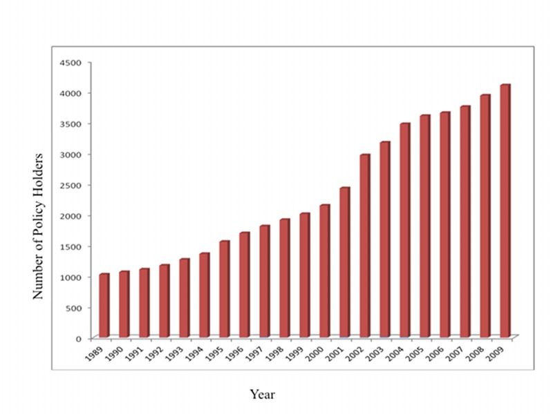
The number of ophthalmologists being insured by OMIC grew steadily from 1,027 in 1989 to 4,107 in 2009 (Figure 1). The number of policyholders doubled between years 2000 and 2009. Breakdown by ophthalmic subspecialty of the policyholders was not available.
FIGURE 1.
The number of Ophthalmic Mutual Insurance Company policyholders from years 1989 through -2009.
From 1989 through December 2009, OMIC had a total of 2,854 closed claims. Of these, 937 claims were related to cataract surgery, and 117 closed claims related to cataract surgery were complicated by retained lens fragments. Therefore, claims related to cataract surgery accounted for 33% of all closed claims during this period, and cataract surgeries complicated by retained lens fragments accounted for 4% of all closed claims and 12.5% of cataract-related claims.
Among 117 closed claims that were related to cataract surgery complicated by retained lens fragments, 9 cases had multiple claims, including 8 cases where both the physician and the OMIC-insured entity were named in the suit and one case where two OMIC-insured physicians were named. For statistical purposes, only the data from the primary surgeon was analyzed in the study. Accounting for these factors, there were 108 unique cataract surgeries that met the inclusion criteria and were the basis for the current analyses.
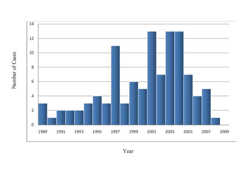
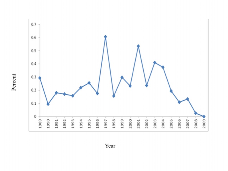
The distribution of the number of closed claims related to the complication of retained lens fragments per year from 1989 through December 2009 is shown in Figure 2. The number peaked in 1997 with 11 cases and again in 2001, 2003, and 2004 with 13 cases each year. Since the number of OMIC-insured ophthalmologists continued to grow each year over this 21-year period, the frequency of closed claims related to retained lens fragments relative to the total number of physicians insured per year was actually the highest in 1997 (Figure 3). Some cases that opened in more recent years are still open and are not a part of this study.
FIGURE 2.
The number of closed claims related to cataract surgery complicated by retained lens fragments each year from 1989 through 2009.
FIGURE 3.
The frequency of claims related to retained lens fragments compared to the number of policyholders for each year from 1989 through 2009.
DEMOGRAPHICS
Physician and Patient Characteristics
Among the 108 cases, two physicians had multiple claims relating to retained lens fragments, with 2 claims each. Each claim was counted separately as a unique case. Of the 108 physician defendants, 94 (87%) were men and 14 (13%) were women. This gender spread was compared with OMIC data on demographics. According to the 2010 report to the OMIC members, approximately 17% of practicing ophthalmologists in the United States are female and 18% of OMIC-insured ophthalmologists are female.17
Physician age ranged from 31 to 72 years (mean, 49 years). Of the 108 defendants, 105 (97%) were cataract surgeons and only 3 (3%) were retinal surgeons. Although there were no cases involving residents, there was one claim against a policyholder ophthalmologist who was overseeing a colleague’s attempt at learning cataract surgery.
Among the 3 claims involving retina surgeons, one claim alleged negligent surgery to remove the dropped nucleus and dislocated IOL, which allegedly led to a subsequent retinal detachment. Another claim alleged that there was a delay in time to pars plana vitrectomy by the retinal surgeon to manage the elevated intraocular pressure. The third claim alleged decreased vision following negligent vitrectomy surgery to manage retained lens fragment. The patient was referred the same day as the complicated cataract surgery to the retina specialist, who performed pars plana vitrectomy on the following day without any complications. The patient’s visual acuity prior to cataract surgery was 20/200 and at the last follow-up, 5 months following vitrectomy, was 20/80. The patient was released to a general ophthalmologist. The patient complained of a black spot with decreased vision 7 months after the cataract and vitrectomy surgery. However, the patient did not show up for appointments, despite being sent “no show” letters. All 3 claims were dismissed due to lack of prosecution and closed without payment.
Among 108 patient claimants, 54 were men and 54 were women. Data on age was available for 101 claimants. The mean age was 69 years (range, 40–90 years). In 6 cases, there was documentation that the defendant had operated on the fellow eye of the claimant previously.
States
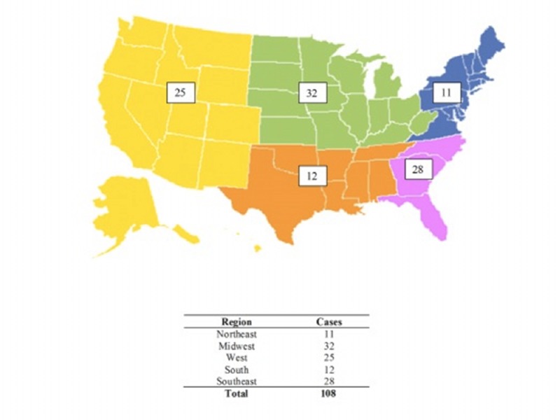
Claims were separated into regions of the United States as seen in Figure 4. There were 11 cases (10%) from the Northeastern states, 32 (30%) from the Midwest, 25 (23%) from the Western states, 12 (11%) from the Southern states, and 28 (26%) from the Southeastern states. The top 5 states in terms of overall frequency of claims in rank order were Illinois (18 cases), Texas (16 cases), California (11 cases), Florida (10 cases), and Louisiana (10 cases). However, these numbers may reflect the states in which OMIC has a major presence, since these are also states in which OMIC has the highest number of insured ophthalmologists.
FIGURE 4.
Distribution of closed claims related to retained lens fragments by region in the United States.
EVALUATION OF CLINICAL FINDINGS
Visual Acuity
Mean preoperative visual acuity of the eye involved in the claim was 20/80 (range, 20/25 to hand motions). Mean final visual acuity was 20/200 (range, 20/20 to no light perception). Mean change in visual acuity between preoperative visual acuity and final visual acuity for all patients was a worsening of 2 lines.
In 91 eyes, preoperative visual acuity was recorded for both eyes. Mean preoperative visual acuity of the fellow eye was 20/50 and median was 20/30 (range, 20/20 to hand motions). In 11 eyes, the operated eye was the better eye.
Preoperative Findings
Among the 108 claims, 107 claims had a record of which eye was operated on; 42 cases (39%) involved the right eye and 65 (61%) involved the left eye. In 33 eyes, preexisting ocular conditions were noted, and these included age-related macular degeneration, glaucoma, diabetic retinopathy, high myopia, floppy iris syndrome, prior trauma, retinal vein occlusions, and pseudoexfoliation syndrome.
Intraoperative Issues
There was a posterior dislocation of nucleus in all except 4 cases, in which the retained lens material was in the anterior segment. In 10 cases, the tear of posterior capsule was not recognized by the cataract surgeon or was not indicated in the operative note and only became apparent during the investigation of the case. In addition to alleged negligent cataract surgery with retained lens fragments, placement of the wrong IOL was cited as a contributing negligence in 3 cases: (1) placement of wrong-powered IOL handed to the surgeon by a nurse; (2) not having the correct type of IOL to insert in the setting of capsular rupture, resulting in increased likelihood of subsequent dislocation of IOL; and (3) placement of wrong-powered IOL due to incorrect transfer of A-scan data by a technician.
In 7 cases, the cataract surgeon documented an intraoperative attempt at retrieval of the lens fragment (Table 2). These manipulations included use of a lens loop, an attempt at impaling the lens with a microvitreoretinal blade, irrigation to float the lens, and pars plana vitrectomy by the cataract surgeon. In all cases, retinal detachment occurred, 5 after the cataract surgery and 2 after pars plana vitrectomy and lensectomy by retinal specialists. In all cases, final visual acuity was 20/200 or worse, including 2 cases of no light perception. In one of the claims, the cataract surgeon, who had some retinal training, attempted retrieval of the posteriorly dislocated lens material. However, he could not complete the surgery and his retinal colleague needed to intervene intraoperatively.
TABLE 2.
CLAIMS WITH A DOCUMENTATION OF INTRAOPERATIVE MANIPULATION BY THE CATARACT SURGEON DURING MANAGEMENT OF POSTERIOR DISLOCATION OF LENS FRAGMENTS
| CASE | PREOP VA | POSTOP VA | INTRAOPERATIVE MANIPULATIONS | TIME TO REFERRAL | ASSOCIATED COMPLICATIONS | DISPOSITION |
|---|---|---|---|---|---|---|
| 1 | 20/60 | LP | Spatula; irrigation; probe posteriorly | Same day | RD × 2; hypotony; corneal decompensation | Plaintiff verdict; $125,000 |
| 2 | 20/70 | NLP | MVR blade to impale the fragment that landed on optic nerve | 9 days | RD × 2 | Settled; $135,000 |
| 3 | 20/40 | HM | Lens loop | 7 days | RD × 2 with the initial RD during PPV by a retina specialist |
Defense verdict |
| 4 | 20/80 | HM | Lens loop; irrigation | 4 days | RD × 2; glaucoma | Settled; $150,000 |
| 5 | 20/50 | NLP | Spatula | 1 day | RD × 2 with the initial RD occurring 1 month after PPV, PPL |
Settled; $25,000 |
| 6 | 20/70 | CF | Irrigation | 7 days | RD × 3 | Plaintiff dismissed |
| 7 | CF | 20/200 | PPV by the cataract surgeon | 9 days | RD; vitreous hemorrhage | Statute of limitation expired; dismissed |
CF, counting fingers; HM, hand motions; LP, light perception; MVR, microvitreoretinal; NLP, no light perception; PPL, pars plana lensectomy; PPV, pars plana vitrectomy; RD, retinal detachment; VA, visual acuity.
Intraocular lens was implanted in 85 (90%) of 94 cases where this was recorded, with 63 (67%) being posterior chamber IOL and 22 (23%) being anterior chamber IOL. The remaining 9 cases (10%) were left aphakic by the cataract surgeon. Even when an IOL was initially placed at the time of complicated cataract surgery, subsequent dislocation of IOL occurred in 6 cases. Overall, IOL had to be removed, sutured, inserted, or exchanged during pars plana vitrectomy by a retinal specialist in 17 (16%) of 108 cases.
Claims Involving Vicarious Liability
In some cases, the cause of capsular tear and resulting complication of retained lens fragment was due to circumstances other than the surgeon’s surgical technique. In one case, the surgical technician failed to securely attach the cystotome to the needle, and the cystotome shot off during injection of the viscoelastic material. The needle impaled the lens and tore the lens capsule. In 10 cases, the tear reportedly occurred as a result of a sudden movement of the patient during surgery. Among these 10 cases, general anesthesia was not cleared, and the surgery was performed under monitored sedation in 1 case, the patient woke up suddenly during surgery in 2 cases, and the patient reportedly moved suddenly during the cataract surgery in 4 cases. In 3 cases, malfunctioning or unavailability of necessary equipment resulting in prolonged cataract surgery time was thought to have contributed to the patient movement and complication of capsular tear.
Associated Postoperative Ocular Complications
The majority of eyes developed one or more ocular complications following surgery, many of which contributed to poor visual outcome. The most common complications were elevated intraocular pressure requiring initiation of pressure-lowering medications and development of visual field damage due to elevated intraocular pressure. Both of these were defined as “glaucoma,” and there were a total of 31 cases. There were 25 cases of retinal detachment, 21 cases of corneal edema or corneal decompensation, and 18 cases of cystoid macular edema. Thirty-four cases had other complications, including endophthalmitis, vitreous hemorrhage, choroidal detachment, macular hole formation, central retinal artery occlusion, uveitis, anterior ischemic optic neuropathy, floaters, and epiretinal membrane. More than one of these complications was noted in 31 cases.
Time to Referral
In 94 cases, a referral was made to a subspecialist. Among these, the patients sought a second opinion and referred themselves in 3 cases. The overwhelming majority of the referrals were to a retina specialist, but referrals also included cornea and glaucoma specialists. Time between cataract surgery and referral to a subspecialist was a median of 7 days, ranging from the same day as the cataract surgery to 15 months after cataract surgery. “Delay in diagnosis” or “delay in referral” was alleged in 12 (11%) of 108 claims. Time to additional surgical procedures such as vitrectomy was at the discretion of the subspecialist.
Additional Surgeries
In addition to the original cataract surgery, patients underwent a mean of 1.3 additional surgeries (range, 0–4) where one or more combined procedures were performed. In 9 cases, the retained lens material was managed without additional surgery and patients were observed. In one additional case, observation was recommended without further surgery because the retina specialist felt that the retinal detachment was inoperable. Four patients declined any further surgery. If these cases are excluded, there was a mean of 1.5 return visits to the operating room among 94 patients who had additional surgical procedures.
The most common additional surgical procedure was pars plana vitrectomy to remove retained lens material or to manage retinal detachment, but procedures to manage IOL, glaucoma, corneal decompensation, and strabismus were also performed (Table 3).
TABLE 3.
ADDITIONAL SURGICAL PROCEDURES PERFORMED TO MANAGE COMPLICATIONS FROM RETAINED LENS FRAGMENTS
| PROCEDURE | NUMBER |
|---|---|
| Pars plana vitrectomy/lensectomy | 113 |
| IOL removal/insertion/exchange/suturing | 17 |
| Scleral buckling procedure (±vitrectomy) | 9 |
| Penetrating keratoplasty | 6 |
| Trabeculotomy/shunt placement | 6 |
| Drainage of choroidal hemorrhage | 2 |
| Management of endophthalmitis | 2 |
| Pneumatic retinopexy | 2 |
| Strabismus surgery | 1 |
IOL, intraocular lens.
CLAIMS AND SUITS
Allegations
The allegations for the claims associated with cataract surgery complicated by retained lens fragments are listed in Table 4. The overwhelming majority of allegations consisted of negligent cataract surgery with or without subsequent complications, followed by delayed diagnosis or referral, and issues related to preoperative discussions such as informed consent. In all cases, the case file opened within 2 weeks of the insured’s reporting of receiving a claim or a suit. The time between the date of cataract surgery and the date of reporting by the insured to OMIC regarding litigation was a mean of 15.5 ± 8.7 months. On average, a claim took 28.8 ± 21.2 months to close.
TABLE 4.
LIST OF ALLEGATIONS IN THE CLAIMS RESULTING FROM CATARACT SURGERY COMPLICATED BY RETAINED LENS FRAGMENTS
| ALLEGATIONS | NUMBER | % OF TOTAL |
|---|---|---|
| Preoperative | ||
| Unnecessary surgery | 5 | 4.24 |
| Lack of informed consent | 3 | 2.54 |
| Intraoperative | ||
| Negligent surgery with: | 47 | 39.83 |
| retinal detachment | 19 | 16.10 |
| loss of vision | 13 | 11.02 |
| cystoid macular edema | 2 | 1.69 |
| dislocated IOL | 2 | 1.69 |
| endophthalmitis | 2 | 1.69 |
| corneal damage | 2 | 1.69 |
| uveitis | 1 | 0.85 |
| aggressive retrieval | 1 | 0.85 |
| formation of a cystic bleb | 1 | 0.85 |
| Wrong IOL | 3 | 2.54 |
| Failure to properly restrain | 1 | 0.85 |
| Improper use of anesthetic | 1 | 0.85 |
| Malfunction of equipment | 1 | 0.85 |
| Postoperative | ||
| Delayed diagnosis/referral | 12 | 10.17 |
| Additional surgery/expense | 2 | 1.69 |
| Total* | 118 | 100 |
IOL, intraocular lens.
More than one allegation in some cases
Claims Outcomes
Among the 108 cases in this study, the final dispositions of the claims were as follows: 12 cases (11%) were resolved by a trial, of which 2 cases (17%) resulted in a verdict in favor of the patient plaintiff and 10 cases (83%) cases with a verdict in favor of the physician defendant; 30 cases (28%) were settled; and 66 cases (61%) were dismissed.
Indemnity payments totaling more than $3,586,000 were made in 32 (30%) of the cases. They ranged from a low of $7,500 to a high of $500,000. The mean payment was $117,688, and the median payment was $90,000. The difference between the mean and median payment reflects the right-skewed payment distribution. Among the 12 claims that resulted in a jury trial, 2 cases resulted in indemnity payment. The first case closed in 1992 for $125,000, and the second case closed in 2002 for $250,000. This is without adjustment for potential differences in dollar amount due to inflationary changes. The remaining 76 claims (70%) closed without any payments.
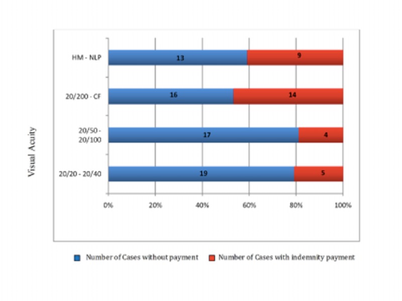
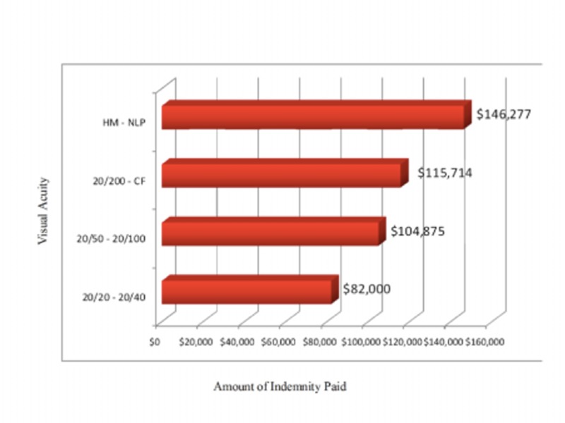
The final visual acuity for claims resulting in indemnity payment vs no payment is shown in Figure 5. Whereas good final visual acuity did not prevent indemnity payment, 23 of 32 claims (72%) with indemnity payment had final visual acuity of 20/200 or worse. Also, claims with worse final visual acuity tended to have higher indemnity payments (Figure 6).
FIGURE 5.
Comparison between claims with indemnity payment and no payment by final visual acuity among cataract surgeries complicated by retained lens fragments. The number of cases in each visual acuity grouping for claims with payment and no payment is also shown. CF, counting fingers; HM, hand motions; NLP, no light perception.
FIGURE 6.
The amount of indemnity payment for each grouping of final visual acuity among cataract surgeries complicated by retained lens fragments. CF, counting fingers; HM, hand motions; NLP, no light perception.
The largest indemnity payment case, with a payment of $500,000, closed in 2005 with a settlement. It involved a 70-year-old female patient who went from preoperative visual acuity of 20/60 to final visual acuity of no light perception. The complication of capsular tear and retained lens fragments was further aggravated by development of corneal wound dehiscence, corneal ulcer, and endophthalmitis. The patient was referred 1 month after the initial cataract surgery to a retina specialist and underwent two pars plana vitrectomy surgeries, corneal wound closure, and intravitreal antibiotic injections. The defense experts felt that the case needed to settle because it was below the standard of care to delay referral by not recognizing endophthalmitis in a timely manner.
Defense Costs
Total cost of defense for all 108 claims was $3,312,688. Average defense costs per claim were $30,692 and ranged from a low of $0 to a high of $190,961. The costs including indemnity payments and defense costs are summarized in Table 5. The mean defense costs were significantly lower in cases that were dismissed but were considerably higher in cases that went on to a trial, even when there was no indemnity paid. The mean defense cost for 12 cases that went on to a trial was $96,464 with a mean defense cost of $97,924 for cases with a defense verdict and $95,004 for cases with a plaintiff verdict; the mean expense for claims that were dismissed was $9,226. Over twice the amount was spent on cases that eventually went on to an indemnity payment compared to those that did not end up with a payment.
TABLE 5.
FINAL DISPOSITION OF CLOSED CLAIMS RESULTING FROM CATARACT SURGERY COMPLICATED BY RETAINED LENS FRAGMENTS
| FINAL CLAIM STATUS | NUMBER OF CLAIMS (%) (TOTAL N=108) | MEAN INDEMNITY PAID | MEAN EXPENSE PAID |
|---|---|---|---|
| No indemnity payment | 76 | $20,897 | |
| Defense verdict | 10 | $97,924 | |
| Dismissed | 66 | $9,226 | |
| Indemnity payment | 32 | $117,688 | $53,985 |
| Plaintiff verdict | 2 | $187,500 | $95,004 |
| Settled | 30 | $107,033 | $51,250 |
Claims Outcomes by State
Of the 12 claims that went on to a trial, there were 5 claims from Illinois, 2 claims from Arizona, and 1 claim each from Colorado, Florida, Kentucky, Rhode Island, and Texas. Of the 30 claims that were settled, there were 6 claims from Illinois; 5 from Florida; 3 from California; 2 claims each from Colorado, Michigan, and New York; and one claim each from Georgia, Louisiana, Missouri, Nevada, Tennessee, Texas, Virginia, Washington, West Virginia, and Wyoming. Of the 66 claims that were dismissed, Texas had the most claims with 14, followed by Louisiana with 9, California with 8, Illinois with 7, Virginia and Florida each with 4, Kentucky and Colorado each with 3, Arizona, Michigan, and Missouri each with 2, and Alabama, Massachusetts, Nevada, North Carolina, Ohio, Pennsylvania, West Virginia, and Washington, DC, each with one claim. Although claims from Illinois, Texas, and California accounted for 42% of all claims, claims from Illinois were more likely to go to trial or settlement, and claims from Texas and California were more likely to be dismissed. Florida and Louisiana each had 10 claims. Claims from Florida were evenly split between those closing with an indemnity payment and those with no payment, whereas the overwhelming majority of claims from Louisiana ended with a dismissal and no payment.
Claims Outcomes by Gender of Physicians
Of the 108 physician defendants, 94 (87%) were men and 14 (13%) were women. When evaluated for indemnity payment or no payment, the male-to-female physician ratios were 27:5 and 66:9, respectively. Of the 12 claims resulting in a trial, 30 claims resulting in a settlement, and 66 claims resulting in a dismissal, the male-to-female physician defendant ratios were 12:0, 25:5, and 57:9, respectively. Gender of the physician was not found to be a significant predictor of indemnity payment of the claims outcomes (Tables 6 and 7).
TABLE 6.
DESCRIPTIVE STATISTICS OF THE ANALYSIS VARIABLES GROUPED BY WHETHER INDEMNITY WAS PAID
| VARIABLE | N | INDEMNITY NOT PAID* | INDEMNITY PAID* | PVALUE† |
|---|---|---|---|---|
| N=75 | N=32 | |||
| Gender of insured: male | 108 | 88% (66) | 84% (27) | 0.61§ |
| Gender of claimant: male | 108 | 51% (38) | 47% (15) | 0.72§ |
| Age of insured‡ | 108 | 42 51 57 | 38 48 56 | 0.40# |
| Age of claimant‡ | 101 | 65 70 77 | 66 71 78 | 0.41# |
| Operated eye: left eye | 107 | 62% (46) | 59% (19) | 0.79§ |
| LogMAR VA of fellow eye‡ | 96 | 0.10 0.18 0.48 | 0.10 0.30 0.49 | 0.53# |
| LogMAR preop VA‡ | 98 | 0.42 0.60 1.14 | 0.48 0.54 0.70 | 0.26# |
| LogMAR final VA‡ | 97 | 0.27 0.65 2.20 | 0.64 1.41 2.30 | 0.04# |
| ΔLogMAR VA‡ | 90 | −0.44 0.00 0.88 | 0.50 1.22 1.83 <0.001# | |
| Retinal detachment | 108 | 20% (15) | 31% (10) | 0.21§ |
| Cystoid macular edema | 108 | 15% (11) | 22% (7) | 0.36§ |
| Glaucoma/elevated IOP | 108 | 24% (18) | 41% (13) | 0.083§ |
| Corneal edema/decompensation | 108 | 15% (11) | 31% (10) | 0.048§ |
| Other causes of poor VA | 108 | 32% (24) | 28% (9) | 0.69§ |
| Type of IOL | 94 | 0.78§ | ||
| PC IOL | 70% (46) | 63% (17) | ||
| AC IOL | 23% (15) | 26% (7) | ||
| Aphakia | 8% (5) | 11% (3) | ||
| Time to referral (days)‡ | 94 | 1 7 28 | 4 12 21 | 0.48# |
| Duration of claim opening to closing (months)‡ | 108 | 14 22 36 | 19 26 39 | 0.15# |
| Duration between surgery and claim occurring (months)‡ | 108 | 8.2 13.0 22.6 | 10.9 17.4 23.8 | 0.18# |
| Year when the claim opened‡ | 108 | 1997 2001 2004 | 1997 2001 2004 | 0.51# |
AC IOL, anterior chamber intraocular lens; IOP, intraocular pressure; PC IOL, posterior chamber intraocular lens; VA, visual acuity.
N is the number of non–missing values. Numbers after percents are frequencies.
The P values evaluate whether the variable is predictive of having an indemnity payment.
For each of these variables, the three numbers given represent the lower quartile, the median, and the upper quartile for continuous variables.
Pearson test.
Wilcoxon test.
TABLE 7.
DESCRIPTIVE STATISTICS OF THE ANALYSIS VARIABLES BY CLAIMSOUTCOME ASSOCIATED WITH RETAINED LENS FRAGMENT
| VARIABLE | N | DISMISSED* | SETTLED* | TRIAL WITH VERDICT* | COMBINED* | PVALUE† |
|---|---|---|---|---|---|---|
| N=66 | N=30 | N=12 | N=108 | |||
| Gender of insured: male | 108 | 86% (57) | 83% (25) | 100% (12) | 87% (94) | 0.58§ |
| Gender of claimant: male | 108 | 53% (35) | 47% (14) | 42% (5) | 50%(54) | 0.4§ |
| Age of insured‡ | 108 | 41 51 58 | 38 48 57 | 44 48 52 | 41 50 57 | 0.26# |
| Age of claimant‡ | 101 | 64 70 76 | 66 71 78 | 66 72 78 | 65 70 77 | 0.26# |
| Operated eye: left eye | 107 | 62% (40) | 57% (17) | 67% (8) | 61% (65) | 0.94§ |
| LogMAR VA of fellow eye‡ | 96 | 0.10 0.24 0.46 | 0.10 0.30 0.51 | 0.10 0.18 0.33 | 0.10 0.18 0.48 | 0.77# |
| LogMAR preop VA‡ | 98 | 0.48 0.70 1.30 | 0.48 0.57 0.70 | 0.35 0.48 0.57 | 0.42 0.60 1.00 | 0.026# |
| LogMAR final VA‡ | 97 | 0.18 0.54 1.30 | 0.77 1.41 2.27 | 1.48 2.25 2.35 | 0.40 1.00 2.20 | <0.001# |
| ΔLogMAR VA‡ | 90 | −0.54 −0.06 0.30 | 0.45 1.21 1.74 | 0.72 1.90 2.00 | −0.22 0.19 1.50 | <0.001# |
| Retinal detachment | 108 | 20% (13) | 27% (8) | 33% (4) | 23% (25) | 0.26§ |
| Cystoid macular edema | 108 | 15% (10) | 23% (7) | 8% (1) | 17% (18) | 0.8§ |
| Glaucoma/elevated IOP | 108 | 20% (13) | 43% (13) | 42% (5) | 29% (31) | 0.014§ |
| Corneal edema/decompensation | 108 | 14% (9) | 30% (9) | 25% (3) | 19% (21) | 0.085§ |
| Other causes of visual loss | 108 | 32% (21) | 27% (8) | 42% (5) | 31% (34) | 0.9§ |
| Type of IOL | 94 | 0.28§ | ||||
| PC IOL | 71%(41) | 64% (16) | 55% (6) | 67% (63) | ||
| AC IOL | 19%(11) | 24% (6) | 45% (5) | 23% (22) | ||
| Aphakia | 10%(6) | 12% (3) | 0% (0) | 10% (9) | ||
| Time to referral (days)‡ | 94 | 1.0 5.0 21.0 | 6.2 14.0 23.2 | 5.0 30.0 37.0 | 1.0 7.5 26.0 | 0.02# |
| Duration of claim opening to closing (Months)‡ | 108 | 14 20 30 | 20 26 37 | 21 37 47 | 15 22 37 | 0.004# |
| Duration between surgery and claim occurring (months)‡ | 108 | 8 13 23 | 11 17 24 | 12 15 19 | 9.6 13.3 23.0 | 0.35# |
| Year when the claim opened‡ | 108 | 1998 2001 2004 | 1997 2001 2004 | 1996 1998 2002 | 1997 2001 2004 | 0.15# |
AC IOL, anterior chamber intraocular lens; IOP, intraocular pressure; PC IOL, posterior chamber intraocular lens; VA, visual acuity.
N is the number of non–missing values. Numbers after percents are frequencies.
The P values evaluate whether the variable ispredictive of a more “severe” outcome.
For each of these variables, the three numbers given represent the lower quartile, the median, and the upper quartile for continuous variables.
Proportional odds likelihood ratio test.
Jonckheere-Terpstra trend test.
Cases with a Plaintiff Verdict
Two cases went on to trial and ended with a verdict in favor of the plaintiff. In the first case, the cataract surgery was performed in 1989. The claim alleged that the physician was inappropriately aggressive in attempting to retrieve the nucleus and that he was not qualified to do so. The claimant was a 74-year-old woman who had been a patient of the plaintiff for 2 years. Her preoperative visual acuity was 20/25 in the right eye and 20/60 in the left eye, which fell to 20/400 with glare testing. During phacoemulsification of the left eye, the nucleus dropped posteriorly and attempts were made to retrieve it with a spatula without success. An anterior vitrectomy was performed. It appeared that the nucleus was resting on the optic nerve. Continuous irrigation was performed, but the nuclear fragment could not be elevated. The vitrectomy probe was inserted in an attempt to aspirate the lens, but the lens could not be aspirated to be removed. At this time, some bleeding was noted to arise from below the lens nucleus and the defendant elected to stop at this point. A retinal surgeon who was called into the operating room was able to remove the nucleus using 3-port pars plana vitrectomy. Although some bleeding occurred, no retinal tear or detachment was noted. The patient was informed of the complication. Postoperatively, the patient developed hypotony and fibrin reaction. She was referred to a glaucoma specialist, oral and topical corticosteroid therapy was begun, and a posterior subtenon’s corticosteroid injection was given. Two weeks later, visual acuity was hand motions. One month later, she developed a tractional retinal detachment, ciliochoroidal detachment, and hypotony. She underwent pars plana vitrectomy, scleral buckling procedure, membrane peeling, removal of IOL, endolaser, and gas-fluid exchange. The retina initially attached and intraocular pressure improved to 10 mm Hg, but the retina detached again 5 months later and corneal decompensation developed. She underwent corneal transplantation, pars plana vitrectomy, membrane peeling, and silicone oil placement. Acuity improved to 20/200, but eventually the eye became phthisical with light perception vision at 19 months after the initial cataract surgery. The claim was reported 2½ years after the cataract surgery and closed 1 year later. The trial verdict was for the plaintiff in the amount of $125,000, although the initial demand was for $450,000. After the trial, the jurors were polled. They disclosed that they felt “it was appropriate to have attempted to remove the lens, but, once he got to the back of the eye and saw what he was dealing with, he should have quit and called the retina surgeon rather than attempting retrieval further.” They believed that he was not experienced enough to proceed as he had. The verdict was 6 for plaintiff and 2 for defendant.
In the second case, a male patient with advanced posterior subcapsular cataract in the left eye underwent cataract surgery with phacoemulsification in 1996, reportedly without complications. During the immediate postoperative period, the visual acuity was 20/40 and the posterior chamber IOL was documented to be in good position. One month after the cataract surgery, the patient called and reported that the vision in the right side of the left eye was “gone.” He was seen on the same day and found to have visual acuity of 20/400 with a superotemporal retinal detachment. He was referred to a retina specialist, who saw him the next day. The retina specialist confirmed that the IOL was well positioned without vitreous in the anterior segment. An opening in the inferior portion of the posterior capsule was seen and retinal detachment was confirmed. Because the patient was receiving warfarin therapy, pneumatic retinopexy was performed. The retina successfully reattached with a final visual acuity of 20/25. Later records indicate that the patient complained of blurry vision from the presence of a vitreous strand. The patient refused laser treatment for vitreolysis. The plaintiff’s expert stated that it is below the standard of care to not notice the posterior tear during cataract surgery and the retained cortex was not removed at the time of surgery. Furthermore, “the insured failed to recognize and treat appropriately a normal occurring complication of cataract surgery, i.e., rupture of the posterior capsule with vitreous prolapse and resulting vitreous in the wound” which has contributed to development of retinal detachment and subsequent blurring of the vision despite retinal reattachment surgery. The defense expert stated that (1) it is unclear as to when the vitreous prolapsed, since it was not noted at the time of postoperative examinations by the cataract surgeon or even by the retina specialist at the initial consultation, (2) the standard of care does not require that every rupture of the posterior capsule be recognized, and (3) following treatment for the retinal detachment, the patient attained a visual acuity of 20/25, which indicated a successful management of this complication. The cataract surgeon felt strongly that he was not at fault and wished to go to a trial rather than settle. The trial was in favor of the plaintiff with a payment of $231,754. There was additional $103,000 in legal expenses. Attempts for post-trial settlement were rejected by the plaintiff. A new trial and correction of the amount of verdict and judgment were all denied by the trial judge. The case was closed with an indemnity payment of $215,000. Interestingly, this physician had another case brought against him 6 years later. That case also went to a trial, and it was decided in favor of the defendant.
Documentation
In one case of alleged delayed referral, the defendant stated that he made a call immediately after the complication to a retina specialist regarding recommendation for the management, but the retina specialist stated that he did not recall the conversation. However, the cataract surgeon did not document having made this call and the case was settled. There were also cases where the defense experts felt that the case was less defensible due to poor office visit documentations that did not include or had illegible notations regarding visual acuity, intraocular pressure, or dilated fundus examination in the setting of declined visual acuity. In one case, the physician’s honesty was questioned when the operative note was dictated 1 week after the incident and appeared to be dictated in a manner to “cover up” the damages. In another case, the operative note was the usual macro for standard cataract surgery and did not seem to take into account the problems encountered during the surgery. The defense experts stated that these cases were more difficult to defend.
FINDINGS FROM STATISTICAL ANALYSIS TO IDENTIFY FACTORS ASSOCIATED WITH INDEMNITY PAYMENT VS NO PAYMENT
Univariate descriptions of the analysis variables grouped by the presence of indemnity payment are shown in Table 6. In the univariate analysis, final visual acuity, development of corneal edema, and the difference between preoperative visual acuity and final visual acuity were found to be statistically significant. All variables significant in the univariate analyses were included in a multivariate logistic regression model. The model was simplified using backward selection keeping all predictors with a P value of .25 or less. In the multivariate analysis, two factors were found to be associated with indemnity payment: (1) the difference between preoperative visual acuity and final visual acuity and (2) the development of corneal edema or corneal decompensation. The estimated effects of each predictor are shown in Table 8. In the table, the estimates give the odds ratio of an indemnity payment when the predictor is changed by one unit for continuous variable (eg, visual acuity change), whereas for categorical variable (eg, corneal edema or decompensation), it means a change from the unlisted group to the listed one. An example of one unit change in visual acuity would be going from 20/20 to 20/200. One unit change between preoperative and final visual acuity (Δ logMAR visual acuity) resulted in a 2.30-fold increase in likelihood of indemnity payment (P=.001).
TABLE 8.
ESTIMATES FROM THE MULTIVARIATE LOGISTIC REGRESSION MODEL FOR INDEMNITY PAYMENT AMONG CATARACT SURGERIES COMPLICATED BY RETAINED LENS FRAGMENTS
| FACTOR | OR | SE (OR) | 95% CI | PVALUE |
|---|---|---|---|---|
| Δ LogMAR visual acuity | 2.30 | 0.25 | 1.40–3.77 | .0010 |
| Corneal edema/decompensation | 3.56 | 0.60 | 1.10–11.55 | .0346 |
CI, confidence interval; OR, odds ratio; SE, standard error.
The amount of indemnity payment according to the final visual acuity and by amount of change between preoperative visual acuity and final visual acuity is summarized in Table 9. Although achieving final visual acuity of 20/20 to 20/40 or improvement of visual acuity after surgeries did not prevent a claim or indemnity payment, the likelihood and the amount of payment were certainly higher for those with worse final visual acuity and the greatest amount of visual acuity decline. For those claims with greater than 2 logMAR worsening in visual acuity, 62% resulted in an indemnity payment averaging $158,500. In comparison, 30% of 108 claims related to retained lens fragments resulted in an indemnity payment with an average payment of $117,688.
TABLE 9.
CLAIMS WITH INDEMNITY PAYMENT BY FINAL VISUAL ACUITY AND CHANGE IN VISUAL ACUITY AMONG CATARACT SURGERIES COMPLICATED BY RETAINED LENS FRAGMENTS
| FACTOR | N | PROPORTION WITH INDEMNITY PAYMENT | AVERAGE INDEMNITY PAYMENT |
|---|---|---|---|
| Final visual acuity | |||
| 20/20–20/40 | 24 | 21% | $82,000 |
| 20/50–20/100 | 21 | 19% | $104,875 |
| 20/200-CF | 30 | 47% | $115,714 |
| HM-NLP | 22 | 41% | $146,277 |
| Δ Visual Acuity | |||
| 1+ LogMAR improvement | 6 | 0% | $0 |
| <1 LogMAR improvement | 32 | 19% | $66,167 |
| <1 LogMAR worsening | 23 | 26% | $150,000 |
| 1–2 LogMAR worsening | 21 | 48% | $115,400 |
| >2 LogMAR worsening | 8 | 62% | $158,500 |
| Overall | 108 | 30% | $117,688 |
CF, counting fingers; HM, hand motion; NLP, no light perception.
FINDINGS FROM STATISTICAL ANALYSIS TO IDENTIFY FACTORS ASSOCIATED WITH TRIAL VS SETTLEMENT VS DISMISSED
Univariate descriptions of the analysis variables and the result of statistical analysis are shown in Table 7. The possible outcomes are assumed to be ordered: “Trial with a verdict” > “Settled” > “Dismissed” and the accompanying P value indicates whether a change in the predictor is associated with a more severe outcome. In this analysis, the following factors were found to be statistically significant for more severe legal outcome: logMAR preoperative visual acuity, logMAR final visual acuity, preoperative to postoperative change in logMAR visual acuity, development of glaucoma or elevated intraocular pressure, time to referral, and the duration between opening and closing of a case. The last variable was not included in the predictive model because it is not a preclaim covariate, but rather a type of outcome.
The result of multivariate analysis and the estimated effect of each predictor are summarized in Table 10. The estimates show the odds ratio of being in a more severe category as opposed to less severe category when the predictor is changed by one unit; for categorical variables (corneal edema and elevated intraocular pressure), it means a change from the unlisted group to the listed one, whereas for continuous variables (visual acuity and time to referral), it means an increase in one unit. Note that time to referral was log2-transformed, so an increase of one unit means doubling of time. In the multivariate analysis, only the amount of change between preoperative and final visual acuity (Δ logMAR visual acuity) was found to be statistically significant in predicting more severe legal outcome. There was a trend toward significance for increasing time to referral, but this was not statistically significant (P=.053). Half of all claims in this study were referred within 1 week of cataract surgery or the same day as detection of the retinal detachment. In these early referral cases, the claim was more likely to be dismissed. In 47 claims where the referral to a specialist was greater than 1week, 47% of claims went on to a trial or a settlement and a total of $1,986,000 were paid to the plaintiff. In contrast, among the 47 cases where referral to a specialist was earlier than 1 week, only 28% went on to a trial or settlement. Therefore, it appears that earlier referral is one of the ways a cataract surgeon can improve risk management.
TABLE 10.
ESTIMATES FROM THE MULTIVARIATE PROPORTIONAL ODDS MODEL FOR THE 3-WAY GROUPING OF THE OUTCOME FOR CATARACT SURGERIES COMPLICATED BY RETAINED LENS FRAGMENTS
| VARIABLE | OR | SE (OR) | 95% CI | PVALUE |
|---|---|---|---|---|
| Δ LogMAR visual acuity | 2.93 | 0.26 | 1.76–4.88 | <.0001 |
| Glaucoma/elevated IOP | 1.99 | 0.53 | 0.70–5.64 | .1952 |
| Corneal edema/decompensation | 1.94 | 0.57 | 0.63–5.93 | .2480 |
| Time to referral | 1.31 | 0.14 | 1.00–1.73 | .0526 |
CI, confidence interval; IOP, intraocular pressure; OR, odds ratio; SE, standard error.
DISCUSSION
During the 21 years from 1989 through 2009, OMIC had 937 closed claims related to cataract surgery, and, of these, 117 closed claims from 108 cataract surgeries were related to the cataract surgery complicated by retained or dropped lens fragments. The estimated incidence of the complication of retained or dropped lens fragment in the literature is 0.1% to 1.6% of cataract surgeries, but in the current study 12.5% of closed claims related to cataract surgery were associated with retained lens fragments. Therefore, while retained lens fragment is an infrequent complication of cataract surgery, this complication has a potentially high likelihood of legal consequences.
The distribution of claims resulting in a trial, settlement, dismissal, and indemnity payment seen in this study compares favorably to the current medical liability market for all medical specialties. According to the Physician Insurers Association of America (PIAA), a large multispecialty liability insurance carrier, the following occurred in 2008: 65% of claims were dropped, dismissed, or withdrawn; 25.7% were settled; 4.5% were decided by alternative dispute mechanism; and 5% were resolved by trial, with the defendant prevailing in 90% of those tried cases.79
It is important to point out that the claim frequency should not be used as an estimate of the error rate or malpractice rate in medicine. As noted already, the majority of claims are dropped, dismissed, or closed without payment. Also, settlement should not be considered admission of malpractice, since some physicians, patients, and insurance carriers may elect to settle in order to avoid prolonged litigation or stress or to minimize legal expenses. Furthermore, a review of closed claims by Studdert and colleagues80 showed that no injury had occurred in 3% of malpractice claims, and there had been no error in another 37% of claims. The same study showed that, in terms of compensation for medical errors, the system “gets it wrong” about equally on both sides such that 27% of claims involving errors were uncompensated and, on the flip side, the same percentage of compensated claims did not involve an error. In another study with anesthesiologists, approximately 40% of the claims did not involve substandard care but 42% of these claims ended with an indemnity payment.81
Therefore, it is difficult to clearly predict which physicians will get sued or what the final outcome of the malpractice suit will be. Physician surveys and actuarial data show that one risk factor for lawsuit is the area of specialty, where surgeons, obstetricians, and gynecologists are sued more often than physicians from nonsurgical specialties.2–5,82 Other studies have shown that the claim frequencies increase with increasing age of the physician, physicians with higher clinical activity, male gender, a previous claims history, and higher frequency of patient complaints. 4,11,79,83–85 Medical liability claims are more common among older physicians than among young, yet inexperienced, physicians, because the older physicians have been in practice for a longer period of time and have had greater “exposure” to the possibility of claims. Physicians with higher clinical activity also may have greater “exposure” or deal with more complex medical situations. Male physicians may have a higher likelihood of being sued because male physicians are historically concentrated in the specialties with the highest levels of claim incidence, such as surgery, and female physicians in those with the lowest incidences, such as pediatrics. Physicians with higher frequency of patient complaints may indicate less attention to interpersonal or technical aspects of patient care.2,86–91 Interestingly, a physician’s credentials, such as board certification, ranking of medical school attended, and foreign medical school graduate vs US graduate, did not have a significant impact on whether a physician was more likely to get sued or not.4 However, various studies indicate that the risk of being sued appears to be related to patients’ dissatisfaction, which may be related to their physician’s ability to establish rapport, provide access, administer care consistent with expectations, and communicate effectively and in a timely fashion.4, 87–91 Other physicians who subsequently manage the patient may also have a role in the patient’s decision to bring a claim.91
This current study did not ask which physicians are more likely to get sued when the cataract surgery is complicated by the retained lens fragment, since all cases in this study were closed claims and do not have a comparison group that encountered the complication but were not sued. However, when there was a claim related to retained lens fragments, this study found that the age and gender of the physician did not affect the legal outcomes. There appeared to be differences in legal outcomes depending on the state where the physician practiced, such that claims from Louisiana were most likely to be dismissed.
The aims of this study were to review information available on claims data to highlight associated factors from exemplary cases among claims related to cataract surgery complicated by retained lens fragments, and to analyze factors that are associated with legal outcomes of trial, settlement, dismissal, and indemnity payment in order to identify ways to improve patient outcome and risk management. In this study, the difference between the preoperative and final visual acuity was found to be the strongest and most consistent predictor of legal outcomes for an indemnity payment and going to a trial. Development of corneal edema was statistically significantly associated with an indemnity payment but not for a trial. Other potential associated factors that were identified in univariate analysis but fell out in multivariate analysis for a trial or resulting in an indemnity payment included the duration between complicated cataract surgery and referral to a specialist, and development of elevated intraocular pressure. Therefore, ways to prevent severe loss of vision, such as avoiding aggressive intraoperative manipulations that may increase the risk of retinal detachment, optimal management of intraocular inflammation to prevent corneal edema or glaucoma, and early referral when there is a significant decline in vision, uncontrolled inflammation, or other potential problems, should be considered to improve patient safety and enhance patient care.
VISUAL ACUITY
It is often believed that patients who achieve good visual outcomes are less likely to be angry and are less likely to sue than patients who experience complications and poor visual outcomes. In this study, 23 (72%) of 32 cases with indemnity payments had final visual acuity of 20/200 or worse. In contrast, 29 (45%) of 65 cases with no indemnity payment had final visual acuity of 20/200 or worse. Previous studies of cataract surgery claims have also shown that the largest group of claims resulting in indemnity payments had poor final visual acuity.10,15 However, another way to look at this finding is that not all cases with poor final visual acuity ended up with a trial or a settlement, nor did good final visual acuity of the patient protect the physician from being sued. Other studies also found that good visual outcomes do not prevent legal actions.10,92
The current study found that the amount of difference between the preoperative visual acuity and the final visual acuity was a more significant predictor of legal outcomes than the final visual acuity alone. The difference between the preoperative visual acuity and the final visual acuity was predictive of an indemnity payment (odds ratio [OR], 2.28; P=.001) and going to a trial (OR, 2.93; P<.001). Each log unit of visual acuity loss resulted in more than a twofold increase in likelihood of a claim going to a trial or resulting in an indemnity payment. Therefore, cases that start out with poor visual acuity and end up with poor final visual acuity are less likely to result in a trial, settlement, or indemnity payment than cases with relatively good preoperative visual acuity that end up with poor final visual acuity. This is understandable, since the impact of poor final visual acuity would be greater for the patients who began with a reasonably good baseline visual acuity, and the degree of dissatisfaction would be greater as well. Conservative management could be considered for eyes with good baseline visual acuity. For patients who have relatively good preoperative visual acuity, additional care should be taken during preoperative discussion and informed consent process and proper documentation should be performed as to the necessity of the surgery. When the complication of a retained lens fragment has been encountered, the cataract surgeon should closely follow the patient and monitor for complications associated with retained lens fragment and consider timely referral to a specialist for management of further complications that may contribute to poor visual acuity outcomes.
POSTOPERATIVE COMPLICATIONS
Postoperative complications with significant inflammation causing corneal edema or corneal decompensation were found to be a potential risk factor for increasing the odds of an indemnity payment by more than threefold (P=.037). Since corneal edema is a common finding in eyes with retained lens fragments, close postoperative monitoring and appropriate treatment are advised.20–22,27 Furthermore, the cataract surgeon should minimize significant trauma to the cornea intraoperatively during an effort to handle posterior dislocation of the lens material.
In addition to corneal edema, inflammation from the lens material can result in elevated intraocular pressure that is significant enough to require pressure-lowering medication or development of glaucoma and potential visual field loss that require additional surgical intervention. Most cases of elevated intraocular pressure can be managed with medication or be resolved with pars plana vitrectomy.20–24,28–34,50,51 However, there were claimants in this study who required glaucoma surgeries to lower intraocular pressure and others who had suffered permanent visual field loss despite improved visual acuity. Therefore, appropriate management of elevated intraocular pressure is necessary to reduce poor patient outcome.
Although not found to be an associated factor for the claim resulting in a trial or an indemnity payment, inflammation from the lens material can also result in poor final visual acuity due to development of cystoid macular edema and chronic uveitis.20,28,52–54 Even when the lens material is retained in the anterior segment, significant and chronic inflammation can occur and may require surgical intervention.55–59
One of the most devastating complications after any ophthalmic surgical procedure that can result in profound visual loss is endophthalmitis. It is important to remember that the eye with retained lens fragments may have significant inflammation not only from the lens material but also from concomitant infectious endophthalmitis.60 Therefore, vigilant follow-up and prompt referral of patients with suspected endophthalmitis is recommended. Malpractice claims involving delayed diagnosis or treatment of endophthalmitis tend to have a high amount of indemnity payments.10 The largest amount of indemnity payment in this study was also for a claim from a patient who developed endophthalmitis in the setting of retained lens fragment but allegedly had a delayed diagnosis and referral for management of endophthalmitis.
Finally, retinal detachment is a frequent adverse event in these eyes and can occur after the complicated cataract surgery or after vitrectomy surgery to remove the lens material.21,28,31,36,38,61–65 Therefore, both the cataract surgeon and the retinal surgeon need to closely follow these patients for retinal detachment. Because visual acuity outcomes are often poor in eyes with associated retinal detachment, and the degree of loss of visual acuity is found to be a significant risk factor for a claim resulting in a trial or a payment, it is important to minimize retinal detachment by avoiding aggressive measures to handle dislocated lens material by the cataract surgeon. Postoperatively, dilated fundus examination should be performed to detect possible retinal detachment, and patients should be promptly referred to a retina specialist when it occurs or if the fundus cannot be visualized well by indirect ophthalmoscopy.15,28,34,61 In this study, there were 3 claims that named the retinal surgeon as the defendant. Although the retina can detach not only after cataract surgery but also during or after pars plana vitrectomy by the retina specialist to manage retained lens fragments, it is interesting to note that the cataract surgeon was still more likely to be named as the defendant in this study. However, as consultants, vitreoretinal surgeons should remember that they are still at a risk for being included in lawsuits directed toward other physicians and may be named as primary defendants due to significant potential for severe visual impairment among the conditions they manage.16
INTRAOPERATIVE MANIPULATION
Retained lens fragments can be successfully managed by the retina specialists in most cases. However, when this complication is associated with retinal detachment, the visual outcome is often poor even after successful reattachment.21,61–63 Development of retinal detachment was not found to be one of the factors associated with the claims outcome in this study, possibly because of small sample size or satisfactory management by the retina specialists even when retinal detachment occurred. However, all claims with a record of aggressive intraoperative manipulation by the cataract surgeon resulted in retinal detachment. All of these cases had a final visual acuity of 20/200 or worse, and 5 of 7 of these claims either went on to a trial or settled. There are reports of using a technique called posterior-assisted levitation by cataract surgeons to attempt removal of posteriorly dislocated lens fragments.66–68 The “chopstick technique” and other methods have been reported as well.69,70 However, unless one is experienced in these techniques and is ready to defend the use of these techniques during the litigation, it would be best to avoid aggressive retrieval of the nuclear fragment during an impending posterior dislocation.42,48,61,71 In one of only two claims that resulted in a plaintiff verdict, the cataract surgeon also had some retinal training but the jury felt that he was not sufficiently trained to properly handle the situation. If a surgeon who had some retinal training was deemed not specialized enough to manage such a case, it may be best for most cataract surgeons to seek expertise of a retina specialist and avoid aggressive retrieval. A recent study recommended that the cataract surgeon perform an anterior vitrectomy and place a posterior chamber IOL if possible, prior to referral to a subspecialist in order to achieve better visual outcome.37 Based on the current study findings, it is recommended that the cataract surgeons avoid aggressive intraoperative manipulations to remove retained lens fragment in order to minimize the risk of retinal detachment. After performing an anterior vitrectomy, the cataract surgeon may consider putting in an IOL at the time of complicated cataract surgery but should have the correct type and power of IOL available in order to avoid poor visual outcome and subsequent allegations. Even when an IOL has been inserted by the cataract surgeon, the retinal surgeon should be prepared to manage subsequent complications of dislocated or malpositioned IOL, as was the case in some of the claims in this study.
TIME TO REFERRAL
The time between the date of cataract surgery and the date of evaluation by a specialist to further manage the complications of retained lens fragments was a median of 7 days (range, same day to 15 months) in this study. However, optimal timing of vitrectomy is unknown, and the effect of vitrectomy timing on clinical outcomes has been highly controversial.20–41,74,78 Therefore, there is currently no clarity in best time to refer to a specialist in cases of cataract surgeries complicated by retained lens fragment or the time between referral to vitrectomy. Some studies found that there was a decreased incidence of retinal detachment, glaucoma, or cystoid macular edema in early vitrectomy group compared to delay of more than 1 week to 1 month.34,37–40 Others found only a trend toward better visual acuity outcome with earlier vitrectomy.32,33,36 Yet others found that there was no difference in terms of the incidence of retinal detachment or glaucoma or visual acuity outcome with the timing of vitrectomy.20–31,35
Furthermore, there is no clear evidence that all patients with retained lens fragments need to be referred or need surgical management. Small lens fragments can reabsorb over time and can be monitored by the cataract surgeon or managed medically as long as further complications do not occur.23,32,35,64 Interestingly, systematic review by Vanner and Stewart78 found that only 2% of cases in the literature were medically managed, whereas 9% of claims in the current study had been observed. This may reflect bias in reporting surgical cases in the literature related to this complication or tendency toward legal actions when the patient feels “not enough was done” with observation alone. However, there is no prospective randomized clinical trial to guide which cases should be referred for surgical management. In one study that did attempt comparison of observation vs vitrectomy, randomization was not possible because of bias toward vitrectomy for larger lens fragments and more severe inflammation.40
Review of the literature indicates that complications associated with retained lens material include inflammation, corneal edema, elevated intraocular pressure, hypotony, subluxation or dislocation of IOL, retinal tears or detachments, vitreous hemorrhage, choroidal hemorrhage, cystoid macular edema, epiretinal membrane, and endophthalmitis. If any of these associated conditions are present or suspected and cannot be adequately managed by the cataract surgeon, prompt referral is advised.
If observation is considered, close follow-up is recommended for timely detection of increased intraocular pressure, cystoid macular edema, or retinal detachment. In vitreous specimens of eyes with a history of retained lens fragments, the amount of inflammation increased starting 3 days after the retained lens fragments and increased substantially in eyes with delayed vitrectomy for more than 30 days when compared to less than 30 days.72 Therefore, ancillary testing such as optical coherence tomography should be used to document absence of cystoid macular edema, which may result from persistent inflammation, and ultrasonography should be considered in the setting of significant inflammation with diminished view to the posterior segment to detect possible retinal detachment. Initiation and compliance with any medications to treat inflammation or increased intraocular pressure should be documented. When intraocular pressure or inflammation cannot be managed adequately or cystoid macular edema is detected, the patient should be definitely referred to a specialist.
While some advocate vitrectomy to be performed on the same day as the day of complicated cataract surgery, many retinal surgeons prefer waiting the first few days after cataract surgery to permit the corneal edema to clear to allow unimpeded visualization of the retina.25,28,53 Vanner and Stewart78 performed a systematic review of 43 studies, including meta-analysis of 27 studies, for timing of vitrectomy for retained lens fragments after cataract surgery. They found significantly better clinical outcomes with earlier vitrectomy for retained lens fragments with regard to visual acuity, retinal detachment, increased intraocular pressure, intraocular infection, and inflammation. Early vitrectomy was considered to be between days 3 and 7 after the cataract surgery in their study. However, they could not eliminate the possibility of the second week of surgery being just as adequate, since this time point could not be analyzed based on the published studies. Their analysis also found that vitrectomy on the same day and up to 2 days after the cataract surgery had poorer visual outcome. Therefore, it appears that same-day vitrectomy is not necessary, and it may be better to allow the eye to recover from the complicated cataract surgery prior to vitrectomy. Ho and colleagues37 recommended that cataract surgeons refer patients with retained lens fragments to a retina specialist within 7 days for consideration of a pars plana vitrectomy to decrease the risk of developing secondary glaucoma.
Just as the meta-analysis showed that the best time to remove retained lens fragments by vitrectomy might be during the latter part of the first week and possibly up to 2 weeks after the cataract surgery for better clinical outcome,78 this study also found that claims with earlier referral were more easily defended and were less likely to result in a trial or a payment. The median time to referral was 1 week in this study. Claims with referral within 1 week of the complicated cataract surgery had a lower amount of indemnity payment and were more likely to be dismissed. Brick’s study on cataract surgery claims also recommends earlier referral if there was a potential for retinal complications.10
DOCUMENTATION
When the complication resulted in a claim, there was an average of 15.5 months between the cataract surgery and opening of the case by the insurance company, which was soon after the insured’s notification of being served with the litigation paper. There was another 29 months on average until the closure of a claim. Given this time lag between the cataract surgery and beginning of litigation and the long duration to resolve a claim, the documentation is the most important supporting material to any case.
The documentation includes informed consent, office examination notes, operative notes, any conversation with the patient before or after the cataract surgery, as well as any discussions with a specialist. A study based on a survey of retina specialists recommended that vitreoretinal surgeons should place an increased importance on the informed consent process and the patient/doctor relationship in order to improve risk management.16 Informed consent is a process rather than a form. Although documentation of informed consent does not prevent a malpractice claim, a better informed decision process may set realistic expectations by a patient, and presence of an appropriate informed consent is crucial when there is a malpractice claim. Sufficient and legible documentations, including visual acuity, intraocular pressure, status of the cornea, IOL position, and dilated fundus examination, are essential for risk management purposes. Review of claims data in this study found that those claims with poor documentation were deemed more difficult to defend by the defense experts.
CLAIMS INVOLVING VICARIOUS LIABILITY
When a claim is associated with preventable causes such as insertion of a wrong IOL, in addition to the complication of retained lens fragments, the claim may be more difficult to defend. Simon and colleagues12 found that the most common surgical confusion in ophthalmology was use of the wrong IOL implants. A study on causes of cataract surgery malpractice claims in England showed that claims relating to biometry errors and wrong IOL power were the second most frequent cause of claims and resulted in payment of damages in 62% of cases.73 In 9% of claims related to retained lens fragments, the capsular tear apparently was due to a sudden or uncontrollable movement of the patient during surgery. Because the surgeon ultimately becomes responsible for the outcome of the surgery, it is important to communicate with the anesthesiologist as well as to oversee and proactively troubleshoot any preventable disasters in the operating room.93
FREQUENCY TREND
The incidence of closed claims for retained lens fragments peaked in 1997, but the actual number of closed claims was the highest for years 2001, 2003, and 2004. In this study, the cataract surgery that was complicated by retained lens fragments had been performed before 1996 in approximately 25% of claims, after 2002 in another 25%, and between 1996 and 2002 in the remaining 50%. This trend may reflect increased popularity and adaptation of phacoemulsification by cataract surgeons in the mid-1990s and increased complication rates during transition period from extracapsular cataract surgery. Cataract surgery with phacoemulsification is a procedure that has an initial steep learning curve, and the complication of retained lens fragment is more likely with phacoemulsification than with extracapsular cataract extraction. Previous studies have shown that the incidence of posterior capsule rupture and posterior dislocation of lens material is higher in cases with residents in training than with cataract surgeons who are experienced at phacoemulsification.94 Although none of the cases in this study resulted from a resident case, one case did involve a cataract surgeon who was overseeing a cataract surgery being performed by his colleague in the transition phase. Furthermore, certain eyes are known to have an increased risk for developing this complication, including eyes with prior trauma, pseudoexfoliation, dense cataract, and history of having had prior vitrectomy surgery.42,49 Therefore, additional care should be taken during the cataract surgery in these eyes. Since it takes over 44 months on average between cataract surgery and close of a claim, there still may be open claims from years 2006 and forward. The lower number of claims in the recent years may indicate increased awareness by the cataract surgeons in optimal management of this complication. Therefore, it would be interesting to continue to monitor closed claim incidence trends of this complication.
COST OF MALPRACTICE CLAIMS
In this study, indemnity payments totaling more than $3,586,000 were made in 32 cases (30%) with the mean payment of $117,688 and the median payment of $90,000. The payment was significantly larger when it was after a trial verdict, with an average of $187,500, whereas average payment for the settled claims was $107,033. In comparison, indemnity payment for all closed claims for OMIC is a mean of $150,000 and median of $75,000. Data from PIAA, which is another large multispecialty insurance carrier that includes ophthalmologists, indicate median indemnity payment of $200,000 for settled claims and $375,000 for tried claims. In another study, the number of claims resulting in indemnity payment for ophthalmology was similar to the number for dermatology, internal medicine, and gastroenterology, and the mean and median payments for ophthalmology claims were slightly less than the mean indemnity payment of $274,887 and the median of $111,749 across 25 specialties.5 Therefore, the claims related to retained lens fragments appear to have lower indemnity payment on average when compared to malpractice claims across all specialties. Another possibility for lower mean and median indemnity payments for retained lens fragments in this study may be the use of OMIC data, since mean and median payments for all closed claims are lower for OMIC-insured physicians compared to others.
Although indemnity payment is one measure of cost of malpractice claims, an additional $3,312,688 was spent on legal expenses. Therefore, the total cost of malpractice claims for these 108 cases was nearly $7 million. The mean defense costs per claim were $30,692. Even when a trial ended in favor of a defendant and no payment was made to the plaintiff, the legal expenses were nearly twice that of claims that settled. Data from the PIAA show that for all medical claims in 2008, average defense costs per claim were $40,649, ranging from a low of $22,163 among claims that were dropped, dismissed, or withdrawn, to a high of over $100,000 for tried cases.79 However, none of the studies, including this study, have addressed additional costs that result from stress and time associated with a lawsuit to the plaintiff or to the defendant.
The issue of malpractice has wide-ranging stakeholders, including our society. In a study by Mello and colleagues, 95 the investigators broke down the costs of malpractice for the United States in 2008 as follows: indemnity payments of $5.72 billion and administrative expenses of $4.13 billion, which included $1.09 billion in fees to defense attorneys and $3.04 billion in overhead expenses. Estimated fees to plaintiffs’ attorneys were $2 billion, which was included in indemnity payments. Furthermore, they estimated that an additional $45.59 billion was spent on defensive medicine, most of which went to pay for tests, procedures, and treatments associated with defensive medicine. Therefore, medical malpractice added over $55 billion to the nation’s total healthcare costs both directly through malpractice claims and indirectly to avoid claims.95 The investigators went on to comment that “even though the vast majority of claims are dropped or decided in favor of physicians, the understandable fear of meritless lawsuits can influence how and where physicians practice, when they retire, and how often they practice wasteful defensive medicine.” Others have implemented medical error disclosure programs and found a subsequent decline in the number of liability claims and legal costs.96 Although this current study was not meant to address ways to decrease costs of malpractice, following the recommendations addressed in the study could reduce legal risks and improve patient safety and outcomes, which may result in fewer claims and legal costs.
This study was not designed to answer whether claims resulted from lack of adherence to practice guidelines or standard of care. It also does not answer whether true negligence and damage were present in these malpractice claims. This study is limited to those claims from a single insurer, which may not be nationally representative, although it is one of the largest insurers of ophthalmologists in the United States. For instance, indemnity payment by OMIC is 21% less than ophthalmic claims payment by the next largest insurer of ophthalmologists when settlement was required.97 Therefore, payment amount in this study using OMIC data would be on the lower side compared to the combined indemnity payment from all insured ophthalmologists. The current study is not inclusive of all claims related to retained lens fragments in the United States that occurred during the study period. Whether the findings of this study are representative depends on whether physicians who were covered by the insurance carrier of this study were more or less likely to be sued than physicians who were insured elsewhere. Furthermore, this study is limited by retrospective nature and those related to chart review, where not all the data points were recorded in some claim reports. Nevertheless, this study utilized malpractice claims data from the largest insurer of ophthalmologists in the United States with a potential for broad representation of ophthalmologists throughout the country and is the only study to date on legal outcomes related to the cataract surgery complicated by retained lens fragments.
In summary, although reported in the literature to be an infrequent complication of cataract surgeries, over 12% of cataract-related closed claims during a 21-year period were found to be associated with the complication of retained lens fragments. However, the majority of the claims were dismissed and did not result in an indemnity payment. The mean and median indemnity payments for this group of claims were similar to mean and median of all ophthalmology-related claims combined for this single specialty insurance company. When there was a trial, the verdict was likely to be in favor of the defendant, similar to most malpractice claims. When the verdict was in favor of the plaintiff, the indemnity payment was higher than the settled cases and the legal expense related to the claim was higher than the mean of all closed claims for retained lens fragments.
There were differences between claims associated with retained lens fragments that went on to a trial vs settled vs dismissed and whether indemnity payment occurred or not. Although the final visual acuity was important, the most important factor associated with going to a trial or resulting in an indemnity payment was found to be the amount of visual acuity loss following cataract surgery complicated by retained lens fragments, such that the greater the difference between the baseline visual acuity and the final visual acuity, the greater the likelihood of a claim resulting in a trial or indemnity payment. Referral to a subspecialist more than 1 week after the cataract surgery and development of inflammation severe enough to affect the cornea and intraocular pressure were additional factors associated with a claim resulting in an indemnity payment. One of the ways to reduce the complication of retained lens fragments could be monitoring and reducing the possibility of a sudden patient movement during surgery. Poorly documented cases were deemed more difficult to defend, whereas claims with aggressive intraocular manipulation by the cataract surgeon resulting in retinal detachment were more likely to result in poor final visual acuity and were more likely to go to a trial or settle. Therefore, ways to improve risk management and enhance patient outcome would include optimal management of intraocular pressure and inflammation, avoidance of aggressive maneuvers intraoperatively that may result in retinal detachment, close follow-up and sufficient documentation, and timely referral to a subspecialist when necessary.
Acknowledgments
Funding/Support: Supported in part by an unrestricted grant from Research to Prevent Blindness, Inc, New York, New York (J.K.) and by grant 1UL1RR031973 from the Clinical and Translational Science Award program of the National Center for Research Resources, National Institutes of Health (A.S.).
Financial Disclosures: Mr Weber is an employee of Ophthalmic Mutual Insurance Company. Dr Kim has been on the advisory board for Alimera Science, Allergan, and Genentech.
Author Contributions: Design and conduct of the study (J.K.); Collection, management, analysis, and interpretation of the data (J.K., P.W., A.S.); Preparation, review, and approval of the manuscript (J.K., P.W., A.S.).
Acknowledgments: J.K. would like to thank Dr Harry Flynn Jr for critical reading of the manuscript, his mentorship, and getting her interested in the topic of retained lens fragments.
REFERENCES
- 1.Expert testimony. ACOG Committee Opinion No.374. American College of Obstetricians and Gynecologists. Obstet Gynecol. 2007;110(2 Pt 1):445–446. doi: 10.1097/01.AOG.0000263921.00566.3b. [DOI] [PubMed] [Google Scholar]
- 2.Kane CK. Medical liability claim frequency: a 2007–2008 snapshot of physicians. Available at: http://www.amaassn.org/ama1/pub/upload/mm/363/prp-201001-claim-freq.pdf. Accessed November 14, 2010.
- 3.Slora EJ, Gonzales ML. Medical professional liability claims and premiums. 1985–1989. In: Gonzalez ML, editor. Socioeconomic Characteristics of Medical Practice 1990/1991. Chicago, IL: American Medical Association, Center for Health Policy Research; 1991. pp. 15–20. [Google Scholar]
- 4.Sloan FA, Mergenhagen PM, Burfield B, Bovbjerg RR, Hassan M. Medical malpractice experience of physicians: predictable or haphazard. JAMA. 1989;262(23):3291–3297. [PubMed] [Google Scholar]
- 5.Jena AB, Seabury S, Lakdawalla D, Chandra A. Malpractice risk according to physician specialty. N Engl J Med. 2011;365(7):629–636. doi: 10.1056/NEJMsa1012370. [DOI] [PMC free article] [PubMed] [Google Scholar]
- 6.Benson JS, Coogan CL. Urological malpractice: analysis of indemnity and claim data from 1985 to 2007. J Urol. 2010;184(3):1086–1090. doi: 10.1016/j.juro.2010.05.034. [DOI] [PubMed] [Google Scholar]
- 7.Spicer J. Professional liability insurance: II The legal environment. N J Med. 2001;98(7):41–46. [PubMed] [Google Scholar]
- 8.Miller KP. Malpractice trends: viewing the data and avoiding the hot seat of litigation. J Nurse Pract. 2009;5(9):662–665. [Google Scholar]
- 9.Day S, Menke AM, Abbott RL. Retinopathy of prematurity malpractice claims: the Ophthalmic Mutual Insurance Company experience. Arch Ophthalmol. 2009;127(6):794–798. doi: 10.1001/archophthalmol.2009.97. [DOI] [PubMed] [Google Scholar]
- 10.Brick DC. Risk management lessons from a review of 168 cataract surgery claims. Surv Ophthalmol. 1999;43(4):356–360. doi: 10.1016/s0039-6257(98)00052-6. [DOI] [PubMed] [Google Scholar]
- 11.Abbott RL. Medical malpractice predictors and risk factors for ophthalmologists performing LASIK and PRK surgery. Trans Am Ophthalmol Soc. 2003;101:239–274. [PMC free article] [PubMed] [Google Scholar]
- 12.Simon JW, Ngo Y, Khan S, Strogatz D. Surgical confusions in ophthalmology. Arch Ophthalmol. 2007;125(11):1515–1522. doi: 10.1001/archopht.125.11.1515. [DOI] [PubMed] [Google Scholar]
- 13.Bettman JW. Seven hundred medicolegal cases in ophthalmology. Ophthalmology. 1990;97(10):1379–1383. doi: 10.1016/s0161-6420(90)32406-5. [DOI] [PubMed] [Google Scholar]
- 14.Kraushar MF, Turner M. Medical malpractice litigation in ophthalmology: the New Jersey experience. Ophthalmic Surg. 1986;17(10):671–674. [PubMed] [Google Scholar]
- 15.Kraushar MF, Robb JH. Ophthalmic malpractice lawsuits with large monetary awards. Arch Ophthalmol. 1996;114(3):333–337. doi: 10.1001/archopht.1996.01100130329019. [DOI] [PubMed] [Google Scholar]
- 16.Kraushar MF. Medical malpractice experiences of vitreoretinal specialists: risk prevention strategies. Retina. 2003;23(4):523–529. doi: 10.1097/00006982-200308000-00013. [DOI] [PubMed] [Google Scholar]
- 17.OMIC members report 2010. Available at: http://www.omic.com/about/financial_info/members_rpt.cfm. Accessed November 1, 2011.
- 18.Leaming DV. Practice styles and preferences of ASCRS members—1994 survey. J Cataract Refract Surg. 1995;21(4):378–385. doi: 10.1016/s0886-3350(13)80524-x. [DOI] [PubMed] [Google Scholar]
- 19.Pande M, Dabbs TR. Incidence of lens matter dislocation during phacoemulsification. J Cataract Refract Surg. 1996;22(6):737–742. doi: 10.1016/s0886-3350(96)80313-0. [DOI] [PubMed] [Google Scholar]
- 20.Gilliland GD, Hutton WL, Fuller DG. Retained intravitreal lens fragments after cataract surgery. Ophthalmology. 1992;99(8):1263–1267. doi: 10.1016/s0161-6420(92)31814-7. discussion 1268–1269. [DOI] [PubMed] [Google Scholar]
- 21.Kim JE, Flynn HW, Jr, Smiddy WE, et al. Retained lens fragments after phacoemulsification. Ophthalmology. 1994;101(11):1827–1832. doi: 10.1016/s0161-6420(94)31094-3. [DOI] [PubMed] [Google Scholar]
- 22.Murat Uyar O, Kapran Z, Akkan F, Cilsim S, Eltutar K. Vitreoretinal surgery for retained lens fragments after phacoemulsification. Eur J Ophthalmol. 2003;13(1):69–73. doi: 10.1177/112067210301300110. [DOI] [PubMed] [Google Scholar]
- 23.Schaal S, Barr CC. Management of retained lens fragments after cataract surgery with and without pars plana vitrectomy. J Cataract Refract Surg. 2009;35(5):863–867. doi: 10.1016/j.jcrs.2008.12.030. [DOI] [PubMed] [Google Scholar]
- 24.Ho SF, Zaman A. Clinical features and outcomes of pars plana vitrectomy in patients with retained lens fragments after phacoemulsification. J Cataract Refract Surg. 2007;33(12):2106–2110. doi: 10.1016/j.jcrs.2007.07.037. [DOI] [PubMed] [Google Scholar]
- 25.Colyer MH, Berinstein DM, Khan NJ, et al. Same-day versus delayed vitrectomy with lensectomy for the management of retained lens fragments. Retina. 2011;31(8):1534–1540. doi: 10.1097/IAE.0b013e31821800fc. [DOI] [PubMed] [Google Scholar]
- 26.Fastenberg DM, Schwartz PL, Shakin JL, Golub BM. Management of dislocated nuclear fragments after phacoemulsification. Am J Ophthalmol. 1991;112(5):535–539. doi: 10.1016/s0002-9394(14)76854-0. [DOI] [PubMed] [Google Scholar]
- 27.Oruc S, Kaplan HJ. Outcome of vitrectomy for retained lens fragments after phacoemulsification. Ocul Immunol Inflamm. 2001;9(1):41–47. doi: 10.1076/ocii.9.1.41.3979. [DOI] [PubMed] [Google Scholar]
- 28.Scott IU, Flynn HW, Jr, Smiddy WE, et al. Clinical features and outcomes of pars plana vitrectomy in patients with retained lens fragments. Ophthalmology. 2003;110(8):1567–1572. doi: 10.1016/S0161-6420(03)00488-3. [DOI] [PubMed] [Google Scholar]
- 29.Margherio RR, Margherio AR, Pendergast SD, et al. Vitrectomy for retained lens fragments after phacoemulsification. Ophthalmology. 1997;104(9):1426–1432. doi: 10.1016/s0161-6420(97)30120-1. [DOI] [PubMed] [Google Scholar]
- 30.Vilar NF, Flynn HW, Jr, Smiddy WE, Murray TG, Davis JL, Rubsamen PE. Removal of retained lens fragments after phacoemulsification reverses secondary glaucoma and restores visual acuity. Ophthalmology. 1997;104(5):787–791. doi: 10.1016/s0161-6420(97)30232-2. discussion 791–792. [DOI] [PubMed] [Google Scholar]
- 31.Borne MJ, Tasman W, Regillo C, Malecha M, Sarin L. Outcomes of vitrectomy for retained lens fragments. Ophthalmology. 1996;103(6):971–976. doi: 10.1016/s0161-6420(96)30577-0. [DOI] [PubMed] [Google Scholar]
- 32.Bessant DA, Sullivan PM, Aylward GW. The management of dislocated lens material after phacoemulsification. Eye. 1998;12(Pt 4):641–645. doi: 10.1038/eye.1998.161. [DOI] [PubMed] [Google Scholar]
- 33.Yang CS, Lee FL, Hsu WM, Liu JH. Management of retained intravitreal lens fragments after phacoemulsification surgery. Ophthalmologica. 2002;216(3):192–197. doi: 10.1159/000059629. [DOI] [PubMed] [Google Scholar]
- 34.von Lany H, Mahmood S, James CR, et al. Displacement of nuclear fragments into the vitreous complicating phacoemulsification surgery in the UK: clinical features, outcomes and management. Br J Ophthalmol. 2008;92(4):493–495. doi: 10.1136/bjo.2007.114637. [DOI] [PubMed] [Google Scholar]
- 35.Ross WH. Management of dislocated lens fragments following phacoemulsification surgery. Can J Ophthalmol. 1993;28(4):163–166. [PubMed] [Google Scholar]
- 36.Hansson LJ, Larsson J. Vitrectomy for retained lens fragments in the vitreous after phacoemulsification. J Cataract Refract Surg. 2002;28(6):1007–1011. doi: 10.1016/s0886-3350(01)01223-8. [DOI] [PubMed] [Google Scholar]
- 37.Ho LY, Doft BH, Wang L, Bunker CH. Clinical predictors and outcomes of pars plana vitrectomy for retained lens material after cataract extraction. Am J Ophthalmol. 2009;147(4):587–594.e1. doi: 10.1016/j.ajo.2008.10.026. [DOI] [PubMed] [Google Scholar]
- 38.Chen CL, Wang TY, Cheng JH, Tai MC, Lu DW, Chen JT. Immediate pars plana vitrectomy improves outcome in retained intravitreal lens fragments after phacoemulsification. Ophthalmologica. 2008;222(4):277–283. doi: 10.1159/000139953. [DOI] [PubMed] [Google Scholar]
- 39.Merani R, Hunyor AP, Playfair TJ, et al. Pars plana vitrectomy for the management of retained lens material after cataract surgery. Am J Ophthalmol. 2007;144(3):364–370. doi: 10.1016/j.ajo.2007.05.027. [DOI] [PubMed] [Google Scholar]
- 40.Rossetti A, Doro D. Retained intravitreal lens fragments after phacoemulsification: complications and visual outcome in vitrectomized and nonvitrectomized eyes. J Cataract Refract Surg. 2002;28(2):310–315. doi: 10.1016/s0886-3350(01)01079-3. [DOI] [PubMed] [Google Scholar]
- 41.Kageyama T, Ayaki M, Ogasawara M, Asahiro C, Yaguchi S. Results of vitrectomy performed at the time of phacoemulsification complicated by intravitreal lens fragments. Br J Ophthalmol. 2001;85(9):1038–1040. doi: 10.1136/bjo.85.9.1038. [DOI] [PMC free article] [PubMed] [Google Scholar]
- 42.Kim IK, Miller JW. Management of dislocated lens material. Semin Ophthalmol. 2002;17(3–4):162–166. doi: 10.1076/soph.17.3.162.14779. [DOI] [PubMed] [Google Scholar]
- 43.Michels RG, Shacklett DE. Vitrectomy for removal of retained lens material. Arch Ophthalmol. 1977;95(10):1767–1773. doi: 10.1001/archopht.1977.04450100069004. [DOI] [PubMed] [Google Scholar]
- 44.Kwok AK, Li KK, Lai TY, Lam DS. Pars plana vitrectomy in the management of retained intravitreal lens fragments after cataract surgery. Clin Experiment Ophthalmol. 2002;30(6):399–403. doi: 10.1046/j.1442-9071.2002.00568.x. [DOI] [PubMed] [Google Scholar]
- 45.Stenkula S, Byhr E, Crafoord S, et al. Tackling the “dropped nucleus. Acta Ophthalmol Scand. 1998;76(2):220–223. doi: 10.1034/j.1600-0420.1998.760219.x. [DOI] [PubMed] [Google Scholar]
- 46.Blodi BA, Flynn HW, Jr, Blodi CF, Folk JC, Daily MJ. Retained nuclei after cataract surgery. Ophthalmology. 1992;99(1):41–44. doi: 10.1016/s0161-6420(92)32003-2. [DOI] [PubMed] [Google Scholar]
- 47.Ross WH. Management of dislocated lens fragments after phacoemulsification surgery. Can J Ophthalmol. 1996;31(5):234–240. [PubMed] [Google Scholar]
- 48.Monshizadeh R, Samiy N, Haimovici R. Management of retained intravitreal lens fragments after cataract surgery. Surv Ophthalmol. 1999;43(5):397–404. doi: 10.1016/s0039-6257(99)00022-3. [DOI] [PubMed] [Google Scholar]
- 49.Aasuri MK, Kompella VB, Majji AB. Risk factors for and management of dropped nucleus during phacoemulsification. J Cataract Refract Surg. 2001;27(9):1428–1432. doi: 10.1016/s0886-3350(01)00784-2. [DOI] [PubMed] [Google Scholar]
- 50.Rosenbaum JT, Samples JR, Seymour B, Langlois L, David L. Chemotactic activity of lens proteins and the pathogenesis of phacolytic glaucoma. Arch Ophthalmol. 1987;105(11):1582–1584. doi: 10.1001/archopht.1987.01060110128046. [DOI] [PubMed] [Google Scholar]
- 51.Greven CM, Piccione K. Delayed visual loss after pars plana vitrectomy for retained lens fragments. Retina. 2004;24(3):363–367. doi: 10.1097/00006982-200406000-00005. [DOI] [PubMed] [Google Scholar]
- 52.Cohen SM, Davis A, Cukrowski C. Cystoid macular edema after pars plana vitrectomy for retained lens fragments. J Cataract Refract Surg. 2006;32(9):1521–1526. doi: 10.1016/j.jcrs.2006.04.019. [DOI] [PubMed] [Google Scholar]
- 53.Lu H, Jiang YR, Grabow HB. Managing a dropped nucleus during the phacoemulsification learning curve. J Cataract Refract Surg. 1999;25(3):447–450. doi: 10.1016/s0886-3350(99)80098-4. [DOI] [PubMed] [Google Scholar]
- 54.Romero-Aroca P, Fernández-Ballart J, Méndez-Marín I, Salvat-Serra M, Baget-Bernaldiz M, Buil-Calvo JA. Management of nucleus loss into the vitreous: long term follow up in 63 patients. Clin Ophthalmol. 2007;1(4):505–512. [PMC free article] [PubMed] [Google Scholar]
- 55.Teo L, Chee SP. Retained lens fragment in the anterior segment as a cause of recurrent anterior uveitis. Int Ophthalmol. 2010;30(1):89–91. doi: 10.1007/s10792-008-9279-2. [DOI] [PubMed] [Google Scholar]
- 56.Hui JI, Fishler J, Karp CL, Shuler MF, Gedde SJ. Retained nuclear fragments in the anterior chamber after phacoemulsification with an intact posterior capsule. Ophthalmology. 2006;113(11):1949–1953. doi: 10.1016/j.ophtha.2006.03.066. [DOI] [PubMed] [Google Scholar]
- 57.Yazici AT, Kaya V, Bozkurt E, Imamoglu S, Yilmaz OF. Intermittent corneal edema due to anterior segment retained lens fragments. J Cataract Refract Surg. 2007;33(6):1136–1137. doi: 10.1016/j.jcrs.2007.02.036. [DOI] [PubMed] [Google Scholar]
- 58.Gedde SJ, Karp CL, Budenz DL. Retained nuclear fragment in the anterior segment. Arch Ophthalmol. 1998;116(11):1532–1533. [PubMed] [Google Scholar]
- 59.Bohigian GM, Wexler SA. Complications of retained nuclear fragments in the anterior chamber after phacoemulsification with posterior chamber lens implant. Am J Ophthalmol. 1997;123(4):546–547. doi: 10.1016/s0002-9394(14)70181-3. [DOI] [PubMed] [Google Scholar]
- 60.Kim JE, Flynn HW, Jr, Rubsamen PE, Murray TG, Davis JL, Smiddy WE. Endophthalmitis in patients with retained lens fragments after phacoemulsification. Ophthalmology. 1996;103(4):575–578. doi: 10.1016/s0161-6420(96)30651-9. [DOI] [PubMed] [Google Scholar]
- 61.Moore JK, Scott IU, Flynn HW, Jr, et al. Retinal detachment in eyes undergoing pars plana vitrectomy for removal of retained lens fragments. Ophthalmology. 2003;110(4):709–713. doi: 10.1016/S0161-6420(03)00020-4. discussion 713–714. [DOI] [PubMed] [Google Scholar]
- 62.Smiddy WE, Guererro JL, Pinto R, Feuer W. Retinal detachment rate after vitrectomy for retained lens material after phacoemulsification. Am J Ophthalmol. 2003;135(2):183–187. doi: 10.1016/s0002-9394(02)01843-3. [DOI] [PubMed] [Google Scholar]
- 63.Smiddy WE, Flynn HW, Jr, Kim JE. Retinal detachment in patients with retained lens fragments or dislocated posterior chamber intraocular lenses. Ophthalmic Surg Lasers. 1996;27(10):856–861. [PubMed] [Google Scholar]
- 64.Stilma JS, van der Sluijs FA, van Meurs JC, Mertens DA. Occurrence of retained lens fragments after phacoemulsification in The Netherlands. J Cataract Refract Surg. 1997;23(8):1177–1182. doi: 10.1016/s0886-3350(97)80312-4. [DOI] [PubMed] [Google Scholar]
- 65.Olsson RB, Ritland JS, Bjørnsson OM, Syrdalen P, Eide N, Overgård R. A retrospective study of patients with retained nuclear fragments after cataract extraction. Acta Ophthalmol Scand. 2000;78(6):677–679. doi: 10.1034/j.1600-0420.2000.078006677.x. [DOI] [PubMed] [Google Scholar]
- 66.Por YM, Chee SP. Posterior-assisted levitation: outcomes in the retrieval of nuclear fragments and subluxated intraocular lenses. J Cataract Refract Surg. 2006;32(12):2060–2063. doi: 10.1016/j.jcrs.2006.07.026. [DOI] [PubMed] [Google Scholar]
- 67.Schutz JS, Mavrakanas NA. Posterior-assisted levitation in cataract surgery. Curr Opin Ophthalmol. 2010;21(1):50–54. doi: 10.1097/ICU.0b013e328333101d. [DOI] [PubMed] [Google Scholar]
- 68.Lifshitz T, Levy J. Posterior assisted levitation: long-term follow-up data. J Cataract Refract Surg. 2005;31(3):499–502. doi: 10.1016/j.jcrs.2004.07.021. [DOI] [PubMed] [Google Scholar]
- 69.Lal H, Sethi A, Bageja S, Popli J. Chopstick technique for nucleus removal in an impending dropped nucleus. J Cataract Refract Surg. 2004;30(9):1835–1839. doi: 10.1016/j.jcrs.2004.01.018. [DOI] [PubMed] [Google Scholar]
- 70.Brazitikos PD, Androudi S, Alexandridis A, Ekonomidis P, Papadopoulos NT. Up-irrigation of dropped nuclear fragments during phacoemulsification with the bimanual irrigation-aspiration system. Acta Ophthalmol Scand. 2003;81(1):76–77. doi: 10.1034/j.1600-0420.2003.00028_1.x. [DOI] [PubMed] [Google Scholar]
- 71.Arbisser LB, Charles S, Howcroft M, Werner L. Management of vitreous loss and dropped nucleus during cataract surgery. Ophthalmol Clin North Am. 2006;19(4):495–506. doi: 10.1016/j.ohc.2006.07.002. [DOI] [PubMed] [Google Scholar]
- 72.Wilkinson CP, Green WR. Vitrectomy for retained lens material after cataract extraction: the relationship between histopathologic findings and the time of vitreous surgery. Ophthalmology. 2001;108(9):1633–1637. doi: 10.1016/s0161-6420(01)00698-4. [DOI] [PubMed] [Google Scholar]
- 73.Ali N, Little BC. Causes of cataract surgery malpractice claims in England 1995–2008. Br J Ophthalmol. 2011;95(4):490–492. doi: 10.1136/bjo.2010.182774. [DOI] [PubMed] [Google Scholar]
- 74.Rofagha S, Bhisitkul RB. Management of retained lens fragments in complicated cataract surgery. Curr Opin Ophthalmol. 2011;22(2):137–140. doi: 10.1097/ICU.0b013e3283436fc5. [DOI] [PubMed] [Google Scholar]
- 75.Boscher C, Lebuisson DA, Lean JS, Nguyen-Khoa JL. Vitrectomy with endoscopy for management of retained lens fragments and/or posteriorly dislocated intraocular lens. Graefes Arch Clin Exp Ophthalmol. 1998;236(2):115–121. doi: 10.1007/s004170050051. [DOI] [PubMed] [Google Scholar]
- 76.Horozoglu F, Yanyali A, Macin A, Nohutcu AF, Keskinbora KH. 23-gauge transconjunctival sutureless vitrectomy for retained lens fragments after complicated cataract surgery. Retina. 2012;32(3):493–498. doi: 10.1097/IAE.0b013e3182252b13. [DOI] [PubMed] [Google Scholar]
- 77.Baker PS, Spirn MJ, Chiang A, et al. 23-gauge transconjunctival pars plana vitrectomy for removal of retained lens fragments. Am J Ophthalmol. 2011;152(4):624–627. doi: 10.1016/j.ajo.2011.04.003. [DOI] [PubMed] [Google Scholar]
- 78.Vanner EA, Stewart MW. Vitrectomy timing for retained lens fragments after surgery for age-related cataracts: a systemic review and meta-analysis. Am J Ophthalmol. 2011;152(3):345–357. doi: 10.1016/j.ajo.2011.02.010. [DOI] [PubMed] [Google Scholar]
- 79.Physicians Insurers Association of America . Claim Trend Analysis. 2009 Edition. Rockville, MD: Physicians Insurers Association of America; 2009. [Google Scholar]
- 80.Studdert DM, Mello MM, Gawande AA, et al. Claims, errors, and compensation payments in medical malpractice litigation. N Engl J Med. 2006;354(19):2024–2033. doi: 10.1056/NEJMsa054479. [DOI] [PubMed] [Google Scholar]
- 81.Cheney FW, Posner K, Caplan RA, Ward RJ. Standard of care and anesthesia liability. JAMA. 1989;261(11):1599–1603. [PubMed] [Google Scholar]
- 82.Gonzalez ML. Medical professional liability claims and premiums, 1986–1996. In: Gonzalez ML, Zhang P, editors. Socioeconomic Characteristics of Medical Practice 1997/98. Chicago, IL: American Medical Association; 1998. [Google Scholar]
- 83.Hickson GB, Federspiel CF, Pichert JW, Miller CS, Gauld-Jaeger J, Bost P. Patient complaints and malpractice risk. JAMA. 2002;287(22):2951–2957. doi: 10.1001/jama.287.22.2951. [DOI] [PubMed] [Google Scholar]
- 84.Baldwin LM, Larson EH, Hart LG, et al. Characteristics of physicians with obstetric malpractice claims experience. Obstet Gynecol. 1991;78(6):1050–1054. [PubMed] [Google Scholar]
- 85.Bovbjerg RR, Petronis KR. The relationship between physicians’ malpractice claims history and later claims: does the past predict the future? JAMA. 1994;272(18):1421–1426. [PubMed] [Google Scholar]
- 86.May M, Stengel B. Who sues their doctors? Law Soc Rev. 1990;24(1):105–120. [Google Scholar]
- 87.Hickson GB, Clayton EW, Githens PB, Sloan FA. Factors that prompted families to file malpractice claims following perinatal injuries. JAMA. 1992;267(10):1359–1363. [PubMed] [Google Scholar]
- 88.Hickson GB, Clayton EW, Entman SS, et al. Obstetricians’ prior malpractice experience and patients’ satisfaction with care. JAMA. 1994;272(20):1583–1587. [PubMed] [Google Scholar]
- 89.Levinson W, Roter DL, Mullooly JP, et al. Physician-patient communication. JAMA. 1997;277(7):553–559. doi: 10.1001/jama.277.7.553. [DOI] [PubMed] [Google Scholar]
- 90.Vincent C, Young M, Phillips A. Why do people sue doctors? Lancet. 1994;343(8913):1609–1613. doi: 10.1016/s0140-6736(94)93062-7. [DOI] [PubMed] [Google Scholar]
- 91.Beckman HB, Markakis KM, Suchman AL, Frankel RM. The doctor-patient relationship and malpractice: lessons from plaintiff depositions. Arch Intern Med. 1994;154(12):1365–1370. [PubMed] [Google Scholar]
- 92.Bhan A, Dave D, Vernon SA, Bhan K, Bhargava J, Goodwin H, Medical Defense Union; Medical Protection Society; Medical and Dental Defense Union of Scotland Risk management strategies following analysis of cataract negligence claims. Eye. 2005;19(3):264–268. doi: 10.1038/sj.eye.6701493. [DOI] [PubMed] [Google Scholar]
- 93.Regan JJ, Regan WM. Medical malpractice and respondeat superior. South Med J. 2002;95(5):545–548. [PubMed] [Google Scholar]
- 94.Schwartz SG, Holz ER, Mieler WF, Kuhl DP. Retained lens fragments in resident-performed cataract extractions. CLAO J. 2002;28(1):44–47. [PubMed] [Google Scholar]
- 95.Mello MM, Chandra A, Gawande AA, Studdert DM. National costs of the medical liability system. Health Aff. 2010;29(9):1569–1577. doi: 10.1377/hlthaff.2009.0807. [DOI] [PMC free article] [PubMed] [Google Scholar]
- 96.Kachalia A, Kaufman SR, Boothman R, et al. Liability claims and costs before and after implementation of a medical error disclosure program. Ann Intern Med. 2010;153(4):213–221. doi: 10.7326/0003-4819-153-4-201008170-00002. [DOI] [PubMed] [Google Scholar]
- 97.OMIC Members Report 2011. Available at: http://www.omic.com. Accessed November 14, 2011


