

Introduction
Unilateral exophthalmos has often been a difficult and exciting subject for surgeons [1]. Its most frequent cause appears to be the mucocele from paranasal sinuses. Mucocele constitutes 7% of all orbital tumors and 2.7% of non-endocrine exophthalmos cases [2]. Its etiology involves blockage of a sinus cell ostium. The blockage may occur spontaneously without sinus abnormality or as a result of trauma, postsurgical injury, tumor growth, chronic inflammation, or infection. The maxillary sinus mucocele can advance and generate exophtalmia, with diplopia and genian swelling. These modifications are caused by the destruction of the maxillary and malar bone. In these circumstances, the unilateral exophthalmos is difficult to treat in order to restore both eye function, a normal facial aspect, and normal temporomandibular masticatory function, ensuring a quality of life as high as possible [3].
CASE PRESENTATION
A 54-year-old man presented to the ophthalmology clinic with right unilateral exophthalmia, resulting in diplopia as a single symptom. He had noticed the onset of diplopia approximately one year before, with increasing intensity one month before presenting to the doctor. The patient had a history of surgery for chronic maxillary sinusitis performed when he was 19 years old. CT scan revealed a cystic lesion that developed starting from the right maxillary sinus and subsequently disrupted the anterior wall, the orbital floor and the zygomatic-malar complex (Figure 1).
Figure 1.

Preoperative external anterior and superior view showing right exophthalmos shown by preoperative axial CT with maxillary region compression by the mucocele
The surgical treatment consisted of cystectomy with preservation of the infraorbital nerve, considering that the removal was extended up to the subcutaneous level in the malar region. The bone defect was restored by using an interfacial flap consisting of two bone fragments of different surface areas, harvested from the parietal bone (both diploe). Each was placed on a distinct facial fragment, having its own vascular pedicle (Figure 2), an important fact that allowed the one-step plasty of the orbital floor using one fragment and of the malar region using the other, larger bone fragment. This resulted in a very precise reconstruction of the orbit, with correct repositioning of the right eye and regaining of normal eye movement (Figure 3).
Figure 2.

Preoperative coronal MRI: note the right lateral expanding and intra-operative view of the tumor and the osteofacial flap from parietal bone.
Figure 3.
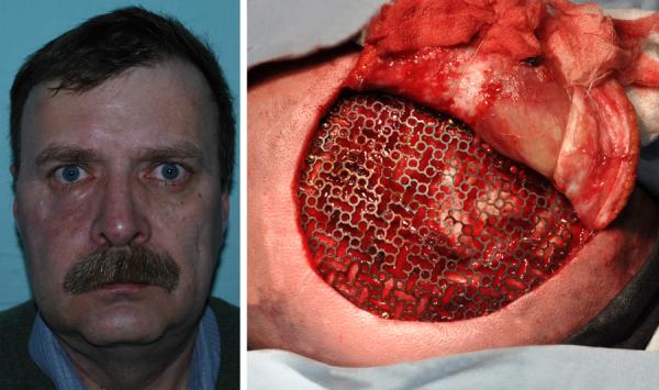
External appearance of the reconstructed orbit with the titanium mesh used for the reconstruction of the calvarial defect.
The flap was harvested from the parietal region, but had to be dissected above the temporal muscle. Therefore a painful restriction in opening the mouth appeared, caused by the scarring of the temporal muscle and the removal of the zygomatic arch. The phenomena diminished only partially by aid of kinesiotherapy and active psychological support. The association of lipostructure with active kinesiotherapy was followed by an increase in the elasticity of the temporal muscle scar and eventually helped to re-establish normal mouth opening. In order to restore the symmetry of the scalp, the reconstruction of the calvarial defect was approached by using a titanium mesh (Figure 3).
The lipostructure combined with cranioplasy further contributed to an increase in comfort by improving esthetics. The cheek lipostructure performed also led to a fast recovery of the infraorbital nerve. Using a minimally traumatizing technique for the patient, the surgical intervention succeeded in correcting the asymmetries at scheletal level, with an excellent final result in terms of esthetics. The patient perceived an increase in skin quality and elasticity; hair growth in the parietal and temporal region returned to the normal appearance prior to surgery.
The symmetry of the facial bones was not perfect but remained stable in time; three years post-surgery the appearance of the calvarial bone in the reconstructed malar region was identical to that of the bone near the harvested area (Figure 4).
Figure 4.

Postoperative external appearance. CT post-operative three year's post-surgery the appearance of the calvarial bone in the reconstructed malar region was identical to that of the bone near the harvested area
After the completion of the surgical part of the treatment, the patient was subject to a complex functional rehabilitation program to regain the shape and mobility of the temporomandibular region. The program included lymphatic drainage massage of the face to help diminish post-surgical edema and increase the elasticity of the scar tissue. Focus was mainly placed on the area affected by the surgery. Again, rehabilitation efforts specifically concentrated on the site of the surgery. The team also provided psychological consultation and follow-up to the patient, to help him cope with the temporary post-surgery disability
Discussion
Maxillary sinus mucocele is a rare condition that generally occurs after sinus surgery, possibly due to sequestration of sinus mucosa [4] or postoperative scarring causing fibrous septation of the antrum [5]. Mucoceles of the paranasal sinuses are cystic lesions lined by respiratory epithelium and can be caused by obstruction of the sinus ostium or obstruction of a mucus-secreting gland, leading to the accumulation of secretions into an expansile mass. They are commonly found in the frontal and ethmoid sinuses. There is an increased incidence of maxillary sinus mucoceles in Japan compared to the United States and Europe, which is thought to be due to an increased number of radical surgeries for sinusitis. The mucosal disruption caused by the intervention sets the stage for mucocele formation. Some suggest that compartmentalisation of the antrum after surgery may leave islands of mucosa without drainage and can eventually lead to mucocele formation.
Treatment consists not only of removing the cyst but also of restoring the initial esthetics and affected function. The shape of the orbit and, at the same time, the structure of the maxillary bone must be reconstructed as precisely as possible. Bullock [6] demonstrated a greater and more rapid resorption of the enchondral bone compared to grafts originated in the intraoral origin or cranial calvarium. The vascularised bone has a greater stability over time, as it is supplied by single or multiple vascular networks and can be raised successfully [7]. The superficial temporal network is better than others because it has a larger arch of rotation and a thin pliable pedicle. In the experienced harvesting of temporoparietal fascial flaps, the resulting traumatism is insignificant and perfectly justified by the great stability in time of the vascularised bone.
When using the pedicle from the superficial temporal fascia, the deformity in the temporal area is less visible because the temporal muscle remains in its original site. The superficial temporal vessel gives perforators to the parietal bone that anastomoses with the branches of the middle meningeal artery in this area. Hence, the full thickness cranial bone can be safely elevated on the superficial temporal vessels. This superficial temporal vascular network should limit the maximal length of the vascularised bone in order to provide enough vascular perforators. The superficial temporal fascia normally has two main vascular branches.
For this reason, in our case it was possible to create two different osteofacial flaps, based on the same vascular pedicle. Billen [8] used a similar technique, with the only difference that he did not divide the fascia between the two bone fragments. Considering the difficulty of reconstructing the malar bone and the orbit floor at the same time by using a single facial fragment, we divided the fascia to allow a precise reconstruction of the orbit. Psillakis [9] used as many as three split calvarial bone flaps with separate pedicles on the same side of the face. He repaired the mandible, the maxilla and the malar region using this method.
It has been stated that a cranial defect of 10 cm2 or less does not need to be reconstructed in the temporal area. However, we performed cranioplasty is with both a protective and an esthetic objective in mind. When harvesting a biocortical graft, duramater exposure requires a reconstruction technique, for example the placement of a titanium mesh. We chose titanium cranioplasty because of its excellent bio-properties and very low rate of associated complications such as adverse healing, wound infections, implant exposure or extrusion [10]. Temporary alopecia is the most common complication in the donor area and is frequently the result of skin flap edema, but it heals spontaneously within 3–6 months. From our experience, lipostructure can also improve the hair density in the affected area.
Moreover, careful skin flap dissection in the subfollicular plan will prevent persistent alopecia. To re-establish facial and skull symmetry while regaining the elasticity of the scarred tissue, a solution can be lipostructure. The adipose tissue is a loose connective tissue composed of adipocytes as well as mesenchymal stem cells, which can differentiate into a variety of connective tissue cell lines.
Accordingly, lipostructure provides not only the restoration of volume but also an amount of mesenchymal stem cells in scarred, atrophied tissue (resulted from the association of surgery with radiotherapy and chemotherapy, when the diagnosis requires). There is a significant improvement in the texture and trophicity of the tegument and of the other tissues in the topographical area of the lipostructure, but there remains an area of paresthesia in this territory.
The injected mesenchymal stem cells transform into young fibroblasts; neovascularization is formed and has beneficial effects on scar tissue by increasing the hydration thereof. With lipostructure, the areolar tissue surrounding the temporalis muscle can be replaced and mandibular mobility can return to normal.
The evaluation of the temporomandibular function and facial esthetics rehabilitation were made according to current methods. The process included psychological consultation and support, as well as a massage program to improve lymphatic circulation, diminish paresthesia and restore the function of the temporomandibular musculature [3].
Conclusions
Although difficult, the treatment of a maxillary sinus mucocele invading the orbit can have very good results in the end. The patient's compliance with the treatment step by step and the joint efforts of a multidisciplinary team are essential to achieve the desired results. The adequate functional rehabilitation program following surgery had a major contribution to the patient's level of psychological, aesthetic and physical comfort.
The treatment often requires complex surgery, which in our case included:
-
-
Reconstruction of both the malar bone and the orbital floor by using a superficial temporal fascia split into two osteofascial flaps and repositioning of the eye globe;
-
-
Aesthetic and functional recovery required the use of a titanium-titanium mesh in the collection area of the calvarial bone and lipostructure;
Surgery was followed by a complex orofacial functional rehabilitation program including psychological consultation and support, massage, physical therapy for restoring the functionality of the ATM from operated region. The satisfaction index of the patient was followed up for evaluation during and after treatment.
Footnotes
Publisher's Disclaimer: This is a PDF file of an unedited manuscript that has been accepted for publication. As a service to our customers we are providing this early version of the manuscript. The manuscript will undergo copyediting, typesetting, and review of the resulting proof before it is published in its final citable form. Please note that during the production process errors may be discovered which could affect the content, and all legal disclaimers that apply to the journal pertain.
References
- 1.Shinder R, Al-Zubidi N, Esmaeli B. Survey of orbital tumors at a comprehensive cancer center in the United States. Head Neck. 2011;33:610–614. doi: 10.1002/hed.21498. [DOI] [PubMed] [Google Scholar]
- 2.Palmer-Hall AM, Anderson SF. Paraocular sinus mucoceles. J Am Optom Assoc. 1997;68:725–733. [PubMed] [Google Scholar]
- 3.Cairns BE. Pathophysiology of TMD pain – basic mechanisms and their implications for pharmacotherapy. Journal of Oral Rehabilitation. 2010;37:391–410. doi: 10.1111/j.1365-2842.2010.02074.x. [DOI] [PubMed] [Google Scholar]
- 4.Kaltreider SA, Dortzbach RK. Destructive cysts of the maxillary sinus affecting the orbit. Arch Ophthalmol. 1988;106:1398–1402. doi: 10.1001/archopht.1988.01060140562023. [DOI] [PubMed] [Google Scholar]
- 5.Som PM, Shugar JM. Antral mucoceles: a new look. J Comput Assist Tomogr. 1980;4:484–488. doi: 10.1097/00004728-198008000-00015. [DOI] [PubMed] [Google Scholar]
- 6.Bullock LJ, Reeves RJ. Unilateral exophthalmos; roentgen graphic aspects. Am J Roentgenol Radium TherNucl Med. 1959;82:290–299. [PubMed] [Google Scholar]
- 7.Choung PH, Nam IW, Kim KS. Vascularized cranial bone grafts for mandible and maxillary reconstruction. J Craniomaxillofac. Surg. 1991;19:235–242. doi: 10.1016/s1010-5182(05)80063-0. [DOI] [PubMed] [Google Scholar]
- 8.Billen BT, Kilinc H, Arslan A, Aslan S. Reconstruction of orbital floor and maxilla with divided vascularized calvarial bone flap in one session. J. Plast. Reconstr. Aesthetic. Surg. 2006;59:1305–1311. doi: 10.1016/j.bjps.2005.12.048. [DOI] [PubMed] [Google Scholar]
- 9.Psillakis JM, Grotting JC, Casanova R, et al. Vascularized outer-table calvarial bone flaps. PlastReconstrSurg. 1986;78:309–317. doi: 10.1097/00006534-198609000-00005. [DOI] [PubMed] [Google Scholar]
- 10.Ducic Y. Titanium mesh and hydroxyapatite cement cranioplasty: a report of 20 cases. J Oral Maxillofac Surg. 2002;60:272–276. doi: 10.1053/joms.2002.30575. [DOI] [PubMed] [Google Scholar]