Abstract
We report on the characterization of 2 mm thick CdTe diode detector with Schottky contacts to be employed in a novel conceptual design of PET scanner. Results at −8°C with an applied bias voltage of −1000 V/mm show a 1.2% FWHM energy resolution at 511 keV. Coincidence time resolution has been measured by triggering on the preamplifier output signal to improve the timing resolution of the detector. Results at the same bias and temperature conditions show a FWHM of 6 ns with a minimum acceptance energy of 500 keV.
These results show that pixelated CdTe Schottky diode is an excellent candidate for the development of next generation nuclear medical imaging devices such as PET, Compton gamma cameras, and especially PET-MRI hybrid systems when used in a magnetic field immune configuration.
Keywords: Solid state detectors; Timing detectors; Spectrometers; Gamma detectors (scintillators, CZT, HPG, HgI etc)
1 Introduction
Cadmium Telluride (CdTe) diode detectors are good candidates to be used in many nuclear medicine applications [1-6]. The aim of this work is to prove the feasibility of the design of a PET scanner presented in [7] at the level of a single detector. A characterization of a CdTe Schottky diode of size 4 mm × 4 mm × 2 mm kept at 5 cm from the radiactive source has been done. It consists of the measurement of the energy resolution of the detector at 511 keV and the measurement of the coincidence time resolution for a configuration with two equal CdTe detectors and a 22Na source placed between them. A bias voltage of −1000 V/mm has been applied to the detector to minimize the drift time and charge trapping in order to achieve a better energy resolution. A low energy threshold has been applied to the preamplifier output signal to obtain a response as fast as possible.
2 Energy resolution
2.1 Experimental setup
The setup consists of one Schottky CdTe detector of 4 mm × 4 mm × 2 mm size, developed by ACRORAD [8], with the pixel electrode made of Au/Ti/Al, and the cathode made of Pt. Its output signal is integrated with an AMPTEK pc250 which has a preamplifier based on the charge sensitive preamplifier A250. DC coupling configuration is used to connect the detector to the board. The output of the AMPTEK pc250 is amplified in a 2 gain buffer and connected to an AMPTEK pc275, which is a shaper based on the pulse amplifier A275. The “5 poles” configuration is used. The VME Peak sensing module CAEN 785N is used to measure the maximum of the signal within a time window that is generated by discriminating the signal coming from the AMPTEK pc250 in a LeCroy quad discriminator 821, with the equivalent threshold set to 25 keV. The pulse width is 4 μs. The CAEN 785N module is controled by the DAQ software developed with Labview [9].
The bias voltage is applied with a Hivolt power supply model J4-3N. To avoid polarization effects, the bias voltage is periodically ramped down and kept at 0 V for 30 seconds before being ramped up again. This process is repeated every 15 minutes.
In order to reduce the leackage current of the detector, the whole setup is kept at −8°C [10] using an industrial fridge. A flux of nitrogen gas continously flushed the detector box inside the fridge in order to keep humidity below condensation point on the detector (Dew Point at −8 °C and 20% humidity is −27°C).
2.2 Results
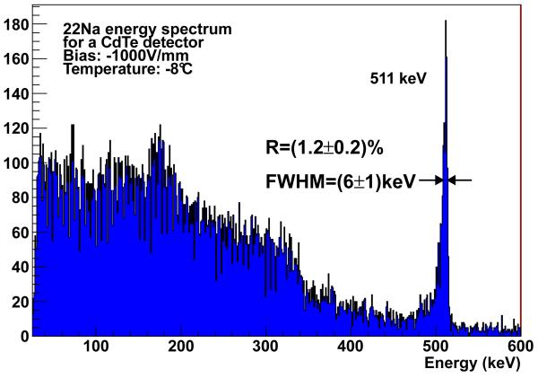
The 22Na energy spectrum obtained by the aforementioned setup is shown in figure 1. It shows an energy resolution of 6 keV FWHM at 511 keV (ΔE/E = 1.2%) which is comparable to the values presented in [1, 5, 11-13] for the same energy and similar conditions.
Figure 1.
Spectroscopy of 22Na isotope at 1000 V/mm at −8 °C. FWHM @ 511 keV = 6 keV (1.2%).
Simulations made with GAMOS [14] show that ~ 2/3 of the events with energy below 350 keV have scattered outside the detector and are detected afterwards, the remaining events are due to photons that underwent a Compton scattering inside the diode before escaping.
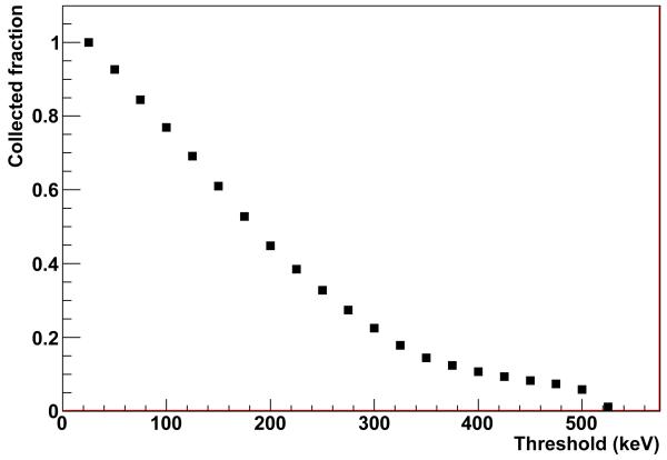
The fraction of collected events for different trigger thresholds is shown in figure 2. The photodetection efficiency decreases with increasing trigger threshold. In the case of a full PET ring, where a non-negligible fraction of 511 keV photons undergo one or more Compton interactions before being finally stopped, the system efficiency depends on the trigger threshold of the single channels. For a 100 keV energy threshold, for example, the efficiency loss of the single detector is of 20% with respect to a 25 keV threshold, whereas for PET events it would be of 62%.
Figure 2.
Fraction of collected events for different offline applied thresholds with respect to the 25 keV one.
3 Coincidence time resolution
3.1 Experimental setup
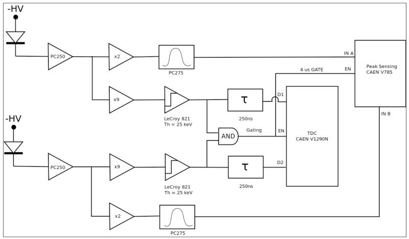
The setup consists of two identical CdTe detectors and readout systems as the ones previously described, an scheme is shown in figure 3. In order to measure the time difference between the time of detection of two photons (one per detector), the time-to-digital-converter VME (TDC) CAEN 1290N module is used.
Figure 3.
Scheme of the setup used to measure the time difference between the signals of two CdTe detectors.
The configuration of the AMPTEK pc250 and the AMPTEK pc275 is the one previously described for every detector readout. The thresholds of LeCroy quad discriminators 821 are set to the equivalent of 25 keV on the preamplifier output signal with an output pulse width of 200 ns. Signals are duplicated in a logic fan-in/fan-out. One set of the triggers are AND by CAEN 3 Fold Unit Logic N405 to enable the TDC. The other two outputs are delayed 250 ns and fed in the TDC inputs channels. The mean trigger rate was about ~1/30 Hz.
For every coincidence, three parameters are measured: the detection time difference, Δ t=ta−tb, the energy in detector A, Ea and the energy in detector B, Eb. The cooling system and the high voltage sources are the same as with the energy spectrum measurements.
3.2 Results
For each coincidence, the time difference between the detection of the two photons has been acquired. The more energetic the photon is, the bigger amount of charge carriers it generates. This leads to the conclusion that it takes less time for more energetic events to reach the threshold level of the discriminator, which makes the response signal faster. Different minimum acceptance energies have been applied in order to get different coincidence time distributions. The FWHM and the width of the region containing 70% and 90% of the coincidences have been measured for each distribution, as shown in figure 4. As expected, the FWHM improves with increasing energy threshold up to the 340 keV Compton threshold. Above such threshold, only statistical fluctuations are observed. Overall, the measured FWHM varies between 12.5 ns and 6 ns. The resulting coincidence time resolution is comparable with findings in [17] and outperforms previous results [1, 5, 15, 16].
Figure 4.
On the left is the FWHM and window width for 70% and 90% of the coincidences for distributions with minimum acceptance energy within 25–500 keV. On the right is the coincidence time measurement for −1000 V/mm at −8°C with trigger threshold at 25 keV. Minimum acceptance energy is 500 keV. FWHM = 6 ns.
4 Conclusions
Spectroscopy results show the excellent energy resolution of CdTe detectors with 1.2% at 511 keV and −8 °C. This would allow one to improve the scattered events rejection of the PET scanner (and hence improve the signal-to-noise ratio) given that state-of-the-art crystal-PMT detectors have a limited energy resolution close to 10% [18].
Results on the coincidence time resolution with a trigger threshold at 25 keV show a FWHM of 6 ns for a minimum acceptance value of the total deposited energy of 500 keV. This result is comparable with the result achieved in [17] with a 0.5 mm thick detector.
Unlike crystal-PMT based scanners, where the maximum dose allowed to avoid pile-up is limited by the processing dead time, the maximum annihilation rate of the VIP-PET is mostly dominated by the coincidence time resolution because of the large number of channels in the detector. With 20 ns coincidence time window no pile-up effect is expected up to 107 Bq activity in the field of view. The system is expected to saturate at 109 Bq [7], well above the typical integral activity of 108 Bq of a brain scan.
This work proves that pixel CdTe detectors are excellent candidates for the developing of next generation high-sensitivity/high-resolution PET scanners.
Acknowledgments
This work has been supported by FP7-ERC-Advanced Grant #250207.
References
- [1].Giboni KL, et al. Coincidence timing of schottky CdTe detectors for tomographic imaging. Nucl. Instrum. Meth. A. 2000;450:307. [Google Scholar]
- [2].Baldazzi G, et al. A radiation detection system for high-energy computerized tomography using CdZnTe detectors. IEEE Trans. Nucl. Sci. 1995;42:575. [Google Scholar]
- [3].Eisen Y, et al. CdTe and CdZnTe gamma ray detectors for medical and industrial imaging systems. Nuclear Instrum. Meth. A. 1999;428:158. [Google Scholar]
- [4].Scheiber C. CdTe and CdZnTe detectors in nuclear medicine. Nucl. Instrum. Meth. A. 2000;448:513. [Google Scholar]
- [5].Bertolucci E, et al. Timing properties of CdZnTe detectors for positron emission tomography. Nucl. Instrum. Meth. 1997;400:107. [Google Scholar]
- [6].Barber HB. Applications of semiconductor detectors to nuclear medicine. Nuclear Instrum. Meth. A. 1999;436:102. [Google Scholar]
- [7].Mikhaylova E, et al. Simulation of pseudo-clinical conditions and image quality evaluation of PET scanner based on pixelated CdTe detector. IEEE Nucl. Sci. Symp. Med. Imaging Conf. Rec. 2011;2011:2716. [Google Scholar]
- [8].ACRORAD webpage: http://www.acrorad.co.jp/.
- [9].Labview support webpage: www.ni.com/labview/.
- [10].Bale G, et al. Cooled CdZnTe detectors for x-ray astronomy. Nucl. Instrum. Meth. A. 1999;436:150. [Google Scholar]
- [11].Takahashi T, et al. High-resolution shottky CdTe diode for hard x-ray and gamma-ray astronomy. Nucl. Instrum. Meth. A. 1999;436:111. [Google Scholar]
- [12].Watanabe S, et al. CdTe stacked detectors for gamma-ray detection. IEEE Trans. Nucl. Sci. 2002;4:2434. [Google Scholar]
- [13].Casali F, et al. Characterization of small CdTe detectors to be used for linear and matrix arrays. IEEE Trans. Nucl. Sci. 1992;39:598. [Google Scholar]
- [14].Arce P, et al. GAMOS: An easy and flexible way to use GEANT4. IEEE Nucl. Sci. Symp. Med. Imaging Conf. Rec. 2011;2011:2230. [Google Scholar]
- [15].Amrami R, et al. Timing performance of pixelated CdZnTe detectors. Nucl. Instrum. Meth. A. 2001;458:772. [Google Scholar]
- [16].Shao Y, et al. Measurement of coincidence timing resolution with CdTe detectors. Proc. SPIE. 2000;4142:254. [Google Scholar]
- [17].Okada Y, et al. CdTe and CdZnTe detectors for timing measurements. IEEE Trans. Nucl. Sci. 2002;4:2429. [Google Scholar]
- [18].Karp JS, et al. Performance of a brain pet camera based on anger-logic gadolinum oxyorthosilicate detectors. J. Nucl. Med. 2003;44:1340. [PubMed] [Google Scholar]


