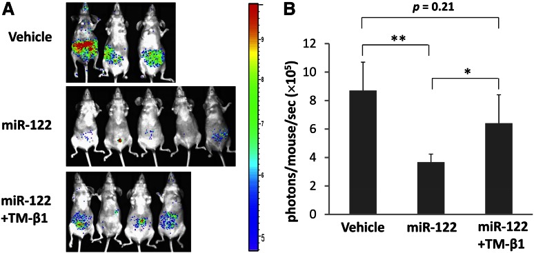
Figure 6.
miRNA stimulation enhances antitumor activity of NK cells in vivo. (A) Ventral bioluminescence imaging of mice bearing A20 lymphoma. Athymic nude mice were injected with 1 × 10⁵ luc-expressing A20 cells via tail veins and subjected to miRNA stimulation alone or combined with TM-β1 treatment according to the schedule described in the Materials and Methods section. The pseudo color indicates the relative signal strength for tumor growth, with strongest in red and weakest in purple. (B) Quantification summary of units of photons per second per mouse from (A). Data are shown as mean ± standard deviation from each group of mice. *P < .05; **P < .01.

