Abstract
A canine distemper virus (CDV) strain, CYN07-dV, associated with a lethal outbreak in monkeys, used human signaling lymphocyte activation molecule as a receptor only poorly but readily adapted to use it following a P541S substitution in the hemagglutinin protein. Since CYN07-dV had an intrinsic ability to use human nectin-4, the adapted virus became able to use both human immune and epithelial cell receptors, as well as monkey and canine ones, suggesting that CDV can potentially infect humans.
TEXT
Signaling lymphocyte activation molecule (SLAM) is a common receptor used by morbilliviruses, such as measles virus (MV), canine distemper virus (CDV), rinderpest virus, and peste des petits ruminants virus, to enter and spread in their hosts (1–3). However, each morbillivirus infects specific animal species (4). They preferentially use the SLAM of their host animals, because there are significant amino acid differences among human, canine, bovine, and goat SLAM (2, 5–7). In part, this receptor specificity generates a species barrier for morbilliviruses. On the other hand, nectin-4, another morbillivirus receptor (8–10), is highly conserved in its amino acid sequence among different mammals and, thus, may play only a minor role in determining the host specificity of morbilliviruses (11, 12). Recently, however, CDV outbreaks have emerged in a variety of mammals, including nonhuman primates, and showed high mortality rates (11, 13–15). The repeated lethal CDV outbreaks in monkeys in recent years clearly demonstrate that CDV is now a real threat for monkeys (11). Our concern is the potential risk of CDV infection in humans (16), since SLAM and nectin-4 both show high homology in their amino acid sequences between monkeys and humans (11). Indeed, MV can spread and cause a measles-like illness in monkeys (17–22). Our recent study demonstrated that a CDV strain, CYN07-dV, which was associated with a lethal CDV outbreak in monkeys in Japan, utilizes macaque SLAM (macSLAM) and macaque nectin-4 (macNectin4) as efficiently as canine SLAM (dSLAM) and canine nectin-4 (dNectin4), respectively (11). In the present study, the potential of CYN07-dV to utilize human SLAM (hSLAM) and human nectin-4 (hNectin4) was analyzed.
Our previous study demonstrated that CYN07-dV utilized hSLAM poorly, even though it utilized macSLAM efficiently (11). However, we could not exclude the possibility that CDV already adapted to macSLAM was isolated in dSLAM-expressing Vero.DogSLAMtag cells (6), resulting in the selection of a CDV with low affinity for hSLAM during CYN07-dV isolation. Therefore, we tried to isolate CDV directly from the tissues of the moribund monkey from which CYN07-dV was isolated (11) using Vero cells expressing hSLAM (Vero/hSLAM) (23). However, no CDV was directly isolated from the monkey specimens in Vero/hSLAM cells, despite the fact that it was easily isolated in Vero.DogSLAMtag cells, indicating that a quasispecies of CDV with high affinity for hSLAM was not circulating in the monkey. Therefore, CDV strains causing a severe disease in monkeys are unlikely to use hSLAM efficiently.
Although no hSLAM-using virus was isolated directly from the monkey tissues, a small number of syncytia were detected in Vero/hSLAM cells at 3 days postinfection (p.i.) when the cells were infected with the isolated CYN07-dV strain (11) at a multiplicity of infection (MOI) of 0.3 (data not shown). At 5 days p.i., the monolayers developed extensive syncytia (data not shown). This experiment was repeated six times. In four of the six trials, hSLAM-using viruses were isolated, showing that CYN07-dV adapted relatively easily to grow in Vero/hSLAM cells. The failure to isolate hSLAM-using CDV directly from the monkey tissues was predicted to arise through insufficient amounts of infectious viruses in the tissues. From the isolates in the six trials, a CDV strain was obtained by a plaque-cloning procedure and passaged twice in Vero/hSLAM cells to produce sufficient amounts of viral stocks. This CDV strain was designated CYN07-hV. The replication kinetics of CYN07-hV were analyzed in Vero cells expressing canine receptors (Vero.DogSLAMtag and Vero/dNectin4) (10), macaque receptors (Vero/macSLAM and Vero/macNectin4) (11), and human receptors (Vero/hSLAM and Vero/hNectin4) (12) and in the parental Vero cells. Although CYN07-hV replicated inefficiently in the parental Vero cells, its replication rates were greatly enhanced in all other cell lines expressing the canine, macaque, and human receptors, including Vero/hSLAM cells (Fig. 1). Thus, CYN07-hV showed significantly different growth kinetics than CYN07-dV in Vero/hSLAM cells (11). The growth kinetics of CYN07-hV in Vero/hSLAM cells were comparable to those in Vero.DogSLAMtag and Vero/macSLAM cells, although the peak titer in Vero/hSLAM cells was higher than those in the other cells (Fig. 1). The peak titer in Vero/hNectin4 cells reached a level comparable to those in Vero/dNectin4 and Vero/macNectin4 cells (Fig. 1). No syncytia were observed in the parental Vero cells and Vero/hSLAM cells infected with CYN07-dV (Fig. 2), as reported previously (11). However, CYN07-hV induced syncytia in Vero/hSLAM cells, as observed in Vero.DogSLAMtag and Vero/macSLAM cells (Fig. 2).
Fig 1.
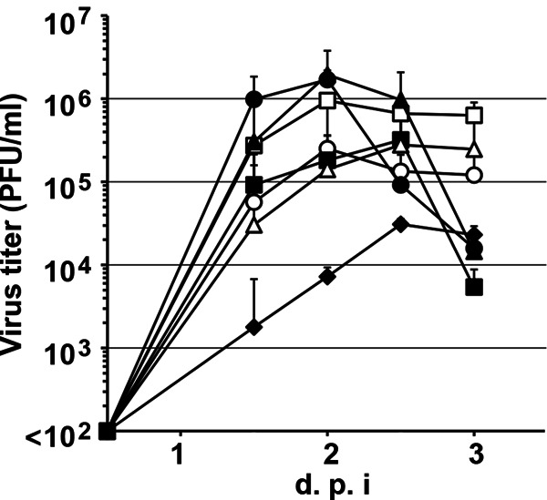
Replication kinetics of CYN07-hV in various cells. Vero.DogSLAMtag, Vero/macSLAM, Vero/hSLAM, Vero/dNectin4, Vero/macNectin4, Vero/hNectin4, and parental Vero cells were infected with CYN07-hV at an MOI of 0.01, and the titers were determined at the indicated time points. Filled circles, squares, triangles, and diamonds indicate the growth kinetics in Vero.DogSLAMtag, Vero/macSLAM, Vero/hSLAM, and parental Vero cells, respectively. Open circles, squares, and triangles indicate the growth kinetics in Vero/dNectin4, Vero/macNectin4, and Vero/hNectin4 cells, respectively. The data shown are the means of three independent assays, with the error bars representing the standard deviations. d.p.i., days postinfection.
Fig 2.
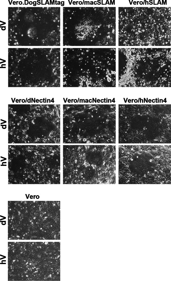
Syncytium induction in various cells upon infection with CYN07-hV and CYN07-dV. Vero.DogSLAMtag, Vero/macSLAM, Vero/hSLAM, Vero/dNectin4, Vero/macNectin4, Vero/hNectin4, and Vero cells were infected with CYN07-hV and CYN07-dV at an MOI of 0.01, cultured, and observed under a phase-contrast microscope. Syncytia induced in Vero.DogSLAMtag, Vero/macSLAM, and Vero/hSLAM cells at 24 h p.i., in Vero/dNectin4, Vero/macNectin4, Vero/hNectin4 cells at 36 h p.i., and in Vero cells at 36 h p.i. are shown.
It became clear that CYN07-hV had adapted to grow efficiently in Vero/hSLAM cells. To reveal the genetic changes in CYN07-hV compared with the parental CYN07-dV sequence, the entire genome nucleotide sequence of CYN07-hV was determined (DDBJ/GenBank accession number AB687721) using previously reported methods (11). There were only three nucleotide differences between the CYN07-dV and CYN07-hV genomes. The differences were uracil-to-adenine, guanine-to-adenine, and cytosine-to-uracil substitutions at nucleotide positions 1798, 8038, and 8699 (U1798A, G8038A, and C8699U), respectively. The latter two changes were located in the hemagglutinin (H) gene. G8038A was a synonymous change, while C8699U was predicted to cause a proline-to-serine change at amino acid position 541 (P541S) in the H protein. U1798A was located in the untranslated region of the phosphoprotein (P) gene, corresponding to the 5′ untranslated region of P mRNA. The C8699U mutation responsible for encoding the P541S change in the H protein of CYN07-hV was likely to have been introduced during the passages of CYN07-dV in Vero/hSLAM cells, since a deep sequencing analysis of CYN07-dV using a Roche GS Junior did not detect any single-nucleotide polymorphisms at that position and direct sequencing of the PCR product of the H gene amplified from the monkey tissues showed C at nucleotide position 8699 (data not shown). Sequence alignments revealed that amino acid position 541 in the CDV H protein corresponds to position 545 in the MV H protein (see Fig. S1 in the supplemental material). Residue 545 of the MV H protein is located at a position proximal to the receptor-binding site (24–26). The proline residue at this position is highly conserved among CDV strains. The MV H protein also possesses a proline residue but not a serine residue (see Fig. S1). No CDV strains with the P541S mutation have been reported to date.
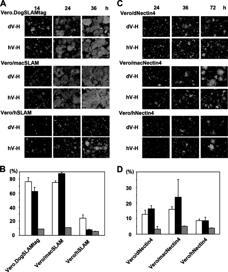
To clarify whether the P541S mutation was responsible for the adaptation to use hSLAM as a receptor, cell-to-cell fusion in various cells was analyzed after transfection of plasmids expressing the F and H proteins, as previously reported (11). Albeit less efficiently, apparent syncytia developed in Vero/hSLAM cells, as well as in Vero.DogSLAMtag and Vero/macSLAM cells, when the CYN07-hV H protein was expressed together with the F protein (Fig. 3A). On the other hand, when the CYN07-dV H protein was expressed instead of the CYN07-hV H protein, no syncytia were observed in Vero/hSLAM cells (Fig. 3A). The sizes of syncytia were similar in cultures with expression of the CYN07-dV or CYN07-hV H protein in Vero.DogSLAMtag and Vero/macSLAM cells (Fig. 3A and B). In Vero/dNectin4, Vero/macNectin4, and Vero/hNectin4 cells, expression of the CYN07-hV H protein together with the F protein induced syncytium formation, although the syncytia in nectin-4-expressing cells were smaller than those in SLAM-expressing cells (Fig. 3C and D). Again, the sizes of syncytia were similar in cultures with expression of the CYN07-dV or CYN07-hV H protein in Vero/dNectin4, Vero/macNectin4, and Vero/hNectin4 cells (Fig. 3C and D). These data demonstrated that the P541S mutation conferred on the CYN07-hV H protein the ability to use hSLAM as a receptor. For further clarification, vesicular stomatitis viruses (VSVs) pseudotyped with the H and F proteins of CYN07-hV and CYN07-dV (VSVΔG*-F-hVH and VSVΔG*-F-dVH, respectively) and with the F protein alone (VSVΔG*-F) were generated as previously reported (11), and the infectivities of these pseudotyped viruses were analyzed in Vero cells expressing SLAM or nectin-4. VSVΔG*-F showed very low infectivity titers (background titers) in all cell types (Fig. 4). VSVΔG*-F-dVH also showed low infectivity titers (similar to the background titers) in Vero and Vero/hSLAM cells (Fig. 4). However, VSVΔG*-F-hVH showed high infectivity titers in Vero/hSLAM cells, as observed in Vero.DogSLAMtag and Vero/macSLAM cells (Fig. 4). VSVΔG*-F-hVH and VSVΔG*-F-dVH showed similar infectivity titers in Vero/macNectin4 cells, Vero/dNectin4 cells, and Vero/hNectin4 cells (Fig. 4). These data confirmed that the CYN07-hV H protein acquired the ability to use hSLAM via the P541S mutation without losing its ability to use SLAM and nectin-4 of other animals.
Fig 3.
Cell-to-cell fusion induction in cells expressing the CDV CYN07-dV or CYN07-hV H and F proteins. Vero.DogSLAMtag, Vero/macSLAM, Vero/hSLAM, Vero/dNectin4, Vero/macNectin4, and Vero/hNectin4 cells were transfected with a mixture of plasmids encoding enhanced green fluorescent protein (EGFP) (pEGFP-C1) and CDV F (pGAGGS-CYN-F) and H (pCAGGS-CYN-hV-H or pCAGGS-CYN-dV-H) proteins, cultured, and observed using a fluorescence microscope. (A, C) Fluorescence microscopic images of SLAM-expressing cells at 14, 24, and 36 h posttransfection (A) and those of nectin-4-expressing cells (C) at 24, 36, and 72 h posttransfection are shown. (B, D) The percentages of fluorescence areas in the total microscopic field were measured by using imageJ software (version 1.36b, NIH). White and black bars indicate data for experiments using pCAGGS-CYN-hV-H and pCAGGS-CYN-dV-H, respectively. Gray bars indicate data without the H-encoding plasmid. The data shown are the means of three independent assays (36 h posttransfection for SLAM-expressing cells [B] and 72 h posttransfection for nectin-4-expressing cells [D]), with the error bars representing the standard deviations.
Fig 4.

Infectivities of pseudotyped VSVs bearing the CDV H and F proteins. Vero.DogSLAMtag, Vero/macSLAM, Vero/hSLAM, Vero/dNectin4, Vero/macNectin4, Vero/hNectin4, and parental Vero cells were infected with EGFP-expressing VSVs pseudotyped with the H and F proteins of CYN07-hV and CYN07-dV (VSVΔG*-F-hVH and VSVΔG*-F-dVH, respectively) and with the F protein alone (VSVΔG*-F). The numbers of cells expressing EGFP were counted at 24 h p.i., and the infectivity titers of each pseudotype in the cells were calculated. The data shown are the means of three independent assays, with the error bars representing the standard deviations.
However, it remained unclear whether the P541S mutation was necessarily required for hSLAM adaptation. Therefore, we analyzed the H protein amino acid sequences of four other hSLAM-using CDV isolates obtained in the six trials described above. One isolate possessed the same P541S change, while the other three showed different amino acid changes in the H protein. Specifically, two isolates exhibited a D540G change and the other had an R519S change. Based on the crystal structure data of the MV H protein (24), amino acid positions 540 and 519 of the CDV H protein were also predicted to be located at positions proximal to or at the SLAM-binding site (data not shown). These data suggest that there are several possible amino acid changes that can confer on the CDV H protein the ability to use hSLAM. To examine the specificity of the CYN07-dV strain, the P541S mutation was introduced into the H protein of two CDV strains, Ac96I and MSA5, isolated from dogs using Vero.DogSLAMtag cells (27). Unlike the CYN07-dV strain, the P541S mutation did not confer hSLAM-using ability on the H proteins of Ac96I and MSA5; however, the mutation instead compromised their abilities to support cell-to-cell fusion, even in Vero.DogSLAMtag cells (Fig. 5). These data indicate a unique potential of the CYN07-dV strain to adapt to humans or that some other mutations in the H protein of Ac96I and MSA5 strains are required to confer hSLAM-using ability.
Fig 5.
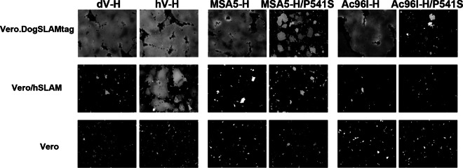
Effects of the P541S substitution on the cell-to-cell fusion-supporting function of different CDV H proteins. Vero.DogSLAMtag, Vero/hSLAM, and parental Vero cells were transfected with a mixture of plasmids encoding EGFP (pEGFP-C1), CDV F protein (pGAGGS-CYN-F), and different H proteins as follows: H proteins of the CYN07-hV, MSA5, and Ac96I strains and those possessing the P541S mutation (the H protein of CYN07-dV possessing the P541S mutation corresponds to the CYN07-hV H protein). The cells were then cultured and observed using a fluorescence microscope. Cell fusion induced at 36 h posttransfection is shown.
Finally, we isolated a CDV strain, CYN07-macV, using Vero/macSLAM cells from the tissues of the same moribund monkey from which CYN07-dV was isolated (11), and analyzed its phenotype. CYN07-macV had the same H protein amino acid sequence as CYN07-dV and showed growth phenotypes similar to those of CYN07-dV (Fig. 6). The CYN07-macV strain did not produce syncytia in Vero/hSLAM cells but did so efficiently in Vero.DogSLAMtag, Vero/macSLAM, Vero/dNectin4, Vero/macNectin4, and Vero/hNectin4 cells (Fig. 6). These data suggest that CYN07-dV could be a representative of the strain that caused a lethal outbreak in monkeys.
Fig 6.
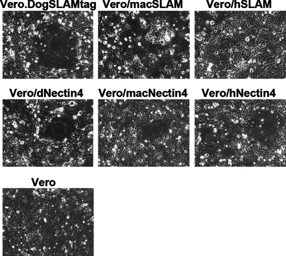
Syncytium induction in various cells upon infection with CYN07-macV. Vero.DogSLAMtag, Vero/macSLAM, Vero/hSLAM, Vero/dNectin4, Vero/macNectin4, Vero/hNectin4, and Vero cells were infected with CYN07-macV at an MOI of 0.01, cultured, and observed under a phase-contrast microscope. Syncytia induced at 36 h p.i. are shown.
CYN07-dV could not efficiently utilize hSLAM (11). Therefore, it is unlikely that CDV strains like CYN07-dV can spread in humans, since SLAM-using ability is critical for MV, and probably also for CDV, to spread in vivo (28–30). Although a previous study by Tatsuo et al. (2) showed that the wild-type CDV HA7 strain uses hSLAM, the SLAM-using ability of the HA7 strain seemed to have been acquired during passages in B95a cells, which express marmoset SLAM (6). The same group later showed that the wild-type CDV strain isolated using Vero.DogSLAMtag cells does not use hSLAM (7). However, the present data clearly demonstrate that CYN07-dV easily adapted to use hSLAM without losing its intrinsic ability to use human nectin-4. Moreover, the P541S mutation in the CYN07-hV H protein did not affect the affinities for the SLAM and nectin-4 of macaque monkeys and dogs, indicating that CDV strains like CYN07-hV are likely to infect monkeys and dogs. Thus, the incompatibility of hSLAM is easily resolvable for CDV strains like CYN07-dV to cross the species barrier to humans when the virus is infected into humans following direct contact with CDV-infected animals. In addition, our recent study suggested that CDV has the potential to counteract the human interferon system and replicate in human cells (12). Accordingly, CDV should be seriously considered a potential threat for humans. We propose that actions for the preparation of measures against CDV infection in humans must be started.
Supplementary Material
ACKNOWLEDGMENTS
We thank Yusuke Yanagi for providing the Vero.DogSLAMtag, Vero/hSLAM, and parental Vero cells and M. A. Whitt for providing the VSV pseudotype system.
This work was partly supported by grants from the Ministry of Education, Culture, Sports, Science and Technology, the Japan Society for the Promotion of Science [Grant-in-Aid for Young Scientists (B), 22700459], and the Ministry of Health, Labor and Welfare (grants H22-shinkou-ippan-006, H22-shinkou-ippan-012, and H24-kokui-shitei-003).
Footnotes
Published ahead of print 17 April 2013
Supplemental material for this article may be found at http://dx.doi.org/10.1128/JVI.03479-12.
REFERENCES
- 1. Tatsuo H, Ono N, Tanaka K, Yanagi Y. 2000. SLAM (CDw150) is a cellular receptor for measles virus. Nature 406:893–897 [DOI] [PubMed] [Google Scholar]
- 2. Tatsuo H, Ono N, Yanagi Y. 2001. Morbilliviruses use signaling lymphocyte activation molecules (CD150) as cellular receptors. J. Virol. 75:5842–5850 [DOI] [PMC free article] [PubMed] [Google Scholar]
- 3. Adombi CM, Lelenta M, Lamien CE, Shamaki D, Koffi YM, Traore A, Silber R, Couacy-Hymann E, Bodjo SC, Djaman JA, Luckins AG, Diallo A. 2011. Monkey CV1 cell line expressing the sheep-goat SLAM protein: a highly sensitive cell line for the isolation of peste des petits ruminants virus from pathological specimens. J. Virol. Methods 173:306–313 [DOI] [PMC free article] [PubMed] [Google Scholar]
- 4. DeLay PD, Stone SS, Karzon DT, Katz S, Enders J. 1965. Clinical and immune response of alien hosts to inoculation with measles, rinderpest, and canine distemper viruses. Am. J. veterinary research. 26:1359–1373 [PubMed] [Google Scholar]
- 5. Tatsuo H, Yanagi Y. 2002. The morbillivirus receptor SLAM (CD150). Microbiol. Immunol. 46:135–142 [DOI] [PubMed] [Google Scholar]
- 6. Seki F, Ono N, Yamaguchi R, Yanagi Y. 2003. Efficient isolation of wild strains of canine distemper virus in Vero cells expressing canine SLAM (CD150) and their adaptability to marmoset B95a cells. J. Virol. 77:9943–9950 [DOI] [PMC free article] [PubMed] [Google Scholar]
- 7. Ohno S, Seki F, Ono N, Yanagi Y. 2003. Histidine at position 61 and its adjacent amino acid residues are critical for the ability of SLAM (CD150) to act as a cellular receptor for measles virus. J. Gen. Virol. 84:2381–2388 [DOI] [PubMed] [Google Scholar]
- 8. Muhlebach MD, Mateo M, Sinn PL, Prufer S, Uhlig KM, Leonard VH, Navaratnarajah CK, Frenzke M, Wong XX, Sawatsky B, Ramachandran S, McCray PB, Jr, Cichutek K, von Messling V, Lopez M, Cattaneo R. 2011. Adherens junction protein nectin-4 is the epithelial receptor for measles virus. Nature 480:530–533 [DOI] [PMC free article] [PubMed] [Google Scholar]
- 9. Noyce RS, Bondre DG, Ha MN, Lin LT, Sisson G, Tsao MS, Richardson CD. 2011. Tumor cell marker PVRL4 (nectin 4) is an epithelial cell receptor for measles virus. PLoS Pathog. 7:e1002240 doi: 10.1371/journal.ppat.1002240 [DOI] [PMC free article] [PubMed] [Google Scholar]
- 10. Pratakpiriya W, Seki F, Otsuki N, Sakai K, Fukuhara H, Katamoto H, Hirai T, Maenaka K, Techangamsuwan S, Lan NT, Takeda M, Yamaguchi R. 2012. Nectin4 is an epithelial cell receptor for canine distemper virus and involved in the neurovirulence. J. Virol. 86:10207–10210 [DOI] [PMC free article] [PubMed] [Google Scholar]
- 11. Sakai K, Nagata N, Ami Y, Seki F, Suzaki Y, Iwata N, Suzuki T, Fukushi S, Mizutani T, Yoshikawa T, Otsuki N, Kurane I, Komase K, Yamaguchi R, Hasegawa H, Saijo M, Takeda M, Morikawa S. 2013. Lethal canine distemper virus outbreak in cynomolgus monkeys in Japan in 2008. J. Virol. 87:1105–1114 [DOI] [PMC free article] [PubMed] [Google Scholar]
- 12. Otsuki N, Sekizuka T, Seki F, Sakai K, Kubota T, Nakatsu Y, Chan S, Fukuhara H, Maenaka K, Yamaguchi R, Kuroda M, Takeda M. 2013. Canine distemper virus with the intact C protein has the potential to replicate in human epithelial cells by using human nectin4 as a receptor. Virology 435:485–492 [DOI] [PubMed] [Google Scholar]
- 13. Qiu W, Zheng Y, Zhang S, Fan Q, Liu H, Zhang F, Wang W, Liao G, Hu R. 2011. Canine distemper outbreak in rhesus monkeys, China. Emerg. Infect. Dis. 17:1541–1543 [DOI] [PMC free article] [PubMed] [Google Scholar]
- 14. Sun Z, Li A, Ye H, Shi Y, Hu Z, Zeng L. 2010. Natural infection with canine distemper virus in hand-feeding Rhesus monkeys in China. Vet. Microbiol. 141:374–378 [DOI] [PubMed] [Google Scholar]
- 15. Yoshikawa Y, Ochikubo F, Matsubara Y, Tsuruoka H, Ishii M, Shirota K, Nomura Y, Sugiyama M, Yamanouchi K. 1989. Natural infection with canine distemper virus in a Japanese monkey (Macaca fuscata). Vet. Microbiol. 20:193–205 [DOI] [PubMed] [Google Scholar]
- 16. Nicolle C. 1931. La maladie du jeune age des chiens est transmissible expérimantalement à l'homme sous forme inapparente. Comp. Rendus Acad. Sci. 193:1069–1071 [Google Scholar]
- 17. de Swart RL. 2009. Measles studies in the macaque model. Curr. Top. Microbiol. Immunol. 330:55–72 [DOI] [PMC free article] [PubMed] [Google Scholar]
- 18. Kobune F, Takahashi H, Terao K, Ohkawa T, Ami Y, Suzaki Y, Nagata N, Sakata H, Yamanouchi K, Kai C. 1996. Nonhuman primate models of measles. Lab. Anim. Sci. 46:315–320 [PubMed] [Google Scholar]
- 19. Auwaerter PG, Rota PA, Elkins WR, Adams RJ, DeLozier T, Shi Y, Bellini WJ, Murphy BR, Griffin DE. 1999. Measles virus infection in rhesus macaques: altered immune responses and comparison of the virulence of six different virus strains. J. Infect. Dis. 180:950–958 [DOI] [PubMed] [Google Scholar]
- 20. Bankamp B, Hodge G, McChesney MB, Bellini WJ, Rota PA. 2008. Genetic changes that affect the virulence of measles virus in a rhesus macaque model. Virology 373:39–50 [DOI] [PubMed] [Google Scholar]
- 21. McChesney MB, Fujinami RS, Lerche NW, Marx PA, Oldstone MB. 1989. Virus-induced immunosuppression: infection of peripheral blood mononuclear cells and suppression of immunoglobulin synthesis during natural measles virus infection of rhesus monkeys. J. Infect. Dis. 159:757–760 [DOI] [PubMed] [Google Scholar]
- 22. McChesney MB, Miller CJ, Rota PA, Zhu YD, Antipa L, Lerche NW, Ahmed R, Bellini WJ. 1997. Experimental measles. I. Pathogenesis in the normal and the immunized host. Virology 233:74–84 [DOI] [PubMed] [Google Scholar]
- 23. Ono N, Tatsuo H, Hidaka Y, Aoki T, Minagawa H, Yanagi Y. 2001. Measles viruses on throat swabs from measles patients use signaling lymphocytic activation molecule (CDw150) but not CD46 as a cellular receptor. J. Virol. 75:4399–4401 [DOI] [PMC free article] [PubMed] [Google Scholar]
- 24. Hashiguchi T, Ose T, Kubota M, Maita N, Kamishikiryo J, Maenaka K, Yanagi Y. 2011. Structure of the measles virus hemagglutinin bound to its cellular receptor SLAM. Nat. Struct. Mol. Biol. 18:135–141 [DOI] [PubMed] [Google Scholar]
- 25. Tahara M, Takeda M, Shirogane Y, Hashiguchi T, Ohno S, Yanagi Y. 2008. Measles virus infects both polarized epithelial and immune cells by using distinctive receptor-binding sites on its hemagglutinin. J. Virol. 82:4630–4637 [DOI] [PMC free article] [PubMed] [Google Scholar]
- 26. Zhang X, Lu G, Qi J, Li Y, He Y, Xu X, Shi J, Zhang CW, Yan J, Gao GF. 2013. Structure of measles virus hemagglutinin bound to its epithelial receptor nectin-4. Nat. Struct. Mol. Biol. 20:67–72 [DOI] [PubMed] [Google Scholar]
- 27. Lan NT, Yamaguchi R, Hirai T, Kai K, Morishita K. 2009. Relationship between growth behavior in Vero cells and the molecular characteristics of recent isolated classified in the Asia 1 and 2 groups of canine distemper virus. J. Vet. Med. Sci. 71:457–461 [DOI] [PubMed] [Google Scholar]
- 28. de Swart RL, Ludlow M, de Witte L, Yanagi Y, van Amerongen G, McQuaid S, Yuksel S, Geijtenbeek TB, Duprex WP, Osterhaus AD. 2007. Predominant infection of CD150(+) lymphocytes and dendritic cells during measles virus infection of macaques. PLoS Pathog. 3:e178 doi: 10.1371/journal.ppat.0030178 [DOI] [PMC free article] [PubMed] [Google Scholar]
- 29. Leonard VH, Hodge G, Reyes-Del Valle J, McChesney MB, Cattaneo R. 2010. Measles virus selectively blind to signaling lymphocytic activation molecule (SLAM; CD150) is attenuated and induces strong adaptive immune responses in rhesus monkeys. J. Virol. 84:3413–3420 [DOI] [PMC free article] [PubMed] [Google Scholar]
- 30. Sawatsky B, Wong XX, Hinkelmann S, Cattaneo R, von Messling V. 2012. Canine distemper virus epithelial cell infection is required for clinical disease but not for immunosuppression. J. Virol. 86:3658–3666 [DOI] [PMC free article] [PubMed] [Google Scholar]
Associated Data
This section collects any data citations, data availability statements, or supplementary materials included in this article.


