

Abstract
INTRODUCTION
An infected urachal cyst is one of a spectrum of presentations of urachal pathology, all of which are rare in adulthood.
PRESENTATION OF CASE
We report the case of a 45-year-old obese Russian lady who presented with a 2-week history of suprapubic pain radiating to the right iliac fossa. Although previously fit and well, she had a history of 17 miscarriages. Both USS and CT suggested a complicated inflammatory mass in the lower abdomen. Ultimately the diagnosis was made by laparotomy, which revealed an abscess of an urachal cyst. The infected cyst and bladder dome were excised. The patient made a good recovery with an uneventful follow up.
DISCUSSION
Urachal cysts are the commonest type of urachal anomaly. Infection is the usual mode of presentation amongst adult cases otherwise the condition usually remains asymptomatic. An infected urachal cyst is an important diagnosis to make as complications include sepsis, fistula formation, and rupture leading to peritonitis. Treatment is by complete excision, however, techniques have been debated.
CONCLUSION
This is a rare but important diagnosis however we recommend that in patients with atypical histories, it should be included in the differential diagnosis.
Keywords: Urachus, Urachal cyst, Infected urachal cyst
1. Introduction
An infected urachal cyst is one of a spectrum of presentations of urachal pathology, all of which are rare in adulthood. Patients tend to present in a heterogeneous fashion, making diagnosis difficult. Ultrasound (USS), computed tomography (CT) and magnetic resonance imaging (MRI) will all assist in making this important diagnosis. Delay in treatment may have serious consequences as complications include sepsis, fistula formation, and rupture leading to peritonitis. We present a case of an infected urachal cyst presenting as an acute abdomen in a middle aged female. We will also discuss the background and review the literature regarding infected urachal cysts and their management (Table 1).
Table 1.
Radiological findings of a urachal cyst.
| Mode | Findings of a non-infected urachal cyst | Findings of a infected urachal cyst |
|---|---|---|
| USS | A smooth edged, cystic lesion in the lower abdominal wall may be found. | A thick, irregular wall with mixed internal echogenicity may be seen. |
| CT | A thin walled, homogeneous and non-enhancing cystic mass between transverse fascia and parietal peritoneum with no connection between cyst and other structures. | Irregular and thickening wall and in homogeneous attenuation higher than water is observed. |
| MRI | A urachal cyst may be seen as a well-defined, thin wall cystic structure with sagittal images helpful in demonstrating anomalies between umbilicus and urinary bladder dome. | |
| For morphologic display, non-infected urachal cysts show homogeneous isointense or hyperintense signals dependent upon image study. Contrast enhanced studies are needed to improve imaging. | ||
2. Presentation of case
A 45-year-old obese Russian lady was admitted via the surgical admissions unit with a 2-week history of worsening suprapubic pain radiating to right iliac fossa. Excluding some intermittent fevers there were no additional symptoms of note. The patient was previously fit and well, however had a previous history of 17 spontaneous miscarriages.
Due to the patient's body habitus abdominal examination was difficult but revealed an exquisitely tender mass in the suprapubic region extending into the right iliac fossa. It was around 8 cm × 6 cm in size and seemed non-fluctuant and mobile. She looked unwell and was clinically septic with pulse rate of 105 bpm, respiratory rate of 28, temperature of 38.8 °C and raised inflammatory markers. At this point the patient was started on wide spectrum intravenous antibiotics.
USS abdomen (see Fig. 1) and CT abdomen and pelvis (see Fig. 2) revealed a 6.4 cm × 6.1 cm sized inflammatory mass in the right lower abdomen which was inseparable from the rectus abdominis (above) and small bowel (below). The mass was multiloculated and heterogeneous in density. The large bowel appeared normal.
Fig. 1.

USS abdomen showing a large multiloculated cyst of mixed echogenicity.
Fig. 2.
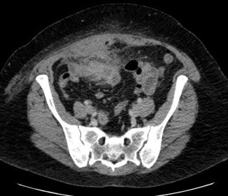
CT abdo/pelvis showing a large inflammatory mass right of midline inseparable from the rectus sheath and small bowel.
Due to its multiloculated nature, the mass was not suitable for aspiration. Given her acute presentation and septic clinical picture the patient was taken for an exploratory laparotomy.
The laparotomy was jointly performed by a general surgeon and urologist and revealed an abscess of an urachal cyst surrounded by an inflammatory mass which extended into the dome of the bladder. The inflammatory mass involved the omentum and small bowel. The infected urachal cyst, and bladder dome were excised and six inches of small bowel was also resected.
The patient had an uneventful recovery and was discharged on day 8. She was reviewed 6 weeks post-operatively and subsequently discharged from any further follow up.
3. Discussion
The urachus (or median umbilical ligament), developmentally is the upper part of the bladder, both of which arise from the ventral part of the cloaca and allantois.1 Descent of the bladder from the 5th month of development into the foetal pelvis pulls the urachus with it resulting in the formation of the urachal canal. This canal progressively obliterates during foetal life, forming a fibrous tract in early adult life with no function.
At the end of development, the urachus lies between the transverse fascia anteriorly and the peritoneum posteriorly (space of Retzius), surrounded by loose areolar tissue attaching the umbilicus to the bladder dome, being 3–10 cm in length and 8–10 mm in diameter.
Histologically, it is composed of 3 layers; an innermost layer of modified transitional epithelium similar to urothelium, a middle fibroconnective tissue layer and an outer layer of smooth muscle continuous with the detrusor.1,2
Incomplete regression of the urachal lumen results in several anomalies:
-
a.
Urachal sinus. The urachus is patent at its umbilical side, but not the bladder, resulting in umbilical mass or inflammation with or without periodic discharge with fever and leucocytosis. A thickened midline tubular structure below the umbilicus can be seen by ultrasound scan (USS).
-
b.
Patent urachus. A persistent communication between bladder lumen and umbilicus, leading to urine leakage from the umbilicus. Diagnosis can be made by fistulography or cystography.
-
c.
Urachal diverticulum. The urachus is patent at the bladder dome. USS can reveal a non-communicating extraluminal fluid-filled structure by the umbilicus. Computerized tomography (CT) demonstrating a midline cystic structure at anterosuperior aspect of the urinary bladder.
-
d.
Urachal cyst. The urachus remains patent between two end points with no connection to the bladder or umbilicus, as shown in our case
Urachal cysts are the commonest type of urachal anomaly. Congenital urachal anomalies are rare (two cases per 300,000 admissions to a paediatric hospital).3 However, their incidence in adults is unknown with reported cases of urachal cysts since 1882 being fewer than 200 cases amongst adults, presenting between ages of 20 and 40 years with reports suggesting it is twice as common in men as women.4,5 This is supported by the study by Risher et al.4 who found only 12 adults who displayed urachal anomalies, with 5 identified as having urachal cysts within a 31-year review.
Infection is the usual mode of presentation amongst adult cases otherwise the condition usually remains asymptomatic.4,6 Various organisms have been implicated including Escherichia coli, Enterococcus faecium and Klebsiella pneumonia.
Patients with infected urachal cysts can present with a wide range of symptoms, most commonly abdominal pain, fever, umbilical discharge and the feeling of a midline mass. Owing to the low incidence and heterogeneous presentation patients can be misdiagnosed.7
Diagnosis is often made following exploratory laparotomy for an unexplained acute abdomen. Complications of infection include sepsis, fistula formation, and rupture leading to peritonitis.8 Necrotizing fasciitis has also been reported as a rare complication of an infected urachal cyst.9
Because of the nature of the condition radiographic evaluation of urachal cyst by USS, CT and/or MRI is essential for confirming diagnosis.
Ultrasound scan can help to make diagnosis in 77% of patients.6 In our case, ultrasound scan was not specific and CT scan was used to make both diagnosis and define its relationship to surrounding structures.
Ultimately, surgical intervention is the treatment of choice but methods have been debated. Complete excision is important because malignant degeneration of the remnant is possible.4,10,11 Traditionally, open excision has been the approach of choice, however, a laparoscopic approach is also an attractive alternative.12–14 In cases where a pre-operative diagnosis has been made, a staged approach with antibiotics followed by surgery has been recommended.8,15
4. Conclusion
This is a rare but important diagnosis however we recommend that in patients with atypical histories, it should be included in the differential diagnosis.
Conflict of interest statement
No conflicting interests.
Funding
None.
Ethical approval
Written informed consent was obtained from the patient for publication of this case report and accompanying images. A copy of the written consent is available for review by the Editor-in-Chief of this journal on request.
Author contributions
Dr. David Maskell and Dr. Colin McMillan did the data collection and writing while Dr. Khalid Qureshi and Chadana Wijewardena wrote the manuscript.
References
- 1.Begg R.C. The urachus: its anatomy, histology and development. Journal of Anatomy. 1930;64:170–183. [PMC free article] [PubMed] [Google Scholar]
- 2.Hammond G.Y., Davis J.E. The urachus, its anatomy and associated fascia. Anatomical Record. 1941:271–274. [Google Scholar]
- 3.Sterling J.A., Goldsmith R. Lesions of urachus which appear in the adult. Annals of Surgery. 1953;137:120–128. doi: 10.1097/00000658-195301000-00021. [DOI] [PMC free article] [PubMed] [Google Scholar]
- 4.Risher W.H., Sarda A., Bolton J. Urachal abnormalities in adults: the Ochsner experience. Southern Medical Journal. 1990;83:1036–1039. doi: 10.1097/00007611-199009000-00014. [DOI] [PubMed] [Google Scholar]
- 5.Ashley R.A., Inman B.A., Routh J.C., Rohlinger A.L., Husman D.A., Kramer S.A. Urachal anomalies: a longitudinal study of urachal remnants in children and adults. Journal of Urology. 2007;178:1615–1618. doi: 10.1016/j.juro.2007.03.194. [DOI] [PubMed] [Google Scholar]
- 6.Yoo K.H., Lee S.J., Chang S.G. Treatment of infected urachal cysts. Yonsei Medical Journal. 2006;47:423–427. doi: 10.3349/ymj.2006.47.3.423. [DOI] [PMC free article] [PubMed] [Google Scholar]
- 7.Ash A., Gujral R., Raio C. Infected urachal cyst initially misdiagnosed as an incarcerated umbilical hernia. Journal of Emergency Medicine. 2012;42:171–173. doi: 10.1016/j.jemermed.2011.05.046. [DOI] [PubMed] [Google Scholar]
- 8.Ekwueme K.C., Parr N.J. Infected urachal cyst in an adult: a case report and review of the literature. Cases Journal. 2009;2:6422. doi: 10.4076/1757-1626-2-6422. [DOI] [PMC free article] [PubMed] [Google Scholar]
- 9.Testerman G.M. Necrotizing fasciitis due to an infected urachal cyst in an adult. Southern Medical Journal. 2010;103:1066–1067. doi: 10.1097/SMJ.0b013e3181ebee2b. [DOI] [PubMed] [Google Scholar]
- 10.Yohannes P., Bruno T., Pathan M., Baltaro R. Laparoscopic radical excision of urachal sinus. Journal of Endourology. 2003;17:475–479. doi: 10.1089/089277903769013612. [discussion 479] [DOI] [PubMed] [Google Scholar]
- 11.Johnson D.E., Hodge G.B., Abdul-Karim F.W., Ayala G. Urachal carcinoma. Urology. 1985;26:218–221. doi: 10.1016/0090-4295(85)90112-8. [DOI] [PubMed] [Google Scholar]
- 12.Groot-Wassink T., Deo H., Charfare H., Foley R. Laparoscopic excision of the urachus. Surgical Endoscopy. 2000;14:680–681. doi: 10.1007/s004640000113. [DOI] [PubMed] [Google Scholar]
- 13.Stone N.N., Garden R.J., Weber H. Laparoscopic excision of a urachal cyst. Urology. 1995;45:161–164. doi: 10.1016/s0090-4295(95)97824-0. [DOI] [PubMed] [Google Scholar]
- 14.Janes V.A., Hogeman P.H., Achten N.B., Tytgat S.H. An infected urachal cyst—a rare diagnosis in a child with acute abdominal pain. European Journal of Pediatrics. 2012;171:587–588. doi: 10.1007/s00431-011-1622-3. [DOI] [PubMed] [Google Scholar]
- 15.Masuko T., Nakayama H., Aoki N., Kusafuka T., Takayama T. Staged approach to the urachal cyst with infected omphalitis. International Surgery. 2006;91:52–56. [PubMed] [Google Scholar]