

Abstract
Eicosanoids, arachidonic acid-derived signaling lipid mediators, are newly formed and nonstorable molecules that have important roles in physiological and pathological processes. EicosaCell is a microscopic assay that enables the intracellular detection and localization of eicosanoid lipid mediator-synthesizing compartments by means of a strategy to covalently cross-link and immobilize eicosanoids at their sites of synthesis followed by immunofluorescent-based localization of the targeted eicosanoid. EicosaCell is a versatile assay which allows analyses of different types of cell preparations, such as cells isolated from humans or harvested cells from in vivo models of inflammation and adherent or suspension cells stimulated in vitro. EicosaCell assays have been successfully used to identify different intracellular compartments of synthesis of prostaglandins and leukotrienes upon cellular activation. This is of particular interest given that over the past decade intracellular compartmentalization of eicosanoid-synthetic machinery has emerged both as a key component in the regulation of eicosanoid synthesis and in delineating functional intracellular and extracellular actions of eicosanoids. This review covers basics of EicosaCell assay including its selection of reagents, immunodetection design as well as some troubleshooting recommendations.
Keywords: Eicosanoids, EicosaCell, Immunofluorescence detection, Bioactive lipid mediators, Heterobifunctional cross-linker
1. Introduction
Among bioactive lipid mediators, eicosanoids – including leukotrienes and prostaglandins – are signaling molecules, derived from the enzymatic oxygenation of arachidonic acid (AA), that control key processes, both intracellularly and extracellularly involving cell–cell communication, such as cell activation, proliferation, apoptosis, metabolism, and migration (1, 2). Therefore, these lipid signals have important roles in physiological and pathological conditions such as tissue homeostasis, host defense, inflammation, and cancer and great efforts have been aimed at understanding the biochemical, cellular, and molecular aspects of their biosynthetic pathway.
Over the past decade, intracellular compartmentalization of eicosanoid-synthetic machinery has emerged as a key component in the regulation of eicosanoid synthesis (reviewed in (3–6)). However, the direct evaluation of specific subcellular locales of eicosanoid synthesis has been elusive, as those lipid mediators are newly formed, not stored, and often rapidly released upon cell stimulation. Indeed, in the majority of studies to date, intracellular sites of eicosanoid synthesis have been inferred based solely on the identified immunolocalization of eicosanoid-forming enzymes. Moreover, the productive synthesis of specific eicosanoids is not merely determined by the availabilities of AA and eicosanoid-forming enzymes, but requires sequential interactions between specific biosynthetic proteins acting in cascade, and may involve very unique spatial interactions. Therefore, just by immunolocalizing eicosanoid-forming enzyme proteins within discrete subcellular structures, one cannot assure that those sites are indeed accountable for the efficient and enhanced eicosanoid synthesis observed during inflammatory responses. The immunolocalization of eicosanoid-forming proteins ascertains neither that the localized protein is functional nor that it is activated to synthesize a specific eicosanoid lipid at an intracellular site. We previously developed a method to capture and localize the eicosanoid, prostaglandin (PG) E2, released extracellularly by a nematode parasite (7). By means of a strategy to covalently cross-link, capture, and localize newly formed eicosanoids at their sites of synthesis, we developed a direct approach to detect the intracellular sites of arachidonic acid (AA)-derived lipid mediator formation in leukocytes and other cell types.
To develop a new strategy for in situ immunolocalization of newly formed eicosanoids to ascertain the intracellular compartmentalization of their synthesis – the EicosaCell assay – modifications of a prior technique was used (7). The EicosaCell rationale relies on the specific features of the heterobifunctional crosslinker 1-ethyl-3-(3-dimethylamino-propyl) carbodiimide (C8H17N3–HCl; EDAC). EDAC immobilizes newly synthesized eicosanoids intracellularly by cross-linking their eicosanoid carboxyl groups to the amines of adjacent proteins localized at eicosanoid- synthesizing sites. Such EDAC-mediated reaction forms a bond without any spacer length between the two molecules, favoring an accurate positioning of the newly synthesized eicosanoid within the cell. In addition, while other cross-linkers formed bonds that often generate foreign molecules, EDAC- driven eicosanoid-bond is homologous to native eicosanoid that allows immunoassays like EicosaCell. Besides the precise positioned coupling of an immuno-detectable eicosanoid at its sites of formation, EDAC enables: (1) the ending of cell stimulation step; (2) cell fixation; (3) cell permeabilization, allowing the penetration of both anti-eicosanoid antibody (Ab) and the secondary detecting fluorochrome-conjugated Ab into cells; and, importantly, (4) the relative preservation of lipid domains, such as membranes and lipid bodies, which dissipate with air drying or commonly used alcohol fixation.
The EicosaCell technique described herein enables one to directly pinpoint the intracellular locales of eicosanoid synthesis by detecting the newly formed lipids and has been successfully able to confirm the dynamic aspect involved in the localization of eicosanoid synthesis, providing direct evidence of compartmentalization within three distinct sites. As schematized in Fig. 1, the perinuclear envelope (8–11), phagosomes (12), or lipid bodies (10, 11, 13–15) were found to compartmentalize eicosanoid synthesis, in accord with specific cell types and stimulatory conditions studied. So far, EicosaCell assays have been used to identify the production of LTC4 (10, 12, 13, 16), LTB4 (14, 17), PGE2 (9, 11, 15), and PGD2 (unpublished observations) in different cell types and under different stimulatory conditions. Moreover, it could in principle be adapted to intracellular detection of other lipid mediators as long as specific antibodies are available.
Fig. 1.
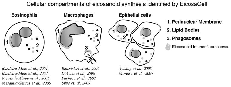
Schematic summary of the EicosaCell applications. EicosaCell-derived reports have identified three distinct intracellular sites of eicosanoid synthesis, including nuclear envelope, cytoplasmic lipid bodies, and zymozan-driven phagosomes within human or murine eosinophils, murine macrophages as well as rat or human epithelial cells (8–17).
Of note the EicosaCell has high sensitivity enabling the detection of low levels of intracellular generated eicosanoids even when extracellular released eicosanoids could not be detected by conventional eicosanoid enzyme immune assay (10, 11). Indeed, it has been shown that besides paracrine/autocrine activities, eicosanoids may display intracrine functions (18, 19). For instance, by employing EicosaCell technique, it has been uncovered that a lipid body-derived LTC4 has intracellular functions in controlling cytokine release from eosinophils (20). Therefore, by identifying compartmentalized levels of eicosanoids, besides providing new insights into the regulation of eicosanoid biosynthesis, EicosaCell assays may contribute to identification of likely intracellular functions of newly synthesized eicosanoids.
2. Materials
2.1. EicosaCell
EDAC: 1-ethyl-3-(3-dimethylamino-propyl) carbodiimide hydrochloride (C8H17N3–HCl) (Sigma-Aldrich Inc., St. Louis, MO; cat no. E7750).
HBSS: Hanks buffered salt solution without calcium chloride and magnesium chloride.
Water bath at 37°C.
Cytocentrifuge.
Glass microscope slides and coverslips.
Mounting medium: Aqua Poly/Mount (Polysciences Inc., Warrington, PA; cat. no. 18606) or Vectashield (Vector, Inc., Burlingame, CA; cat. no. H 1000).
2.2. Double-Labeling Applications
DAPI (4′,6-Diamidino-2-phenylindole dihydrochloride).
TO-PRO-3 (642/661) – 1 mM solution in dimethylsulfoxide (DMSO) (Molecular Probes, Eugene, OR).
Rat monoclonal IgG2a against lysosome-associated membrane protein (LAMP)-1 (BD Pharmingen, San Jose, CA).
BODIPY® 493/503 (4,4-difluoro-1,3,5,7,8-pentamethyl-4-bora-3a,4a-diaza-s-indacene) (Molecular Probes, Eugene, OR; cat no. D-3922). Molecular weight: 262. To prepare BODIPY stock solution, BODIPY should be dissolved in DMSO (1 mM), aliquoted in small Eppendorf tubes (~10 μl/tube) and stored −20°C protected from light. BODIPY working solution should be diluted fresh 1,000× in HBSS−/− and kept from light.
Mouse monoclonal antibody (mAb) to adipose differentiation-related protein (ADRP) (Fitzgerald Industries Intl., Concord, MA; cat no. 10R-A117ax) or guinea pig polyclonal Ab to ADRP (reactivity human/mouse/rat/bovine) (Fitzgerald Industries Intl., Concord, MA; cat no. 20R-AP002).
3. Methods
3.1. Preparation of EDAC Solution for EicosaCell
Water soluble EDAC hydrochloride should be diluted in HBSS−/−. Refer to Note 1 (below) for EDAC solution handling.
The working solution should have twice concentration of the final concentration with cells.
-
EDAC final concentration with cells varies according cell type and protocol used (see next subheadings), as exemplified below:
-
Example 1
With purified human eosinophils stimulated as a cell suspension, EDAC final concentration within eosinophil suspensions should be 0.1% in HBSS−/−, therefore the EDAC working solution should be 0.2%.
-
Example 2
With adherent macrophages stimulated in 6-well plates, EDAC final concentration should be 0.5% in HBSS−/−, therefore the EDAC working solution should be 1.0%.
-
Example 1
3.2. EicosaCell with Cells in Suspension
EicosaCell can be easily performed with various cell types in suspension, such as purified human blood leukocytes, cultured cell lines, as well as peritoneal, pleural, or bronchoalveolar animal cells.
After in vivo or in vitro stimulation of these cell populations, incubation with EDAC instantaneously immobilizes eicosanoids at their sites of synthesis within the cell, just before cytospin slides are prepared to allow microscopic analysis.
As schematically illustrated in Fig. 2, after preparing a cell suspension from, for instance, a mouse peritoneal cavity, EDAC working solution should be added to cell suspension and incubated for a period of time to ensure cell fixation, eicosanoid immobilization, and cell permeabilization (refer to Note 2 for details).
As suggested in the scheme (Fig. 2), slide preparations can be done by cytocentrifuging the cells onto slides. Labeling of newly formed eicosanoids can be done with a various already tested antibodies, as already published elsewhere (10, 14–16).
At the end of the staining procedure, cytospun cells should be always extensively washed with HBSS−/− and then mounted in an aqueous medium to be visualized with 100× objective by both phase-contrast and fluorescence microscopy (see Note 3 for details).
The specificity of the eicosanoid immunolabeling using the EicosaCell system should be always ascertained by including some mandatory control conditions: (1) nonstimulated EDAC-treated cells labeled with the proper anti-eicosanoid Ab; (2) the incubation (1 h before EDAC) with eicosanoid synthesis inhibitors, such as cPLA2-α inhibitor (e.g., pyrrolidine- 2; 1 μM), COX inhibitor (e.g., indomethacin; 1 μg/ml), FLAP inhibitor (e.g., MK886; 50 μg/animal or 10 μM for in vitro incubations), or 5-LO inhibitor (e.g., zileuton; 50 μg/animal or 10 μM for in vitro incubations) to inhibit eicosanoid synthesis and guarantee the specificity of the observed reaction; and (3) the use of an irrelevant Ab control. Optionally, other suitable controls to check specificity and performance of EicosaCell are: (1) to use, instead of EDAC, paraformadehyde which will not immobilize the newly synthesized eicosanoid within cells; (2) in parallel, to carry out the EicosaCell in a different cell type that lacks the ability to synthesize the targeted eicosanoid (for instance, to use neutrophils to check specificity of LTC4 immunodetection by EicosaCell); or (3) to analyze mixed populations of responsive plus unresponsive cells to a specific stimulus, so you can reassure that the targeted eicosanoid is specifically detected only within stimulated cells.
Fig. 2.

Schematic illustration of EicosaCell methodology. EicosaCell preparations, which undergo EDAC-dependent capturing and fixation of newly formed eicosanoids at their sites of synthesis, are analyzed by phase-contrast and fluorescent microscopy and can employ cytospun or adherent cells.
As shown in Fig. 3, EicosaCell system was successfully employed on macrophages recovered from peritoneal cavities of oxidized LDL-stimulated or control mice (17). Briefly, cells obtained 4 h after stimulation with oxidized LDL and controls were recovered from the pleural cavity with 500 μl of HBSS−/−, and immediately mixed with 500 μl of EDAC (1% in HBSS−/−). After 30 min incubation at 37°C with EDAC, leukocytes were then washed with HBSS−/−, cytospun onto glass slides, and incubated with mouse anti-LTB4 (Cayman Chemical) in 0.1% normal goat serum. BODIPY 493/503 and TO-PRO-3 were added together with the secondary Ab for 1 h to distinguish cytoplasmic lipid bodies and nuclei, respectively (see Subheading 2.2). Cells were washed twice and incubated with Cy2-labeled anti-rabbit IgG secondary Ab for 1 h. The slides were washed (three times, 10 min each) and mounted with aqueous mounting medium. Cells were analyzed by confocal laser microscopy. As a control for LTB4 specificity of detection, one group of oxLDL-stimulated animals was treated with MK886 (50 μg/animal) 2 h before sacrificing the animals for cell recovery, and the treatment with MK886 was maintained throughout the subsequent incubation steps at a concentration of 10 μM for MK886.
Fig. 3.
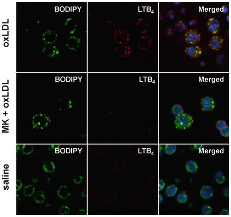
Leukotriene B4 detection in leukocytes. Leukocytes were obtained from in vivo oxLDL- or saline-stimulated mice and EicosaCell for LTB4 was performed as described in (17). Cells were fixed and permeabilized by carbodiimide and stained with bodipy 493/503 (green) for lipid body visualization (left panels). Upper panels show peritoneal macrophages isolated from oxLDL-stimulated animals that were incubated with anti-LTB4 Ab (red) for detection of newly formed LT. Middle panels, oxLDL-stimulated animals were treated with MK886 (50 μg/animal) 2 h prior to sacrifice to inhibit FLAP-dependent LT synthesis and labeling. Lower panel, show peritoneal leukocytes from nonstimulated animals and incubated with anti-LTB4 Ab (red). TO-PRO-3 nuclei staining are shown in blue. Slides were analyzed by laser scanning confocal microscopy.
The EicosaCell technique is particularly suitable to distinguish intracellular sites of eicosanoid synthesis. As mentioned before, EicosaCell studies have provided direct evidence of compartmentalized eicosanoid synthesis within three distinct sites, including perinuclear envelope (8–11), phagosomes (12), or lipid bodies (10, 11, 13–15). As exemplified in Fig. 4, while, eosinophils stimulated with eotaxin for 1 h showed punctate cytoplasmic immunoreactive LTC4 colocalized at lipid bodies, LTC4 biosynthesis after cross-linking of eosinophil membrane-expressed CD9 was found exclusively as a perinuclear immunolabelling (8).
Fig. 4.
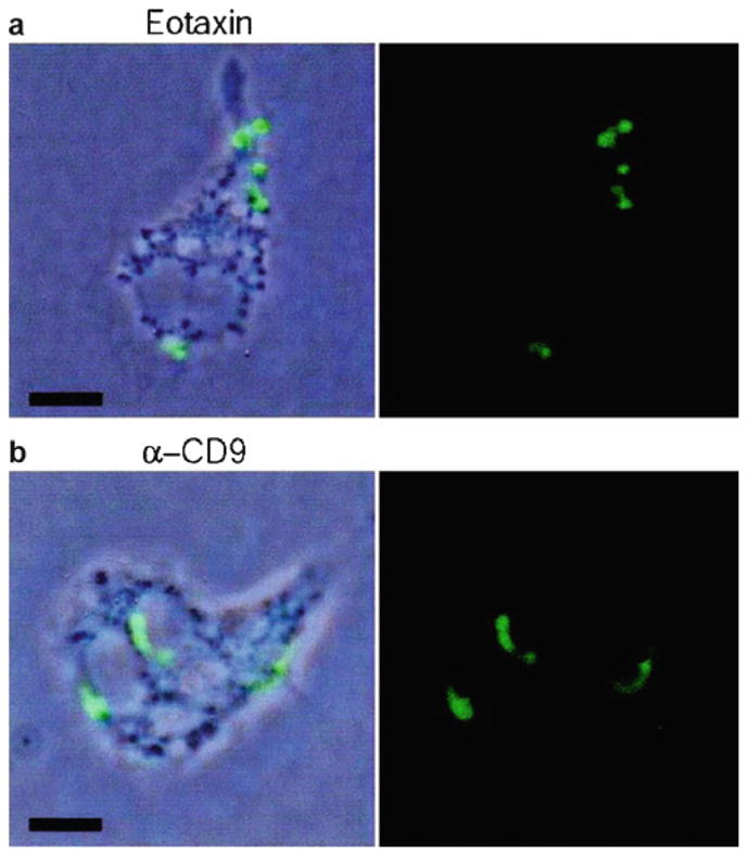
Intracellular localization of LTC4 biosynthesis within human eosinophils. Eosinophils were stimulated with 12 nM eotaxin (a) or 2.5 μg/ml mAb to CD9 (b) for 1 h and EicosaCell for LTC4 was performed as described in (8). Eosinophils were fixed and permeabilized by carbodiimide and stained with Alexa 488-labeled anti-cysteinyl LT mAb. To facilitate intracellular localization, anti-LTC4 immunoreactive sites (green staining) were overlaid on phase-contrast images. Figure reproduced with permission from (8) (Bar 5 μm).
3.3. EicosaCell with Adherent Cells
To study the intracellular compartmentalization of eicosanoid synthesis by EicosaCell in adherent cells, extra care should be taken to ensure the conservation of cell adherence and morphology during the EDAC step. As published, EicosaCell assays have succeeded to immunolocalize PGE2 within at least three distinct adherent cell types: plated murine macrophages (D’Avila et al., unpublished) and two lineages of intestinal cells (as illustrated in Fig. 1), CACO-2 (a human colon adenocarcinoma cell line) (9), and IEC-6 (a rat epithelial cell line) (11).
While adherent CACO-2 cells (on glass coverslips), for instance, should be incubated during 1 h at 37°C with EDAC at 0.5% in HBSS−/− to cross-link PGE2 carboxyl groups to amines in adjacent proteins without affecting cell morphology, IEC-6 cells must be incubated for at most 30 min (at same concentration; 0.5% in HBSS−/−) to retain reasonable cell morphology and PGE2 immunodetection at synthesizing compartments (refer to the original articles (9, 11) for details of blocking and staining conditions with anti-PGE2 mAb (Cayman Chemicals) and proper secondary Abs).
At the end of the staining procedure, coverslip-adherent cells should be always extensively washed with HBSS and then mounted in an aqueous mounting medium to be visualized with 100× objective by both phase-contrast and fluorescence microscopy. As for cytospun cells, the specificity of the eicosanoid immunolabeling using EicosaCell system should be ascertained by including mandatory controls as listed in Subheading 3.2, step 6.
3.4. Double-Labeling Procedures to Identify Eicosanoid-Synthesizing Intracellular Sites
3.4.1. Nuclear Localization
To better visualize perinuclear eicosanoid synthesis by EicosaCell, a double labeling with DAPI or TO-PRO-3 is recommended.
After EDAC and Ab incubation steps, EicosaCell slides preparations should be extensively washed in HBSS−/− and then incubated with DAPI (DAPI working solution 100 ng/ml or 300 nM, see Note 4) for 5 min before aqueous mounting medium application.
The morphology of the cells’ nuclei is observed using a fluorescence microscope at excitation wavelength 350 nm.
3.4.2. Phagosomal Localization
As performed by Balestrieri et al. (12), phagosome involvement in eicosanoid synthesis can be ascertained by colocalizing the phagosomal protein marker LAMP-1 in EicosaCell preparations.
After incubation at 37°C with EDAC, cells should be washed with HBSS−/−, cytospun onto glass slides, and incubated with mouse anti-eicosanoid Ab and rat mAb against LAMP-1 (2.5 μg/ml) in blocking buffer (5% normal donkey serum) for 2 h at room temperature.
Negative control cells are instead incubated for 2 h with rat IgG (Jackson Immunoresearch).
After 2 h, the cells are washed extensively with HBSS−/− and incubated for 1 h at room temperature with fluorescent-labeled secondary Ab to detect the murine anti-eicosanoid Ab and Cy3-conjugated donkey anti-rat IgG (1:200).
The cells should be washed five times with HBSS−/− and then mounted in aqueous mounting medium.
3.4.3. Lipid Body Localization
To investigate the roles of lipid bodies in eicosanoid synthesis by EicosaCell assay, two double-labeling strategies can be employed: (1) BODIPY 493/503 lipid probe is an effective dye for staining neutral lipids and, for this reason it is very efficient for lipid body staining, or (2) ADRP immunostaining. ADRP is a structural protein of lipid bodies considered essential for lipid storage and metabolism that is ubiquitously expressed in lipid bodies containing cells (for further information on lipid body labeling refer to (21). Both approaches can be used for adherent, suspension, or agarose-embedded cells.
To employ BODIPY 493/503 for lipid body labeling, incubate EicosaCell preparations (coverslips or slides) with 1 μm BODIPY 493/503 (working solution) for 45–60 min at 37°C (water bath) simultaneously with secondary Ab incubation.
To remove non-incorporated BODIPY 493/503 after incubation, EicosaCell preparations should be washed at least twice in HBSS−/− before aqueous mounting medium application and coverslip attachment to slides.
Alternatively with ADRP to visualize lipid bodies, ADRP immunolabeling may be performed. In this case, slides are incubated for 1 h with guinea pig anti-ADRP Ab together with the primary anti-eicosanoid Ab to localize lipid bodies within leukocytes. The cells are washed with HBSS for 10 min (3×) and incubated with Cy3-labeled anti-guinea pig secondary antibodies for 1 h.
5. Troubleshooting
Some common problems and nonobvious features found in immunofluorescent-detection of eicosanoids in EDAC preparations by EicosaCell are shown in Table 1 with their possible explanations and potential solutions.
Table 1.
Troubleshooting eicosanoid detection
| Problems | Common causes | Solutions |
|---|---|---|
| Lack of eicosanoid detection (when few or no eicosanoid specific immunostaining is observed, but expected) |
Improper fixation (when the eicosanoid is not cross-linked by EDAC, but washed-out from cells turning detection impossible) Inefficient cell stimulation |
Slight increase in EDAC (by adjusting both EDAC concentration and/or incubation time with cells) Positive control (mandatory inclusion of a well known agonist) |
| Eicosanoid detection within nonstimulated cells (eicosanoids are nonstorable in the cell and newly formed upon stimulation, therefore nonstimulated cells should not show any immunostaining for the targeted eicosanoid) |
Improper stimulation (cell activation during the procedures including cell incubation at 37°C or cell fixation/permeabilization with EDAC can lead to spontaneous, stimulus-independent eicosanoid synthesis) Nonspecific detection |
Negative control (nonstimulated cells should always be included as an important negative control. Care is needed to ensure that cells are not mechanically, chemically, or immunologically stimulated) (Discussed below) |
| Poor preservation of cell morphology | EDAC cell toxicity (during EDAC incubation step, cell appearance may change from unimportant to severe modification of typical cell morphology) | Slight decrease in EDAC (by adjusting both EDAC concentration and incubation time with cells) |
| Non-specific detection (antibodies may non-specifically bind to other cellular structures or to other lipids found within cells) |
Nonspecific binding to cell (since cross-linking properties of EDAC may favor the tendency for cells to be sticky) Nonspecific binding to other eicosanoids |
Irrelevant antibody control (a proper control using host/isotype-matched antibodies in stimulated cells should always be included) Eicosanoid synthesis inhibitor control (stimulated cells treated with a synthesis inhibitor of the targeted eicosanoid is a mandatory control condition that should show no immune-labeling confirming specific detection of targeted eicosanoid) |
| High non-specific binding (when non-specific staining is too high with >10 % positive cells) | Improper detecting antibody conditions |
Change detecting antibody dilution and/or host Incubation of detecting antibody with an adsorbing reagent Incubation of EDAC-treated cells with a normal serum (to effectively block out nonspecific sites) Centrifugation of the detecting antibody (to eliminate aggregates) |
| Losing cell adherence with EDAC (the ability of cells to stay adhered to coverslips or other substrates can be affected by EDAC incubation) | EDAC cell toxicity | Slight decrease in EDAC (previous careful setting of EDAC incubation step is obligatory and should be adjusted for each cell type) |
Acknowledgments
The work of the authors is supported by PRONEX-MCT, Conselho Nacional de Desenvolvimento Cientifico e Tecnológico (CNPq, Brazil), PAPES-FIOCRUZ, Fundação de Amparo à Pesquisa do Rio de Janeiro (FAPERJ, Brazil), and NIH grants (AI022571, AI020241, AI051645). The authors are indebted to Dr. Adriana Vieira de Abreu for the contributions to the figures used in the manuscript.
Footnotes
EDAC working solution should be prepared fresh, keep protected from light, and discarded after each experiment.
Incubation of cells with EDAC can be carried out on either cell in suspension or with the cells already cystopun onto slides by dropping EDAC on top of cells. Even though the latter method is less costly, some differences in preservation of cell morphology, cell permeabilization, and eicosanoid detection may occur and should be analyzed with care.
Fluorescent microscopic analyses of EicosaCell preparations should be performed as soon as the slides are mounted, inasmuch as immunofluorescent labeling is usually not stable for a long period and may exhibit bleaching over time. Even though freezing may preserve fluorescence overnight, EDAC-treated cells may display altered cell appearance after freezing-thawing cycle.
Prepare DAPI stock solution by dissolving 1 mg/ml of powder in distilled water. Aliquots should be stored at −20°C protected from light.
References
- 1.Yaqoob P. Fatty acids as gatekeepers of immune cell regulation. Trends Immunol. 2003;24:639–645. doi: 10.1016/j.it.2003.10.002. [DOI] [PubMed] [Google Scholar]
- 2.Wymann MP, Schneiter R. Lipid signalling in disease. Nat Rev Mol Cell Biol. 2008;9:162–176. doi: 10.1038/nrm2335. [DOI] [PubMed] [Google Scholar]
- 3.Peters-Golden M, Brock TG. Intracellular compartmentalization of leukotriene synthesis: unexpected nuclear secrets. FEBS Lett. 2001;487:323–326. doi: 10.1016/s0014-5793(00)02374-7. [DOI] [PubMed] [Google Scholar]
- 4.Mandal AK, Skoch J, Bacskai BJ, Hyman BT, Christmas P, Miller D, et al. The membrane organization of leukotriene synthesis. Proc Natl Acad Sci U S A. 2004;101:6587–6592. doi: 10.1073/pnas.0308523101. [DOI] [PMC free article] [PubMed] [Google Scholar]
- 5.Bandeira-Melo C, Bozza PT, Weller PF. The cellular biology of eosinophil eicosanoid formation and function. J Allergy Clin Immunol. 2002;109:393–400. doi: 10.1067/mai.2002.121529. [DOI] [PubMed] [Google Scholar]
- 6.Bozza PT, Magalhães KG, Weller PF. Leukocyte lipid bodies – biogenesis and functions in inflammation. Biochim Biophys Acta. 2009 doi: 10.1016/j.bbalip.2009.01.005. [DOI] [PMC free article] [PubMed] [Google Scholar]
- 7.Liu LX, Buhlmann JE, Weller PF. Release of prostaglandin E2 by microfilariae of Wuchereria bancrofti and Brugia malayi. Am J Trop Med Hyg. 1992;46:520–523. doi: 10.4269/ajtmh.1992.46.520. [DOI] [PubMed] [Google Scholar]
- 8.Tedla N, Bandeira-Melo C, Tassinari P, Sloane DE, Samplaski M, Cosman D, et al. Activation of human eosinophils through leukocyte immunoglobulin-like receptor 7. Proc Natl Acad Sci U S A. 2003;100:1174–1179. doi: 10.1073/pnas.0337567100. [DOI] [PMC free article] [PubMed] [Google Scholar]
- 9.Accioly MT, Pacheco P, Maya-Monteiro CM, Carrossini N, Robbsm BK, Oliveira SS, et al. Lipid bodies are reservoirs of cyclooxygenase-2 and sites of prostaglandin-E2 synthesis in colon cancer cells. Cancer Res. 2008;68:1732–40. doi: 10.1158/0008-5472.CAN-07-1999. [DOI] [PubMed] [Google Scholar]
- 10.Bandeira-Melo C, Phoofolo M, Weller PF. Extranuclear lipid bodies, elicited by CCR3-mediated signaling pathways, are the sites of chemokine-enhanced leukotriene C4 production in eosinophils and basophils. J Biol Chem. 2001;276:22779–22787. doi: 10.1074/jbc.M101436200. [DOI] [PubMed] [Google Scholar]
- 11.Moreira LS, Piva B, Gentile LB, Mesquita-Santos FP, D’Avila H, Maya-Monteiro CM, et al. Cytosolic phospholipase A2-driven PGE2 synthesis within unsaturated fatty acids-induced lipid bodies of epithelial cells. Biochim Biophys Acta. 2009;1791:156–165. doi: 10.1016/j.bbalip.2009.01.003. [DOI] [PubMed] [Google Scholar]
- 12.Balestrieri B, Hsu VW, Gilbert H, Leslie CC, Han WK, Bonventre JV, et al. Group V secretory phospholipase A2 translocates to the phagosome after zymosan stimulation of mouse peritoneal macrophages and regulates phagocytosis. J Biol Chem. 2006;281:6691–6698. doi: 10.1074/jbc.M508314200. [DOI] [PMC free article] [PubMed] [Google Scholar]
- 13.Mesquita-Santos FP, Vieira-de-Abreu A, Calheiros AS, Figueiredo IH, Castro-Faria-Neto HC, Weller PF, et al. Cutting edge: prostaglandin D2 enhances leukotriene C4 synthesis by eosinophils during allergic inflammation: synergistic in vivo role of endogenous eotaxin. J Immunol. 2006;176:1326–1330. doi: 10.4049/jimmunol.176.3.1326. [DOI] [PubMed] [Google Scholar]
- 14.Pacheco P, Vieira-de-Abreu A, Gomes RN, Barbosa-Lima G, Wermelinger LB, Maya-Monteiro CM, et al. Monocyte chemoattractant protein-1/CC chemokine ligand 2 controls microtubule-driven biogenesis and leukotriene B4-synthesizing function of macrophage lipid bodies elicited by innate immune response. J Immunol. 2007;179:8500–8508. doi: 10.4049/jimmunol.179.12.8500. [DOI] [PubMed] [Google Scholar]
- 15.D’Avila H, Melo RC, Parreira GG, Werneck-Barroso E, Castro-Faria-Neto HC, Bozza PT. Mycobacterium bovis bacillus Calmette-Guerin induces TLR2-mediated formation of lipid bodies: intracellular domains for eicosanoid synthesis in vivo. J Immunol. 2006;176:3087–3097. doi: 10.4049/jimmunol.176.5.3087. [DOI] [PubMed] [Google Scholar]
- 16.Vieira-de-Abreu A, Assis EF, Gomes GS, Castro-Faria-Neto HC, Weller PF, Bandeira-Melo C, et al. Allergic challenge-elicited lipid bodies compartmentalize in vivo leukotriene C4 synthesis within eosinophils. Am J Respir Cell Mol Biol. 2005;33:254–261. doi: 10.1165/rcmb.2005-0145OC. [DOI] [PMC free article] [PubMed] [Google Scholar]
- 17.Silva AR, Pacheco P, Vieira-de-Abreu A, Maya-Monteiro CM, D’Alegria B, Magalhães KG, et al. Lipid bodies in oxidized LDL-induced foam cells are leukotriene-synthesizing organelles: a MCP-1/CCL2 regulated phenomenon. Biochim Biophys Acta. 2009;1791:1066–1075. doi: 10.1016/j.bbalip.2009.06.004. [DOI] [PubMed] [Google Scholar]
- 18.Devchand PR, Keller H, Peters JM, Vazquez M, Gonzalez FJ, Wahli W. The PPAR-alpha-leukotriene B4 pathway to inflammation control. Nature. 1996;384:39–43. doi: 10.1038/384039a0. [DOI] [PubMed] [Google Scholar]
- 19.Kliewer SA, Lenhard JM, Willson TM, Patel I, Morris DC, Lehmann JM. A prostaglandin J2 metabolite binds peroxisome proliferator-activated receptor gamma and promotes adipocyte differentiation. Cell. 1995;83:813–819. doi: 10.1016/0092-8674(95)90194-9. [DOI] [PubMed] [Google Scholar]
- 20.Bandeira-Melo C, Woods LJ, Phoofolo M, Weller PF. Intracrine cysteinyl leukotriene receptor-mediated signaling of eosinophil vesicular transport-mediated interleukin-4 secretion. J Exp Med. 2002;196:841–850. doi: 10.1084/jem.20020516. [DOI] [PMC free article] [PubMed] [Google Scholar]
- 21.Melo RCN, Bozza PT, Weller PF. Imaging lipid bodies within leukocytes with different light microscopy techniques. Methods Mol Biol. 2009;689:149–161. doi: 10.1007/978-1-60761-950-5_9. [DOI] [PMC free article] [PubMed] [Google Scholar]