
Figure 1. SkE treatment induces cell death of CML cell lines and primary CML CD34+ cells.
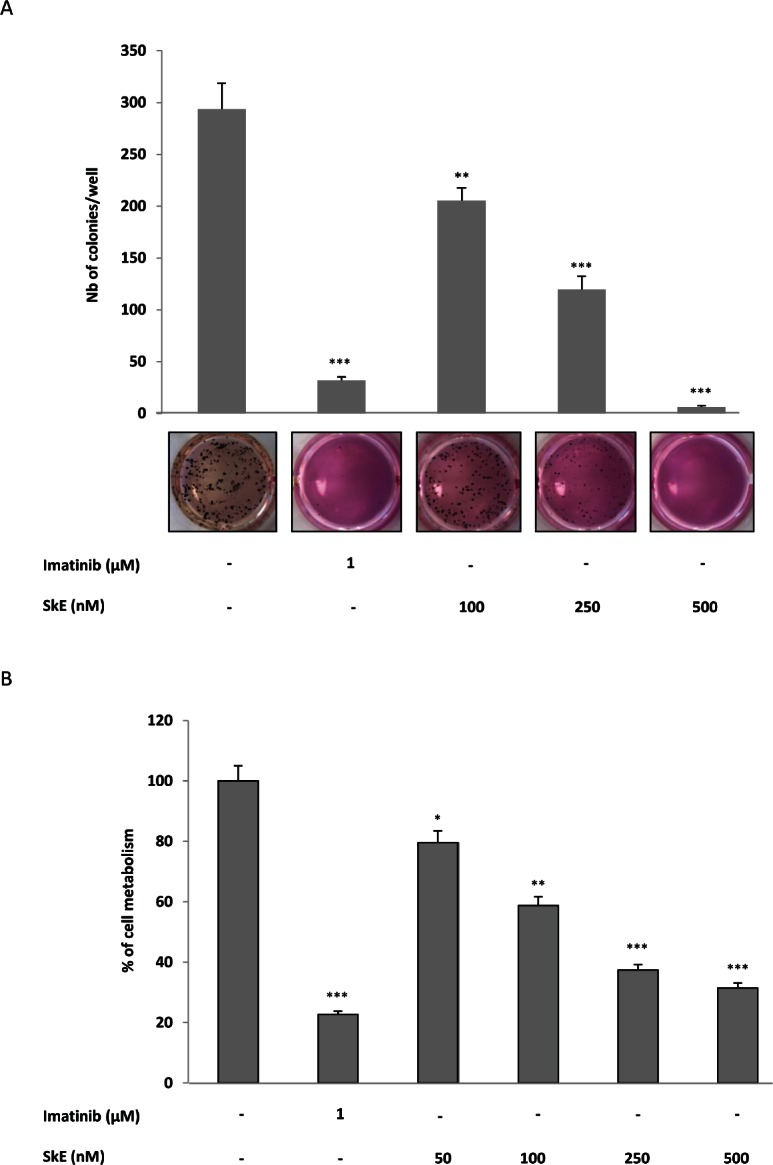
(A) SkE in the 100-500 nM range was added to K562 CML cell lines growing in semi-solid methyl cellulose medium (0.5'103 cells/ml). Imatinib (1 μM) was used as an internal control. Colonies were detected after 10 days of culture by adding 1 mg/ml of MTT reagent and were scored by Image J quantification software. Results are expressed as the number of colony forming cells per well after drug treatment. Results are given as the mean ± SD of 3 different determinations made in triplicate. Error bars = 95% confidence intervals. (B) Primary CML CD34+ cells were incubated for 48 h at 37°C with increasing concentrations of SkE. The cell metabolism was measured by the XTT assay, as described in the Materials and Methods section. Results are given as the mean ± SD of 3 different determinations made in triplicate. Error bars = 95% confidence intervals.