Sir,
We report a long-term effective treatment for drusenoid pigment epithelial detachment.
Case report
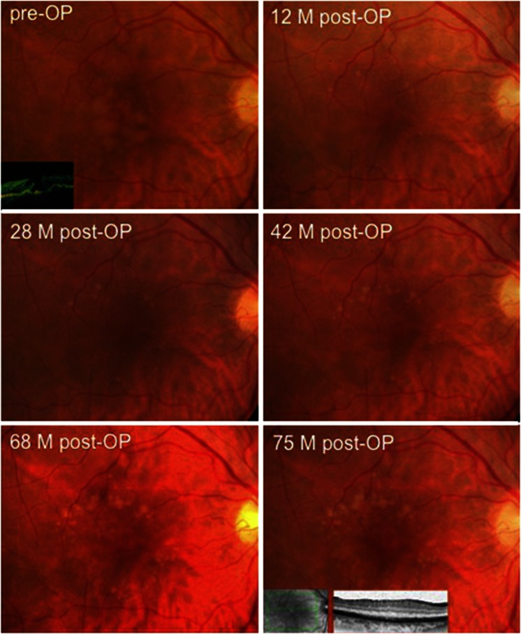
A 66-year-old woman presented with central confluent drusen, incipient cataract OU, and macular hole OD (Figure 1). BCVA was 20/200 OD and 20/30 OS. The patient underwent macular hole surgery including cataract surgery, vitrectomy, posterior vitreous detachment (PVD), peeling of epiretinal membrane plus internal limiting membrane (ILM) after ICG staining, and gas tamponade. Before ILM staining with ICG, the macular hole was covered with a small PFCL-bubble to avoid any contact of ICG with the RPE. Postoperatively, the macular hole was closed and the confluent drusen almost completely disappeared. Only few small drusen were present 12 months after surgery (BCVA 20/60). Comparison of pre- and postoperative photographs suggests that these drusen were newly formed. The drusen in the untreated left eye increased in size. During the further course of >6 years (75 months, Figure 1) BCVA OD stabilized at 20/25. New drusen developed slowly in a ring-shaped pattern almost completely sparing the center. No signs of atrophy occurred. In contrast, OS developed CNV (BCVA 20/100).
Figure 1.
A 66-year-old patient with central confluent drusen and additional macular hole OD. The follow-up of 75 months demonstrate resorption of central confluent drusen postoperatively and slow new drusen formation in a ring-shaped pattern sparing the central area where compact drusen material had been present preoperatively. BCVA increased and stabilized from 20/200 preoperative to 20/25 75 months postoperative.
Comment
Central confluent soft drusen, also called ‘drusenoid pigment epithelial detachment' (DPED) has a poor prognosis and 42% of eyes progressed to advanced ARMD within 5 years.1 No effective and overall accepted treatment of DPED is available so far. Regression of drusen has been shown after laser photocoagulation,2 coincidental rhegmatogenous retinal detachment,3 and intravitreal anti-VEGF therapy.4, 5, 6 However, there is no evidence that laser photocoagulation reduces the risk of developing CNV, geographic atrophy, or visual acuity loss2 and larger studies about intravitreal anti-VEGF treatment for DPED are desirable. The here reported patient with pronounced DPED showed remarkable resorption of almost all drusen without developing atrophic changes or CNV after surgery and with stable good vision for >6 years. Spontaneous resorption of drusen has been reported, however, not to the here shown extent and usually correlating with atrophic changes. While the drusen disappeared in the operated eye, DPED progressed OS. Resorption of drusen after macular hole surgery has been reported only once before.7 The surgical procedure might have stimulated phagocytosis of drusen by macrophages. Stimulating factors might be the surgically induced PVD, peeling of epiretinal membranes, gas tamponade, and increased oxygenation following vitrectomy.8 The additional ILM peeling in the here reported case might have enhanced the stimulation, resulting in an almost complete phagocytosis of drusen. Remarkably, postoperative new drusen formation spared the central area where compact drusen material had been present preoperatively.
The authors declare no conflict of interest.
Footnotes
Case report was presented at annual meeting of the German Society of Ophthalmology 2012 (DOG 2012).
References
- Cukras C, Agrón E, Klein ML, Ferris FL, Chew EY, Gensler G, et al. Natural history of drusenoid pigment epithelial detachment in age-related macular degeneration: Age-Related Eye Disease Study Report No. 28. Ophthalmology. 2010;117:489–499. doi: 10.1016/j.ophtha.2009.12.002. [DOI] [PMC free article] [PubMed] [Google Scholar]
- Parodi MB, Virgili G, Evans JR.Laser treatment of drusen to prevent progression to advanced age-related macular degeneration Cochrane Database Syst Rev 2009(Issue 3)(Art. No. CD006537)doi: 10.1002/14651858.CD006537.pub2 [DOI] [PubMed]
- Margolis R, Ober MD, Freund KB. Disappearance of drusen after rhegmatogenous retinal detachment. Retinal Cases & Brief Reports. 2010;4:254–256. doi: 10.1097/ICB.0b013e3181af5582. [DOI] [PubMed] [Google Scholar]
- Gallego-Pinazo R, Suelves-Cogollos AM, Dolz-Marco R, Arevalo JF, García-Delpech S, Mullor JL, et al. Intravitreal ranibizumab for symptomatic drusenoid pigment epithelial detachment without choroidal neovascularization in age-related macular degeneration. Clin Ophthalmol. 2011;5:161–165. doi: 10.2147/OPTH.S15832. [DOI] [PMC free article] [PubMed] [Google Scholar]
- Kishore K, Jain S, Sharma YR, Kashyap B.Disappearance of drusen after intravitreal anti-VEGF injections for submacular hemorrhage (SMH) secondary to neovascular macular degeneration IOVS 201253(ARVO e abstract 2912). [Google Scholar]
- Krishnan R, Lochhead J. Regression of soft drusen and drusenoid pigment epithelial detachment following intravitreal anti-vascular endothelial growth factor therapy. Can J Ophthalmol. 2010;45 (1:83–84. doi: 10.3129/i09-187. [DOI] [PubMed] [Google Scholar]
- Holz FG, Staudt S. Disappearance of soft drusen following macular hole surgery. Retina. 2001;21:184–186. doi: 10.1097/00006982-200104000-00018. [DOI] [PubMed] [Google Scholar]
- Stefánsson E. Physiology of vitreous surgery. Graefes Arch Clin Exp Ophthalmol. 2009;247 (2:147–163. doi: 10.1007/s00417-008-0980-7. [DOI] [PubMed] [Google Scholar]


