Abstract
The response of Desulfovibrio vulgaris to Cu(II) and Hg(II) was characterized. Both metals increased the lag phase, and Cu(II) reduced cell yield at concentrations as low as 50 μM. mRNA expression was analyzed using random arbitrarily primed PCR, differential display, and quantitative PCR. Both Cu(II) and Hg(II) (50 μM) caused upregulation of mRNA expression for an ATP binding protein (ORF2004) and an ATPase (ORF856) with four- to sixfold increases for Hg(II) and 1.4- to 3-fold increases with Cu(II). These results suggest that D. vulgaris uses an ATP-dependent mechanism for adapting to toxic metals in the environment.
Sulfate-reducing bacteria (SRB) are known to be involved in metal bioremediation and for this purpose are often encouraged to grow in engineered systems. They can directly reduce and precipitate many metals, including uranium (11), technetium (9, 10), arsenate (12), and chromium (23), or precipitate metal cations, as metal sulfides (4, 24). Sulfide itself is also known to reduce certain oxidized metals (20), resulting in precipitation. Bioremediation processes may be conducted in bioreactors (4, 24, 25) or in situ (17). While SRB are capable of varied metal transformations, metals can also inhibit their growth (14-16). However, we are not aware of previous research aimed at addressing mechanisms of resistance to divalent metal ions by SRB.
Differential display using random arbitrarily primed PCR (RAP-PCR) has been used to identify genes induced by growth under different conditions (1, 2, 8). These techniques were recently used by our group with Desulfovibrio desulfuricans subsp. aestuarii to identify differentially transcribed genes during growth on lactate or hydrogen (22). In the present study, we have used a similar approach to identify genes involved in metal resistance. After first determining metal concentrations needed to inhibit growth of Desulfovibrio vulgaris, we used RAP-PCR to identify genes whose transcription was enhanced in the presence of Cu2+ or Hg2+. These transcripts were subsequently quantitated in metal-treated cultures and control cultures and were identified through genetic database analysis.
Effects of metals on growth.
Initial studies were aimed at determining suitable metal concentrations for differential expression experiments. D. vulgaris Hildenborough was grown anaerobically on lactate sulfate medium (LSM) and pyruvate medium (PM) without sulfide, bicarbonate, and CO2. LSM was prepared as previously described (7) and contained sodium dl- lactate (50 mM), Na2SO4 (50 mM), yeast extract (0.1%), vitamins, minerals, trace metals, and resazurin. D. vulgaris was also cultivated fermentatively in a similar pyruvate medium with pyruvate (20 mM) and MgCl2 instead of MgSO4 · 7H2O and no other sources of sulfate.
For the preparation of inocula, cultures were grown to mid-exponential phase and harvested by centrifugation (4,650 × g for 20 min). The cell pellet was washed twice and finally resuspended in fresh medium (without bicarbonate-sulfide). Cultures for metal inhibition and expression experiments (described below) were inoculated with 1% of their volume of washed cells. Incubation was static at 37°C. Although the media were initially oxidized (as determined by the color of resazurin), as growth occurred, the media became reduced. Cultures grew to maximum optical densities at 600 nm (OD600) that were similar to those grown on LSM with sulfide, bicarbonate, and CO2 added (data not shown).
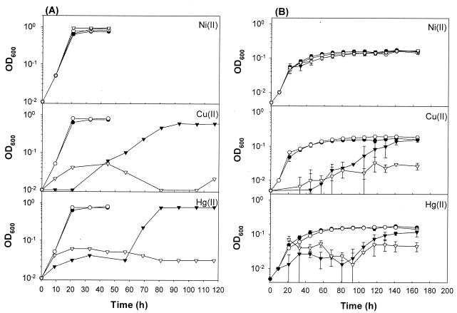
Because of the low solubility of many metal complexes at neutral pH, sodium nitrilotriacetate (NTA) was used in metal stock solutions. We determined the influence of NTA on the growth of D. vulgaris by transferring cultures to similar media with and without NTA addition (0.1 g/liter). At this concentration, NTA had no influence on the growth curve of D. vulgaris in either LSM or PM (data not shown). D. vulgaris grew at concentrations of up to 300 μM Pb(II) (PbCl2; Alfa Aesar, Ward Hill, Mass.) and 1,000 μM Ni(II) (NiCl2; Fisher Scientific, Fair Lawn, N.J.) in LSM (Fig. 1A). Growth curves with Pb(II) are not shown, as PbS precipitation prevented an accurate determination of OD in all cases except at 10 μM, where no difference was observed relative to the control. Cu(II) (CuCl2; Aldrich, Milwaukee, Wis.) and Hg(II) (HgCl2; Sigma, St. Louis, Mo.) partially inhibited growth at 100 μM and completely inhibited growth at 1 mM under the same conditions (Fig. 1). Partial inhibition was determined by lower biomass formation [Cu(II)] or longer lag times [Cu(II) and Hg(II)]. These two phenomena have been previously described in another species of Desulfovibrio (15, 16), although the latter work showed toxicity at lower Cu(II) concentrations. This difference is likely due to the presence of the ligands, NTA, and phosphate in our media. Although measurement of OD was straightforward in cultures that produced no sulfide (did not grow), formation of metal sulfides in active cultures grown on LSM, especially with Pb(II) and Ni(II), likely caused an overestimation of the final growth yield.
FIG. 1.
Effects of metals on the growth of D. vulgaris under sulfate respiration (A) and pyruvate fermentation (B) conditions. D. vulgaris was cultivated in serum tubes containing 10 ml of LSM or PM with NiCl2, CuCl2, and HgCl2 (0 mM [•], 0.01 mM [○], 0.1 mM [▾], and 1 mM [▿]). The means ± standard deviations (error bars) of three independent cultures are shown.
To be certain that we were obtaining a clear picture of metal toxicity, we also grew D. vulgaris fermentatively on PM with and without metals. Cells grown on LSM were transferred four times on PM prior to the start of these experiments. Although growth on PM was always slower and the final OD600 was always less than that with LSM (Fig. 1), toxicity results followed similar trends with PM as with LSM (Fig. 1B). However, only with Pb(II) was any metal precipitation observed. This was likely due to sulfate contamination of PbCl2 added to the medium.
When D. vulgaris was treated with 100 μM Cu(II) or Hg(II), growth was observed but the lag time was prolonged (Fig. 1). We further studied growth at both 50 and 100 μM in order to determine the metal concentration that would have the strongest influence on physiological processes yet still allow growth. Growth curves were generated by measuring cell proteins by the biuret method (5) in order to avoid potential interference of metal sulfides on OD measurements (data not shown). At 50 and 100 μM, the lag period increases as the metal concentration increases. However, there was no effect of Hg(II) on the growth rate. Cu(II), on the other hand, decreased both the growth rate and the final growth yield as concentration increased.
Growth yield determination.
These observations on inhibition by metals led us to speculate on the response of these organisms to toxic metals in the growth medium. Molecular studies (described below) were used to identify genes whose transcription was induced in the presence of metals. We also performed growth experiments to determine the impact of metals on sulfate consumption and growth yield (Table 1). Sulfate was analyzed by ion chromatography (17). With all metals except Cu(II), sulfate consumption was unaffected by metals, but growth yield was decreased by 10 to 20%. Cu(II) decreased both growth yield and growth rate, resulting in similar yields (gram of cell protein per mole of sulfate) to other metal-treated cultures. These results suggested that all metals tested somehow decrease the efficiency of cell growth at inhibitory concentrations.
TABLE 1.
Difference in yields of D. vulgaris grown on LSM with and without metal treatment
| Metal | Concn | Amt of cell protein (g/liter) | Amt of sulfate consumed (mM) | Yielda (g/mol) |
|---|---|---|---|---|
| None | 0 | 0.340 ± 0.008 | 16.87 ± 0.45 | 20.15 |
| Cu(II) | 50 | 0.264 ± 0.021 | 14.25 ± 0.78 | 18.52 |
| Cu(II) | 100 | 0.142 ± 0.004 | 8.08 ± 0.25 | 17.57 |
| Hg(II) | 100 | 0.314 ± 0.001 | 16.91 ± 1.12 | 18.56 |
| Pb(II) | 100 | 0.300 ± 0.005 | 17.96 ± 0.25 | 16.70 |
| Ni(II) | 100 | 0.293 ± 0.016 | 17.92 ± 0.20 | 16.35 |
Yield equals the amount of cell protein/amount of sulfate consumed.
RAP-PCR of metal-exposed cultures.
Cells were cultivated on LSM (15 ml) or PM (20 ml). When cultures had reached the early exponential phase (OD600 of 0.05 to 0.1 for PM and 0.1 to 0.2 for LSM), cells were harvested, and total RNA was isolated. We typically inoculated 10 tubes of media [with and without 50 μM Cu(II) or Hg(II)] from 10 independently grown cultures for each treatment. Cells were washed (by centrifugation) with fresh media prior to inoculation.
Because of the variable lag phase, we harvested four to seven cultures for RNA extraction. Total RNA was isolated by a modified version (22) of the method of Shepard and Gilmore (18). The integrity of RNA was determined by electrophoresis (0.8% agarose at 70 V for 1 h) in TBE (Tris-borate-EDTA) buffer, and concentration was determined by measuring the A260/A280 ratio spectrophotometrically.
RAP-PCR was performed on cellular RNA by the method of Shepard and Gilmore (18). The primer sequence used in separate RAP-PCR experiments was AATCTAGAGCTCCCTCCA. Extracted RNA appeared as three major bands on the agarose gel (likely the 23S, 16S, and 5S rRNAs; data not shown). These total RNAs were then used to run the RAP-PCR. RAP-PCR products were visualized on a denaturing polyacrylamide gel (6%), run at 1,200 V for 10 h to achieve maximum separation. The gel was treated using a fixation solution (5% methanol and 5% acetic acid solution) for 20 min, transferred to 3MW paper (Midwest Scientific, Valley Park, Mo.), and dried under vacuum at 80°C for 3 h. Gels were exposed to Kodak BioMax MR film and to a PhosphorImager screen (Molecular Dynamics, Sunnyvale, Calif.) for 18 h at room temperature.
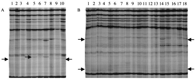
For each growth condition, we observed cDNAs from the metal-exposed cultures that were not present in the non-metal-exposed cultures (Fig. 2). In one case (Fig. 2A), a single DNA fragment was observed in the control lanes, perhaps from a transcript downregulated by the presence of metals. Cu(II)-treated and Hg(II)-treated samples shared the appearance of the same DNA fragments.
FIG. 2.
cDNA products derived from RAP-PCR of total RNA from independent cultures of D. vulgaris. (A) D. vulgaris grown on LSM and treated with 50 μM Cu(II) (lanes 1 to 4) or with 50 μM Hg(II) (lanes 8 to 10) or not treated with metal (lanes 5 to 7) (controls). (B) D. vulgaris grown on PM and treated with 50 μM Cu(II) (lanes 1 to 7) or with 50 μM Hg(II) (lanes 14 to 18) or not treated with metal (lanes 8 to 13) (controls). Differentially expressed cDNA products which were cut from the gels, cloned, and sequenced are indicated by the arrows. A paired control reaction without RT in the first-strand synthesis reaction indicated that no differently transcribed products were derived from contaminating genomic DNA.
Sequence identification and confirmation of differentially expressed RAP-PCR products.
Putative differentially transcribed fragments were excised from the gel, eluted with elution buffer (0.5 M ammonium acetate, 10 mM magnesium acetate, 1 mM EDTA, 0.1% sodium dodecyl sulfate), precipitated with 100% ethanol, and resuspended in 10 μl of sterile water. The cDNA was then reamplified with PCR parameters used in second-strand synthesis and resolved on a denaturing polyacrylamide gel with the original RAP-PCR product for comparison to ensure that the correct DNA fragment was isolated. The candidate PCR product was ligated into pCR4-TOPO vector and transformed into chemically competent One Shot TOP10 Escherichia coli (Invitrogen). For each fragment excised from the polyacrylamide gel, 10 clones were picked. Plasmid was isolated from 3-ml liquid cultures of candidate clones using a plasmid isolation kit (Qiaprep Spin Miniprep kit; QIAGEN Inc., Valencia, Calif.) according to the manufacturer's directions. After isolation, plasmids were digested with EcoRI and run on a 0.8% (wt/vol) agarose gel to compare inserts.
DNA sequencing was performed at the Oklahoma Medical Research Foundation Core Sequencing Facility (Oklahoma City, Okla.). Candidate insert sequences were initially compared to the whole genome sequence of D. vulgaris in the The Institute for Genomic Research (TIGR) BLAST Search Engine for Unfinished Microbial Genomes (UFMG). Interestingly, some of the inserts possessed two different transcripts, both derived from the D. vulgaris genome. All of the mRNAs obtained were derived from chimeric inserts, consisting of both an mRNA and 23S or 16S rRNA (3). Table 2 shows the open reading frames (ORFs) of D. vulgaris genes that showed DNA sequence homology with differentially transcribed fragments. GenBank sequence comparison was then done using both nucleotide and protein BLAST searches. Search results with ORF2004 showed high homology with MRP (multidrug resistance protein) (GenBank protein accession no. P21590) of E. coli (lowest-sum probability score = 6.9 e−46) and related proteins in other organisms. The other transcripts present during growth with metals coded for an ATPase (ORF856) and two hypothetical conserved proteins (ORF1445 and ORF2581).
TABLE 2.
DNA sequence homology of differently transcribed genes obtained from RAP-PCR procedure
| Substrate | Size (bp) of cloned fragment | Protein (ORF of D. vulgaris) | Lowest-sum probability score | Confirmed by real-time PCR (% increasea)
|
Databaseb | |
|---|---|---|---|---|---|---|
| Cu(II) treated | Hg(II) treated | |||||
| Lactate | 127 | ATP binding protein (ORF2004) | 3.2 × 10⁻²² | Yes (190) | Yes (314) | TIGR UFMG |
| Lactate | 43 | Histidine kinase-, DNA gyrase B-, phytochrome-like ATPase (ORF856) | 0.31 | Yes (41) | Yes (485) | NCBI BLAST |
| Pyruvate | 446 | Hypothetical conserved (ORF2581) | 7.1 × 10−84 | Yes (61) | Yes (45) | |
| Pyruvate | 422 | Hypothetical conserved (ORF1445) | 3.3 × 10−75 | No | No | |
Percent increase is the increase relative to the untreated culture and is determined as follows: [(amount of mRNA or rRNA from metal-treated culture)/(amount of mRNA or rRNA from non-metal-treated culture) − 1] × 100.
TIGR UFMG database (http://tigrblast.tigr.org/ufmg/index.cgi?database=d_vulgaris/seq) and National Center for Biotechnology Information (NCBI) BLAST (http://www.genome.ou.edu/blast/GDV_SEQ.html) websites.
To confirm that transcriptional levels for these mRNAs had increased on exposure to Cu or Hg, real-time reverse transcriptase PCR (RT-PCR) was then employed and the specific mRNAs were quantitated. Primers of real-time RT-PCR were designed to yield PCR products of approximately 100 bp. We designed one set of primers based on the RAP-PCR product sequence and another two sets based on the corresponding (somewhat longer) D. vulgaris ORF sequences obtained from TIGR. Primer sets were tested using D. vulgaris chromosomal DNA as the template. Chromosomal DNA was isolated using the Easy-DNA kit (Invitrogen) according to the manufacturer's directions. PCR mixtures contained 1 μg of DNA diluted in nuclease-free water, 6 μl of primer mixture (10 mM [each] forward and reverse primers), and 12.5 μl of SYBR green PCR master mix (Applied Biosystems, Foster City, Calif.), with the total volume adjusted to 25 μl. The reaction mixture was incubated at 95°C for 10 min, and 45 cycles of PCR were performed, with 1 cycle consisting of 15 s at 95°C, 30 s at 55°C, and 30 s at 60°C, with a final extension step of 10 min at 60°C. PCR products were resolved on a 1.5% (wt/vol) agarose gel (120 V, 30 min). One primer set for each sequence was selected on the basis of the yield of a single intense band of the appropriate size.
For each RT-PCR experiment, the reverse transcription reaction mixture contained 100 ng of total RNA, 1.25 μl of reverse primer (100 mM), 2.5 μl of RT buffer (10×), 5.5 μl of MgCl2 (25 mM), 5 μl of a mixture of deoxynucleoside triphosphates (each 2.5 mM), and 0.5 μl of RNase block (50 U/liter) in a volume of 24.4 μl. After gentle mixing and brief centrifugation, each reaction mixture was heated to 85°C for 10 min, 65°C for 5 min, and 56.5°C for 5 min. Then 0.6 μl of Moloney murine leukemia virus RT (20 U/liter) was added to the reaction mixture and incubated for 30 min at 48°C. The cDNA from each reaction mixture was either used immediately or stored at −20°C for less than 48 h.
For PCR amplification, 1 μl of a 1:20 dilution of first-strand cDNA product was mixed with 24 μl of PCR mix containing 3.5 μl of primer mix (8.5 mM forward primer and 1.42 mM reverse primer), 8 μl of sterile water, and 12.5 μl of SYBR green PCR master mix (Applied Biosystems). Conditions were as described above except with a real-time PCR machine (Smart Cycler; Cepheid, Sunnyvale, Calif.). The intensities of PCR products were analyzed by using Smart Cycler software, and the threshold cycle value was used to compare the relative quantities of PCR products. At least three individually extracted RNA samples from each Cu(II)- or Hg(II)-treated and control cultures were tested. Reactions lacking RT were used as negative controls, and 23S rRNA was used as a reference so that changes in expression were determined relative to 23S rRNA concentration. Genes for an ATP binding protein (ORF2004) and an ATPase (ORF856) were transcriptionally upregulated with both the Cu(II) and Hg(II) treatments (Table 2).
Metals including Hg(II) and Cu(II) may enter cells through energy-dependent uptake systems, as occurs in other microorganisms (6, 13). Several bacterial metal resistance systems are also energy dependent (19), typically using an energy-dependent efflux mechanism. For example, copA and copB are involved in Cu uptake and efflux systems in microorganisms (21). It is plausible that the genes in D. vulgaris whose expression was upregulated may be involved in metal transport. However, it is worth noting that gene expression in a specific situation does not necessarily mean that that gene plays an active role in the response process.
Two other sequences were observed, both designated hypothetical conserved proteins. We were able to confirm that one of these genes (ORF2581) was upregulated in the presence of the two metals (Table 2). Even though significant upregulation of several genes (ORF2004, ORF856, and ORF2581) was observed in both Cu(II)- and Hg(II)-treated samples, background transcriptional activities were also observed in control samples (not treated with metal). This may be a result of metals typically present in the culture medium. The basal medium used to cultivate anaerobic microorganisms contains dissolved metals as nutrients.
Acknowledgments
This research was supported in part by a grant from the Natural and Accelerated Bioremediation Research (NABIR) program of the Office of Biological and Environmental Research of the Office of Science of the U.S. Department of Energy.
REFERENCES
- 1.Abu Kwaik, Y., and L. L. Pederson. 1996. The use of differential display-PCR to isolate and characterize a Legionella pneumophila locus induced during the intracellular infection of macrophages. Mol. Microbiol. 21:543-556. [DOI] [PubMed] [Google Scholar]
- 2.Chakrabortty, A., S. Das, S. Majumdar, K. Mukhopadhyay, S. Roychoudhury, and K. Chaudhuri. 2000. Use of RNA arbitrarily primed-PCR fingerprinting to identify Vibrio cholerae genes differentially expressed in the host following infection. Infect. Immun. 68:3878-3887. [DOI] [PMC free article] [PubMed] [Google Scholar]
- 3.Chang, I. S., J. D. Ballard, and L. R. Krumholz. 2003. Evidence for chimeric sequences formed during random arbitrarily primed PCR. J. Microbiol. Methods 54:427-431. [DOI] [PubMed] [Google Scholar]
- 4.Chang, I. S., P. K. Shin, and B. H. Kim. 2000. Biological treatment of acid mine drainage under sulphate-reducing conditions with solid waste materials as substrate. Water Res. 34:1269-1277. [Google Scholar]
- 5.Herbert, D., P. J. Phipps, and R. E. Strange. 1971. Analysis of microbial cells. Methods Microbiol. 5B:209-344. [Google Scholar]
- 6.Kanamaru, K., S. Kashiwagi, and T. Mizuno. 1993. The cyanobacterium, Synechococcus sp. PCC7942, possesses two distinct genes encoding cation transporting P-type ATPases. FEBS Lett. 330:99-104. [DOI] [PubMed] [Google Scholar]
- 7.Krumholz, L. R., and M. P. Bryant. 1986. Eubacterium oxidoreducens sp. nov. requiring H2 or formate to degrade gallate, pyrogallol, phloroglucinol and quercetin. Arch. Microbiol. 144:8-14. [Google Scholar]
- 8.Liang, P., and A. B. Pardee. 1992. Differential display of eukaryotic messenger RNA by means of the polymerase chain reaction. Science 257:967-971. [DOI] [PubMed] [Google Scholar]
- 9.Lloyd, J. R., H. F. Nolting, V. A. Sole, K. Bosecker, and L. E. Macaskie. 1998. Technetium reduction and precipitation by sulfate-reducing bacteria. Geomicrobiol. J. 15:45-58. [Google Scholar]
- 10.Lloyd, J. R., J. Ridley, T. Khizniak, N. N. Lyalikova, and L. E. Macaskie. 1999. Reduction of technetium by Desulfovibrio desulfuricans: biocatalyst characterization and use in a flowthrough bioreactor. Appl. Environ. Microbiol. 65:2691-2696. [DOI] [PMC free article] [PubMed] [Google Scholar]
- 11.Lovley, D. R., and E. J. Phillips. 1992. Reduction of uranium by Desulfovibrio desulfuricans. Appl. Environ. Microbiol. 58:850-856. [DOI] [PMC free article] [PubMed] [Google Scholar]
- 12.Macy, J. M., J. M. Santini, B. V. Pauling, A. H. O'Neill, and L. I. Sly. 2000. Two new arsenate/sulfate-reducing bacteria: mechanisms of arsenate reduction. Arch. Microbiol. 173:49-57. [DOI] [PubMed] [Google Scholar]
- 13.Odermatt, A., H. Suter, R. Krapf, and M. Solioz. 1993. Primary structure of two P-type ATPases involved in copper homeostasis in Enterococcus hirae. J. Biol. Chem. 268:12775-12779. [PubMed] [Google Scholar]
- 14.Poulson, S. R., P. J. S. Colberg, and J. I. Drever. 1997. Toxicity of heavy metals (Ni, Zn) to Desulfovibrio desulfuricans. Geomicrobiol. J. 14:41-49. [Google Scholar]
- 15.Sani, R. K., G. Geesey, and B. M. Peyton. 2001. Assessment of lead toxicity to Desulfovibrio desulfuricans G20: influence of components of lactate C medium. Adv. Environ. Res. 5:269-276. [Google Scholar]
- 16.Sani, R. K., B. M. Peyton, and L. T. Brown. 2001. Copper-induced inhibition of growth of Desulfovibrio desulfuricans G20: assessment of its toxicity and correlation with those of zinc and lead. Appl. Environ. Microbiol. 67:4765-4772. [DOI] [PMC free article] [PubMed] [Google Scholar]
- 17.Senko, J. M., J. D. Istok, J. M. Suflita, and L. R. Krumholz. 2002. In-situ evidence for uranium immobilization and remobilization. Environ. Sci. Technol. 36:1491-1496. [DOI] [PubMed] [Google Scholar]
- 18.Shepard, B. D., and M. S. Gilmore. 1999. Identification of aerobically and anaerobically induced genes in Enterococcus faecalis by random arbitrarily primed PCR. Appl. Environ. Microbiol. 65:1470-1476. [DOI] [PMC free article] [PubMed] [Google Scholar]
- 19.Silver, S., and L. T. Phung. 1996. Bacterial heavy metal resistance: new surprises. Annu. Rev. Microbiol. 50:753-789. [DOI] [PubMed] [Google Scholar]
- 20.Smillie, R. H., K. Hunter, and M. Loutit. 1981. Reduction of chromium(VI) by bacterially produced hydrogen sulfide in a marine environment. Water Res. 15:1351-1354. [Google Scholar]
- 21.Solioz, M., A. Odermatt, and R. Krapf. 1994. Copper pumping ATPases: common concepts in bacteria and man. FEBS Lett. 346:44-47. [DOI] [PubMed] [Google Scholar]
- 22.Steger, J. L., C. Vincent, J. D. Ballard, and L. R. Krumholz. 2002. Desulfovibrio sp. genes involved in the respiration of sulfate during metabolism of hydrogen and lactate. Appl. Environ. Microbiol. 68:1932-1937. [DOI] [PMC free article] [PubMed] [Google Scholar]
- 23.Tebo, B. M., and A. Y. Obraztsova. 1998. Sulfate-reducing bacterium grows with Cr(VI), U(VI), Mn(IV), and Fe(III) as electron acceptors. FEMS Microbiol. Lett. 162:193-198. [Google Scholar]
- 24.White, C., and G. M. Gadd. 1996. Mixed sulphate-reducing bacterial cultures for bioprecipitation of toxic metal: factorial and response-surface analysis of the effects of dilution rate, sulphate and substrate concentration. Microbiology 142:2197-2205. [Google Scholar]
- 25.White, C., A. K. Sharman, and G. M. Gadd. 1998. An integrated microbial process for the bioremediation of soil contaminated with toxic metals. Nat. Biotechnol. 16:572-575. [DOI] [PubMed] [Google Scholar]


