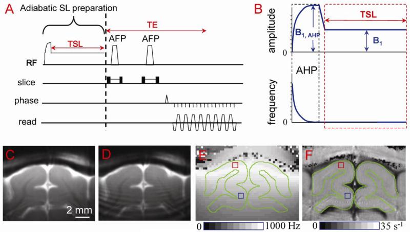
Fig. 1. Surface-coil T1ρ pulse sequence (A, B) and applications to cat primary visual cortex studies (C-F).
The pulse sequence (A) is a double spin-echo EPI acquisition with a non-selective spin-locking (SL) preparation (B). Adiabatic half passage (AHP, 2 ms in length with amplitude B1, AHP) ensures that spins nutate into the transverse plane over a relatively large volume of B1 inhomogeneity. The RF amplitude is then immediately ramped to the desired SL level (B1) and held constant for the spin-lock time (TSL); the transmit frequency of the pulse remains the same during the short ramp and TSL. Following the spin-lock preparation, transverse spins are refocused by two adiabatic full-passage (AFP) pulses with slice-selection gradients. High quality T1ρ-weighted image (ω1 = 500 Hz and TSL = 40 ms) can be obtained when adiabatic SL condition is satisfied (C), but signal oscillations appear otherwise (D). (E) The spin-locking nutation frequency ω1 map shows B1 inhomogeneity. For our conditions, ω1 at an area proximal to the coil (red square) is ~600 Hz, while at a distal area (blue square) it is ~ 400 Hz. Gray matter areas are outlined in green. (F) The map of R1ρ (=1/T1ρ) shows little spatial variations within visual cortex because the T1ρ dispersion is very small within the narrow ω1 range (e.g., 400-600 Hz).

