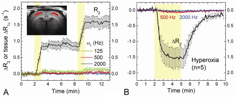
Fig. 5. Effects of intravascular susceptibility variation and hyperoxia challenge on extravascular water R1ρ.
In order to detect the contribution of intravascular susceptibility changes to tissue R1ρ, the blood signal was suppressed with the injection of 5 mg/kg MION before experiments. Dynamic changes in tissue R2 and R1ρ were obtained during two injections of 1 mg/kg MION (A, n = 4 rats) and 3 minutes inhalation of 60% O2 (B, n = 5 rats) indicated by the yellow shaded regions. Time courses were obtained from the red pixels within the cortex (Inset). When spin-locking frequencies were measured at ≥500 Hz, a variation in intravascular susceptibility does not contribute to tissue R1ρ measurements.

