Summary
For neck protection technique to prevent migration or protrusion of GDCs, non detachable balloons or microcatheters/wires are used. Non detachable balloon has good protective effect with great effect to the flow in the parent artery which is risky for safe procedure. Microcatheter has little effect to the parent flow and poor protective effect. We have developed retrievable microbasket for neck protective technique which can be delivered and retrieved through standard 18 catheters. These baskets have good protective effect in aneurysm model.
Key words: large neck aneurysms, remodelling technique
Introduction
By development of GDC 1,2, intracranial berry aneurysms are safely treated by endovascular procedure. However, wide neck aneurysms are difficult for this technique. The reason is, of course, more possibility of protrusion or migration of GDCs. Recently, neck protective technique such as non-detachable balloons or catheter/guide wire are introduced to prevent coil protrusion3,4,5.
Non-detachable balloons are very effective for neck protection because of its wide or complete coverage of aneurysm orifice. But this technique has possible risks. One is distal occlusion before aneurysm treatment. Sudden shift of the balloon by blood flow occlude distal parent artery to elevate intraaneurysmal pressure and possibly rupture the aneurysm. The other is possible ischemia of distal territory of the parent artery if prolonged protection. Guidewire or catheter do not have such risks because these materials have poor effect to the blood flow. But using these devices, the coverage of the orifice is not enough and resisting force toward the protruding coils are not enough, either. Here, we consider if the basket shaped device which can be delivered and retrieved through standard microcatheters, this device might be effective for neck protection with smaller risk.
Material and Methods
The basket system was designed to have four to eight arms and the arms were mounted at the tip of delivery wire. The system had radiopaque and shapable distal tip with radiopaque proximal marker. The basket itself was made of stainless steel and platinum. These materials were selected to check radiopacity.
As the evaluation how the basket works, glass model of wide neck aneurysm was made. The diameter of dome and neck were 7 mm and the diameter of parent structure were also 7 mm. The model was placed in normal saline bath. At first, radiopacity was evaluated using a DSA unit. Baskets were placed at the forehead of a phantom skull and digital anigography was taken.
Smooth passage of the basket was checked using curved tube.
Then protection was tested. The tip of standard 18 microcatheter was placed within the lumen of aneurysm model following the second microcatheter was placed in the parent artery. The basket was delivered through the second microcatheter. The basket was exposed in the lumen of parent artery to cover the orifice of the experimental aneurysm by pulling the second catheter while leaving the basket delivery wire there. Then 7 mm Interlocking Detachable Coil (IDC) was delivered in the aneurysm. After delivery of IDC, the basket was retrieved by advancing the microcatheter leaving basket at the orifice. The protective effect was checked by VTR and observed.
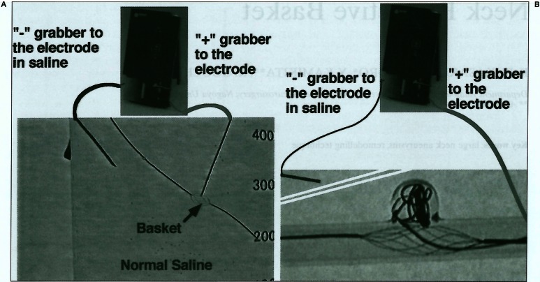
To evaluate the tolerance toward the electric current, the stainless basket was touched with platinum rod in a bath filled with normal saline. Positive (red) electrode of GDC power supply was connected to the platinum rod and the negative (black) one was connected to the platinum rod placed in the bath (figure 1A). The damage was observed macroscopically. As the second test for tolerance, IDC basket and IDC were delivered as the same manner as protection test (figure 1B). After delivery, power unit for GDC was connected to the delivery wire of IDC and to an electrode in the normal saline bath.
Figure 1.
A) Tolerance test in normal saline bath. Basket was placed in normal saline bath freely and platinum electrode touched the basket filament. B) Tolerance test in the glass model of aneurysm. Basket was place in the parent artery of the model. “+” grabber was connected with the delivery wire of IDC and “-” grabber was connected with electrode in normal saline. Basket and delivery wire had no direct connection with the circuit.
The GDC power unit worked for 45 minutes to evaluate whether the electric current for detachment of GDC damaged the basket or not. VTR was taken while the power unit working. After the electric current, the basket was examined macroscopically and using scanning electron microscope (SEM).
Results
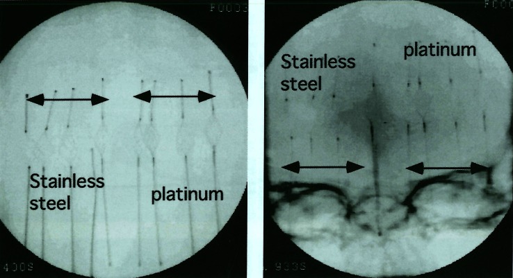
Radiopacity of the baskets were examined by DA film. The basket arms made from stainless steel filament was hard to observe and platinum arms were difficult. Of course better than stainless steel but there were no big difference (figure 2). So, we decided to use stainless wire to make baskets for further assessment.
Figure 2.
Stainless steel baskets were almost invisible. At the same time, platinum baskets were visible but very unclear.
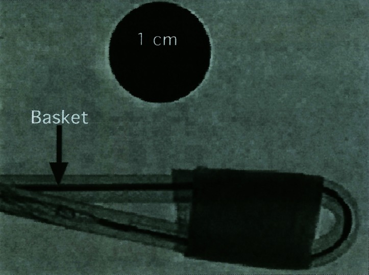
The basket could be delivered smoothly through standard 18 catheters such as Tracker series or Transit series, (figure 3) However, at the junction of arms, we felt some resistance. At the same time, the basket could be easily retrieved within the catheter lumen by pushing the catheter. After introducing 18 microcatheter in the 7 mm curve plastic tube, the basket could pass thought the catheter without large resistance.
Figure 3.
The basket could pass the sharp angled model vessel and standard 18 catheter, The black circle is 1 cm in diameter.
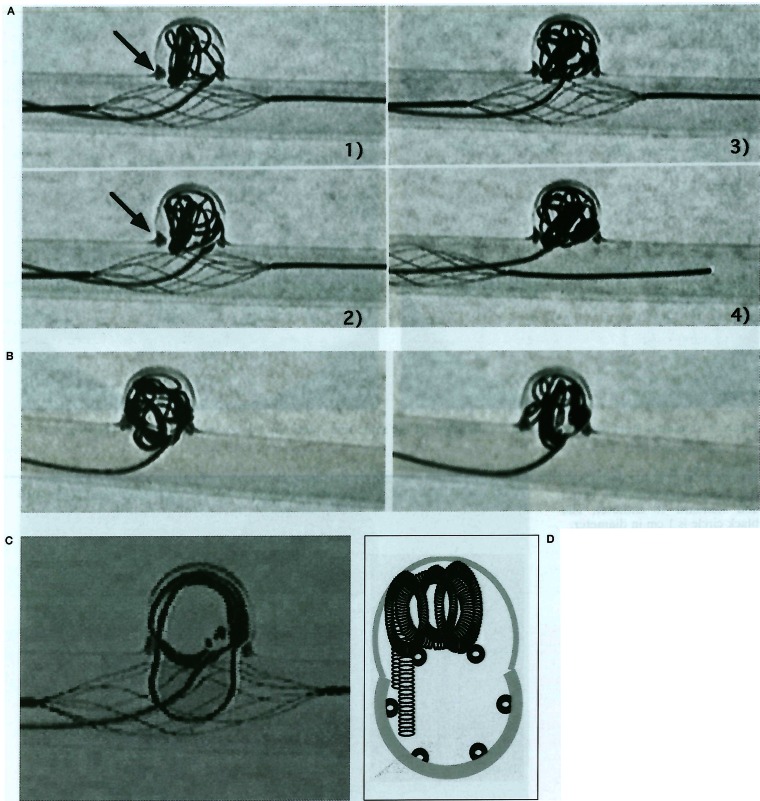
The protective effect of the basket was checked by glass model of aneurysm. The orifice of the model aneurysm could be covered with two or one of the arms. Because these basket systems had support at the opposite side of orifice, these baskets could resist big protruding force while delivery of coils. The basket could push back the IDC after deformity of themselves.
We would rather worry about rupture of aneurysm by over packing (figure 4A). while our observation, these protrusion was finally replaced in the aneurysm. Without our basket, IDC could not stay safely within the aneurysm lumen (figure 4B). Some loops protruded into the parent artery. If the protrusion of GDC occur in parallel with the basket arms, protection was not enough (figure 4C, D).
Figure 4.
A) With neck protective basket protrusion of coil was controllable. The basket deformed by protrusion, however, the basket pushed back the coil. After retrieve of the basket, IDC was placed in the aneurysm stably. [From 1 to 4] B) Without protective system, IDC protruded into the parent artery. C) Tʼne basket could not control the protrusion in parallel with its arms, completely. This loop in the parent artery was finally replaced in the aneurysm by continuing to push delivery wire, only. D) Schematic drawing of cross section of figure (C).
Tolerance to the electric current while detachment was checked in bath and in model. While examination in the bath, no macroscopic damage were observed. And examination in glass model of aneurysm, no microscopic erosion were noticed. However, scanning electron microscope revealed mild erosion the arm (figure 5).
Figure 5.
After 30 minutes electric current (1 mA, 3 V), SEM disclosed erosive change on the surface of arms (A). Macroscopically, the erosion was not noticed. After coating (Teflon) erosive change was not observed (B).
Discussion
Balloon neck protection or balloon remodeling technique has created new horizon of GDC treatment.
But this technique has possible risks. One isdistal occlusion by the balloon itself to lead sudden elevation of intraaneurysmal pressure and the other is possible ischemia at the distal territory. And catheter or wire protection has possible risk, smaller than non-detachable balloon because of smaller affect to the flow of parent artery. At the same time, protective effect is smaller, too. Our basket system has more effect than wire or catheter and less risk than the balloon system because wire filament basket has less effect to the flow in the parent artery and stronger resistance than wire or catheter protection because of wider coverage of orifice and supporting filaments at the opposite side.
SEM disclosed the damage of the bare arm. But the basket could be retrieved without migration or detachment of itself. This basket could be retrieved after 40 minutes current. We used IDC in stead of GDC because if GDC, the coil is detached frequently so we need a lot of GDC for test. Recent advance of quick release of GDC, we can detach 10 or more coils while 40 minutes. And electrolysis affects the weakest point at first. So, the detach zone of GDC is destructed for detachment at first and after detachment, the current stops immediately. Here, the possibility of erosion might be smaller comparing our test using IDC and prolonged currency. And we can preserve this risk by retrieving the basket before detachment. After pulling back the basket system within the microcatheter, electric current can not affect the basket. After the experience of erosion, we modified the basket to have coating. After coating, erosion was not observed by SEM (figure 5).
Recently, combination use of GDCs and metallic stents were reported6,7,8,9. Stents were made from stainless steel, titanium or Ergyroy, etc. According to our experience, stainless steel stents has the same possible risk of erosion while detachment of GDC. Concerning battery effect, we had better more be much more cautious for these combination use.
While our trial, the coils did not intertwine with the arm of basket. But this kind of intertwining is a considerable risk. We must retrieve the basket before detachment and if we felt resistance, the coil must be retrieved. This basket system has spindle shape. Usual protective balloon systems have spindle or sausage shape, too. These protective systems which have long axis can not protect aneurysm orifice at the bifurcation. Some spherical shaped protective device and/or 3D coil should be used in such occasion.
For much more safe basket, we are renewing this device to have #1 more arms of more effective protection and #2 coating of the arm for more resistance for electric current. We are planning further clinical use of this device to cluck thrombus at acute ischemic stroke and retrieving foreign materials in the vascular lumen.
Conclusions
Retrievable basket system was assembled. The basket could be delivered through standard 18 size catheters and was very effective to prevent migration of GDCs.
The basket could be dissolved if prolonged electric current.
References
- 1.Guglielmi G, Viñuela, et al. Electrothrombosis of saccular aneurysms via endovascular approach. Part 1: Electrochemical basis, technique, and experimental results. J Neurosurg. 1991;75:1–7. doi: 10.3171/jns.1991.75.1.0001. [DOI] [PubMed] [Google Scholar]
- 2.Guglielmi G, Viñuela, et al. Electrothrombosis of saccular aneurysms via endovascular approach. Part 2: Preliminary clinical experience. J Neurosurg. 1991;75:8–14. doi: 10.3171/jns.1991.75.1.0008. [DOI] [PubMed] [Google Scholar]
- 3.Moret J, Cognard C, et al. Reconstruction technic in the treatment of wide-neck intracranial aneurysms. Longterm angiographic and clinical results. Apropos of 56 cases. J Neuroradiol. 1997;24(1):30–44. [PubMed] [Google Scholar]
- 4.Lefkowitz MA, Gobin YP, et al. Ballonn-assisted Guglielmi detachable coiling of wide-necked aneurysma: Part II clinical results. Neurosurgery. 1999;45:531–537. doi: 10.1097/00006123-199909000-00024. [DOI] [PubMed] [Google Scholar]
- 5.Mericle RA, Wakhloo AK, et al. Temporary balloon protection as an adjunct to endosaccular coiling of widenecked cerebral aneurysms: technical note. Neurosurgery. 1997;41:975–978. doi: 10.1097/00006123-199710000-00045. [DOI] [PubMed] [Google Scholar]
- 6.Turjman F, Massoud TF, et al. Combined stent implantation and endosaccular coil placement for treatment of experimental wide-necked aneurysms: a feasibility study in swine. Am J Neuroradiol. 1994;15:1087–1090. [PMC free article] [PubMed] [Google Scholar]
- 7.Mericle RA, Lanzino G, et al. Stenting and secondary coiling of intracranial internal carotid artery aneurysm: technical case report. Neurosurgery. 1998;43:1229–1234. doi: 10.1097/00006123-199811000-00130. [DOI] [PubMed] [Google Scholar]
- 8.Lanzino G, Wakhloo AK, et al. Efficacy and current limitations of intravascular stents for intracranial internal carotid, vertebral, and basilar artery aneurysms. J Neurosurg. 1999;91:538–546. doi: 10.3171/jns.1999.91.4.0538. [DOI] [PubMed] [Google Scholar]
- 9.Higashida RT, Smith W, et al. Intravascular stent and endovascular coil placement for a ruptured fusiform aneurysm of the basilar artery. Case report and review of the literature. J Neurosurg. 1997;87:944–949. doi: 10.3171/jns.1997.87.6.0944. [DOI] [PubMed] [Google Scholar]


