Abstract
Spindle cell lipoma (SCL) is a benign lipomatous tumor predominantly occurring at the posterior neck and shoulder area. Face, forehead, scalp, cheek, perioral area, and upper arm are less common sites. In oral cavity, it is a relatively uncommon neoplasm, particularly in tongue, which is relatively devoid of fat cells. We present a case report of SCL located on the left lateral border of the tongue in a 64-year-old Caucasian female patient with diabetes mellitus type 2 and arterial hypertension.
Keywords: Oral cavity, spindle cell lipoma, tongue
INTRODUCTION
The first case reported of lingual lipoma was credited to Barling in 1858 and was cited by Guillou et al.[1] Lipomas are slow growing benign mesenchymal neoplasms.[2] composed of mature adipocytes, commonly surrounded by a thin fibrous capsule that originate in mature fat cells.[3,4] Most oral lipomas are composed of mature fat cells that do not differ microscopically from normal fat cells;[5] however, differ metabolically from normal fat cells.[2] Although lipoma can occur in any part of the body that contains fat tissue, it is not commonly found in the oral cavity.[1]
Lipoma of the tongue is not a common occurrence and accounts for 0.3% of all lingual tumors.[6] It is described traditionally as well-circumscribed, submucosal mass of less than 1 cm in size, and located on the lateral border of the dorsal anterior two-thirds of the tongue.[1] Based on histopathologic features, lipomas can be classified as simple lipoma which constitutes 80% of lipomas, whilst other variants such as fibrolipomas, angiolipomas, intramuscular lipomas, pleomorphic lipomas, spindle-cell lipomas (SCLs), sialolipomas, myxoid lipomas and atypical lipomas[7] make up the remaining 20% of all lipomas.
SCLs are composed of bland mitotically inactive spindle cells arranged in parallel registers between the fat cells, associated with thick rope-like collagen bundles.[8] It was described previously in the oral cavity and tongue, and remains a rare entity in these sites.[9–13] The etiology and pathogenesis of lipomas is still unclear.[5] Most patients are around 40 years or older and it is extremely rare in children.[5,14] Moreover, lipomas elsewhere in the body are reported to be twice as common in females than in males, but oral lipomas are characterized by a more balanced sex distribution.[5] The histological variant identified in this case was SCL of the tongue.
CASE REPORT
A 64-year-old Caucasian female patient sought dental care due to injury in a swelling on the left lateral border of the tongue. The patient noticed a swelling about 3 years back which was progressively increasing in size and presented as an enlarged tongue. At present, the patient reports difficulty in speech and swallowing. Her medical history revealed type 2 diabetes mellitus (T2DM) and hypertension for almost a decade. She was taking medication such as metformin, gliclazide, and beta blockers for her systemic pathologies. She has no history of smoking or alcohol usage. Clinical intraoral examination revealed the patient to be a removable denture wearer with good oral hygiene status. A characteristic nodular lesion which was elastic in consistency was noticed on the left lateral border on the anterior two-thirds of the tongue [Figure 1].
Figure 1.
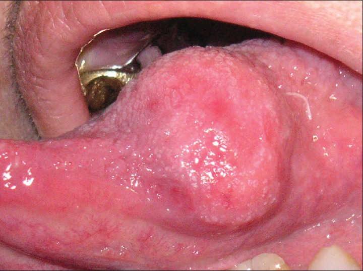
Swelling in the left lateral border of the tongue
An ultrasonography of the lesion was requested which showed a solid area about 20 × 20 × 13 mm in its largest diameter, located in the left lateral border of the tongue. It showed regular, well-defined margins with homogeneous texture. A Doppler study showed some peripheral vessels associated with the lesion [Figure 2].
Figure 2.
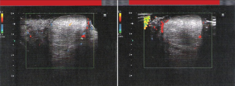
The ultrasonography with Doppler study showing a solid rounded lesion associated with peripheral vessels
The patient was referred to her physician for pre-operative routine examination following which, the patient underwent excisional biopsy under local anesthesia. The lesion was removed after dilatation and dissection of the surrounding tissues [Figure 3].
Figure 3.
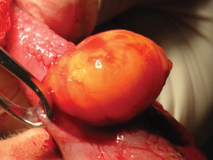
The excised lesion
After irrigation, the surgical wound was sutured with 4.0 vicryl suture material. The patient received appropriate postoperative care and was discharged. The specimen was sent for pathological examination. Macroscopic examination revealed a nodular lesion measuring approximately 2.2 × 1.8 × 1.3 cm in its greatest dimension covered by a smooth, shiny, yellowish translucent capsule [Figure 4].
Figure 4.
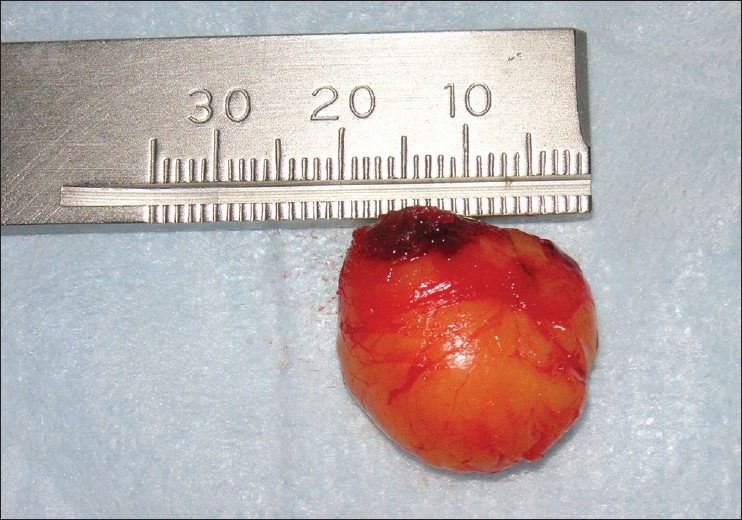
Macroscopic examination revealing a nodular lesion with smooth external surface
Microscopic examination revealed expansive mesenchymal neoplasm composed of sheets of mature fat cells with typical fine vascular network, thick fibrous beams, and spindle cells permeating the sheets of fat cells. The subcapsular margins were free of tumor invasion. The features were consistent with a SCL [Figure 5].
Figure 5.
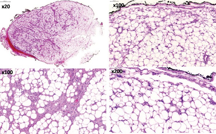
The histological features revealed sheets of mature fat cells, fine vascular network, spindle cells and thick fibrous beams permeating the sheets of fat cells (H and E stain)
The post-operative follow-up demonstrated regression of the lesion with good tissue repair. No complaint in mastication and speech was noted.
DISCUSSION
Lipomas of oral cavity are not frequent and commonly occur as well circumscribed lesions in the buccal mucosa, tongue, lips and floor of the mouth.[7,15–17] It is a benign condition, especially in tongue, which accounts for 0.3% of tongue neoplasms.[6,18–20] It is diagnosed often in adult patients at the mean age of 50.2-62 years.[7,17,18,21] The etiology of lipomas remains unclear. Trauma, chromosomal abnormality, hereditary, chronic irritation, hormonal imbalance and metabolic conditions are some of causative factors in the pathogenesis of this neoplasm.[22,23] In the present case, the patient had a systemic disease T2DM that is considered a predisposing condition and also a possibility of traumatic injury (as she was wearing removable dentures) could be considered as a cause for the lesion. There was no family history of lipoma. Previously, there were very few reports of SCL variant of oral lipomas.[17,20] Furlong et al,[21] in 2004 reported 59 cases of SCL in 125 lipomas in the oral and maxillofacial region, showing a common occurrence than previously estimated.
SCL is an asymptomatic benign neoplasm and was first described in 1975 by Enzinger and Harvey[24] as an uncommon variant of lipoma accounting about 1.5% of all adipose tissue neoplasms.[11,25] The majority of these tumors, in terms of histology, are characterized by an admixture of mature adipocytes, uniform spindle cells, mucoid material and bundles of collagen associated with spindle cells.[20,22,24]
The oral cavity and the tongue are previously described as common sites of SCLs, but are rarer than ordinary lipomas,[9–13] specially the occurrence of multiple SCLs of the tongue.[25]
The clinical features of the case reported are suggestive of lipoma or a related lesion. Hematoxylin and eosin stained sections showed an expansive mesenchymal neoplasm composed of mature typical fat cells, fine vascular network in between, spindle cells and thick fibrous bundles permeating the fat cells. The spindle cells have little cytoplasm and elongated nuclei that are arranged parallelly. No lipoblastic activity was observed in SCL and mitotic figures were seldom seen.
The differential diagnosis of SCL is important because it can easily be misdiagnosed as a malignant lesion such as a liposarcoma. Liposarcoma in sites of easily accessible areas such as tongue should be classified as atypical lipomas whilst similar tumors arising in inaccessible sites where complete excision of the lesion is difficult to perform should be designated as liposarcoma.[26]
Mitotic activity, pleomorphism and the amount of vasculature should be accurately evaluated to rule out malignancy. SCL are composed of bland spindle cells with minimal polymorphism and mitoses.[22] These bland features are important in differentiating SCL from a liposarcoma.
Immunohistochemistry (IHC) was not performed in this case. The cluster of differentiation (CD) 34 expression plays a key role in the differential diagnosis of some lesions and others can help to resolve diagnostic problems.[27] Although it is known that immunoreactivity for CD34 in spindle cell component is usually observed, it may be helpful only in some cases.[13,25,27,28] On the other hand, this marker can be found in a wide variety of both normal and neoplastic cells of the human body, hence diagnosis on a normal hematoxylin and eosin stained section is more reliable.[27] Immunohistochemistry (IHC) plays a relatively minor role in diagnosis and might not be useful as a tool for the diagnosis of SCL[12] but contributes to understand the pathology of the lesion.
Moreover, tumors indistinguishable from SCLs have occasionally be reported under special names.[27,28]
The treatment of choice of all histological variants is local surgical excisions.[2,3,6,7,9–12,14,23,26] The prognosis is usually good with rare recurrence,[10,18] especially in case of infiltrating lipomas that tend to invade surrounding muscles.[19] The SCL is not usually encapsulated.[10,11,22] However, this case reveals an encapsulated mass of fat, which was easily removed from the surrounding tissue. Although the growth of oral lipomas is usually limited,[7] in the present case, SCL was considerably larger in dimension that was interfering with speech and mastication.
In conclusion, SCL is an asymptomatic benign soft-tissue neoplasm that is an uncommon variant of lipoma. The lesion is typically of slow growing nature, encapsulated and usually symptom free. Surgical excision is the elective treatment of choice in cases where the lesion is encapsulated and can be easily removed from surrounding tissues. The relapse of this variant is an uncommon, but long-term follow-up is mandatory.
Footnotes
Source of Support: Nil.
Conflict of Interest: None declared.
REFERENCES
- 1.Guillou L, Dehon A, Charlin B, Madarnas P. Pleomorphic lipoma of the tongue: Case report and literature review. J Otolaryngol. 1986;15:313–6. [PubMed] [Google Scholar]
- 2.Sekar B AD, Murali S. Lipoma, a rare intraoral tumor: A case report with review of literature. J Oral Maxillofac Pathol. 2011;2:174–7. [Google Scholar]
- 3.Adoga AA, Nimkur TL, Manasseh AN, Echejoh GO. Buccal soft tissue lipoma in an adult Nigerian: A case report and literature review. J Med Case Rep. 2008;2:382. doi: 10.1186/1752-1947-2-382. [DOI] [PMC free article] [PubMed] [Google Scholar]
- 4.de Visscher JG. Lipomas and fibrolipomas of the oral cavity. J Maxillofac Surg. 1982;10:177–81. doi: 10.1016/s0301-0503(82)80036-2. [DOI] [PubMed] [Google Scholar]
- 5.Neville BW, Damm DD, Allen CM, Bouquot JE. Soft tissue tumors. In: Neville BW, Damm DD, Allen CM, Bouquot JE, editors. Oral and Maxillofacial Pathology. Philadelphia PA: WB Saunders Company; 2002. pp. 452–4. [Google Scholar]
- 6.Roles DM. Lipoma of the tongue. Br J Oral Maxillofac Surg. 1995;33:196–7. doi: 10.1016/0266-4356(95)90307-0. [DOI] [PubMed] [Google Scholar]
- 7.Bandéca MC, de Pádua JM, Nadalin MR, Ozório JE, Silva-Sousa YT, da Cruz Perez DE. Oral soft tissue lipomas: A case series. J Can Dent Assoc. 2007;73:431–4. [PubMed] [Google Scholar]
- 8.Miettinen MM, Mandahl N. Spindle cell lipoma/pleomorphic lipoma. In: Fletcher CD, Unni K, Mertens F, editors. Pathology and Genetics of Tumours of Soft Tissue and Bone, WHO Classification of Tumours. Lyon: IARC Press; 2002. pp. 31–2. [Google Scholar]
- 9.Lombardi T, Odell EW. Spindle cell lipoma of the oral cavity: Report of a case. J Oral Pathol Med. 1994;23:237–9. doi: 10.1111/j.1600-0714.1994.tb01120.x. [DOI] [PubMed] [Google Scholar]
- 10.Tosios K, Papanicolaou SI, Kapranos N, Papadogeorgakis N. Spindle cell lipoma of the oral cavity. Int J Oral Maxillofac Surg. 1995;24:363–4. doi: 10.1016/s0901-5027(05)80493-x. [DOI] [PubMed] [Google Scholar]
- 11.Dutt SN, East DM, Saleem Y, Jones EL. Spindle-cell variant of intralingual lipoma: Report of a case with literature review. J Laryngol Otol. 1999;113:587–9. doi: 10.1017/s0022215100144561. [DOI] [PubMed] [Google Scholar]
- 12.Kaku N, Kashima K, Daa T, Nakayama I, Kerakawauchi H, Hashimoto H, et al. Multiple spindle cell lipomas of the tongue: Report of a case. APMIS. 2003;111:581–5. doi: 10.1034/j.1600-0463.2003.1110507.x. [DOI] [PubMed] [Google Scholar]
- 13.Billings SD, Henley JD, Summerlin DJ, Vakili S, Tomich CE. Spindle cell lipoma of the oral cavity. Am J Dermatopathol. 2006;28:28–31. doi: 10.1097/01.dad.0000189615.13641.4b. [DOI] [PubMed] [Google Scholar]
- 14.Venkateswarlu M, Geetha P, Srikanth M. A rare case of intraoral lipoma in a six year-old child: A case report. Int J Oral Sci. 2011;3:43–6. doi: 10.4248/IJOS11008. [DOI] [PMC free article] [PubMed] [Google Scholar]
- 15.de Freitas MA, Freitas VS, de Lima AA, Pereira FB, Jr, dos Santos JN. Intraoral lipomas: A study of 26 cases in a Brazilian population. Quintessence Int. 2009;40:79–85. [PubMed] [Google Scholar]
- 16.Manor E, Sion-Vardy N, Joshua BZ, Bodner L. Oral lipoma: Analysis of 58 new cases and review of the literature. Ann Diagn Pathol. 2011;15:257–61. doi: 10.1016/j.anndiagpath.2011.01.003. [DOI] [PubMed] [Google Scholar]
- 17.Fregnani ER, Pires FR, Falzoni R, Lopes MA, Vargas PA. Lipomas of the oral cavity: Clinical findings, histological classification and proliferative activity of 46 cases. Int J Oral Maxillofac Surg. 2003;32:49–53. doi: 10.1054/ijom.2002.0317. [DOI] [PubMed] [Google Scholar]
- 18.Epivatianos A, Markopoulos AK, Papanayotou P. Benign tumors of adipose tissue of the oral cavity: A clinicopathologic study of 13 cases. J Oral Maxillofac Surg. 2000;58:1113–7. doi: 10.1053/joms.2000.9568. [DOI] [PubMed] [Google Scholar]
- 19.Colella G, Lanza A, Rossiello L, Rossiello R. Infiltrating lipoma of the tongue. Oral Oncol Extra. 2004;40:33–5. [Google Scholar]
- 20.Said-Al-Naief N, Zahurullah FR, Sciubba JJ. Oral spindle cell lipoma. Ann Diagn Pathol. 2001;5:207–15. doi: 10.1053/adpa.2001.26973. [DOI] [PubMed] [Google Scholar]
- 21.Furlong MA, Fanburg-Smith JC, Childers EL. Lipoma of the oral and maxillofacial region: Site and subclassification of 125 cases. Oral Surg Oral Med Oral Pathol Oral Radiol Endod. 2004;98:441–50. doi: 10.1016/j.tripleo.2004.02.071. [DOI] [PubMed] [Google Scholar]
- 22.Weiss SW, Goldblum JR, Enzinger FM. Benign lipomatous tumors Enzinger and Weiss's Soft Tissue Tumors. 4th ed. St Louis: Mosby; 2001. pp. 590–648. [Google Scholar]
- 23.Srinivasan K, Hariharan N, Parthiban P, Shyamala R. Lipoma of tongue: A rare site for a rare site for a common tumour. Indian J Otolaryngol Head Neck Surg. 2007;59:83–4. doi: 10.1007/s12070-007-0027-0. [DOI] [PMC free article] [PubMed] [Google Scholar]
- 24.Enzinger FM, Harvey DA. Spindle cell lipoma. Cancer. 1975;36:1852–9. doi: 10.1002/1097-0142(197511)36:5<1852::aid-cncr2820360542>3.0.co;2-u. [DOI] [PubMed] [Google Scholar]
- 25.Imai T, Michizawa M, Shimizu H, Imai T, Yamamoto N, Yura Y. Bilateral multiple spindle cell lipomas of the tongue. Oral Surg Oral Med Oral Pathol Oral Radiol Endod. 2008;106:264–9. doi: 10.1016/j.tripleo.2008.02.026. [DOI] [PubMed] [Google Scholar]
- 26.Moore PL, Goede A, Phillips DE, Carr R. Atypical lipoma of the tongue. J Laryngol Otol. 2001;115:859–61. doi: 10.1258/0022215011909198. [DOI] [PubMed] [Google Scholar]
- 27.Tardío JC. CD34-reactive tumors of the skin. An updated review of an ever-growing list of lesions. J Cutan Pathol. 2008;35:1079–92. doi: 10.1111/j.1600-0560.2008.01124.x. [DOI] [PubMed] [Google Scholar]
- 28.Suster S, Fisher C. Immunoreactivity for the human hematopoietic progenitor cell antigen (CD34) in lipomatous tumors. Am J Surg Pathol. 1997;21:195–200. doi: 10.1097/00000478-199702000-00009. [DOI] [PubMed] [Google Scholar]


