Abstract
Background:
Many studies in literature have supported the role of wrist arthroscopy as an adjunct to the stable fixation of unstable intraarticular distal radial fractures. This article focuses on the surgical technique, indications, advantages, and results using wrist arthroscopy to assess articular reduction and evaluates the treatment of carpal ligament injuries and triangular fibrocartilage complex (TFCC) injuries in conjunction with the stable fixation of distal radial fractures.
Materials and Methods:
We retrospectively evaluated 27 patients (16 males and 11 females), who underwent stable fixation of intraarticular distal radial fractures with arthroscopic evaluation of the articular reduction and repair of associated carpal injuries. As per the AO classification, they were 9 C 1, 12 C2, 2 C3, 3 B 1, and 1 B2 fractures. The final results were evaluated by modified Mayo wrist scoring system. The average age was 41 years (range: 18-68 years). The average followup was of 26 months (range 24-52 months).
Results:
Five patients needed modification of the reduction and fixation after arthroscopic joint evaluation. Associated ligament lesions found during the wrist arthroscopy were TFCC tears (n=17), scapholunate ligament injury (n=8), and luno-triquetral ligament injury (n=1). Five patients had combined injuries i.e. included TFCC tear, scapholunate and/or lunotriquetral ligament tear. There were 20 excellent, 3 good, and 4 fair results using this score.
Conclusion:
The radiocarpal and mid carpal arthroscopy is a useful adjunct to stable fixation of distal radial fractures.
Keywords: Distal radius fractures, intercarpal injuries, wrist arthroscopy
INTRODUCTION
Anatomic reduction of the articular surface of distal radius is the primary goal while treating intraarticular fractures of distal radius.1 Restoration of the articular congruency plays an important role in the prevention of posttraumatic arthritis.1,2 Nearly 91% of the fractures with some degree of residual step-off and 100% of those with a step-off of 2 mm or more develop radio-carpal arthritis.1 The risk of this complication decreases when the final step-off after reduction is less than 1 mm.2,3 Appropriate assessment of intraarticular reduction is difficult while performing open reduction and internal fixation without opening the joint capsule. Although radiographs have been the traditional method to assess intraarticular step-off, these studies represent a two-dimensional evaluation of a three-dimensional structure in such a way that fragments and step-offs seem to be reduced in the antero-posterior view, while they are displaced in the lateral projection. Fluoroscopy can be more reliable on a three-dimensional assessment; however, the resolution and quality of the images may be poor.4 Furthermore, fluoroscopy as the only method to evaluate articular reduction is inadequate, especially in the presence of significant comminution.5 It has been demonstrated that almost 33% of the apparent good reductions obtained with fluoroscopy and plain films actually have an intraarticular displacement of more than 1 mm confirmed by arthroscopy.6
Arthroscopic evaluation provides excellent direct visualization of the entire articular surface, ligaments, and triangular fibrocartilage complex (TFCC), thereby facilitating joint debridement and correction of gaps and step-offs with minimal disruption of soft tissues.4,7,8,9,10,11,12 Under the arthroscopic and fluoroscopic guidance, the fragments can be elevated, reduced, and fixed; moreover, concomitant ligamentous injuries and TFCC injuries can also be treated in the same sitting.7,8,9,10,11,12,13,14,15,16 Blood, debris, and small loose bodies that are undetectable on standard films can be identified and removed with arthroscopy. Associated injuries are commonly found in intraarticular distal radius fractures: 68% to 98% involve wrist ligaments and 32% present with chondral lesions.17,18 TFCC along with the ulnar styloid process (26-78%) is most commonly injured while scapho-lunate ligament (19-54%) and luno-triquetral ligament (9%) are also common.17,18,19 Concomitant lesions are present in 21% of the patients.17 Studies have shown that there is no correlation between the fracture pattern and the type of ligament injury,18 however, other authors have observed that ligament injuries are commonly seen when the lunate facet is involved.17 This study focuses on technique and results after arthroscopic-assisted stable fixation for distal radial fractures and also delineates the advantages of using arthroscopy as an adjunct.
MATERIALS AND METHODS
We retrospectively evaluated 27 consecutive patients with distal radial fractures, who satisfied the inclusion criteria of “articular injuries in young and high-demand patients” and who were managed with arthroscopic-assisted stable fixation of distal radius. As per the AO Classification,20 there were 9 C1, 12 C2 [Figures 1a and 2a], 2 C3, 3 B1, 1 B2. There were 16 men and 11 women with an average age of 41 (range: 18-68 years). Right and left wrists were involved in 15 and 12 patients, respectively. The dominant extremity was involved in 13 patients. Average followup was 26 months (range: 24-52 months). Mechanism of injury was fall on an outstretched hand (n = 14), motor vehicle accident (n = 6), fall from height (n = 3), and sport related injuries (n = 4). Different treatment strategies were chosen as per the fracture morphology and associated injuries found during wrist arthroscopy. Open reduction and internal fixation with a Volar fixed angle locking plate (DVR plate, Depuy Orthopaedics) was done in 17 cases [Figures 1b and 2b]. Three patients with severe comminution were managed with an external fixator and unthreaded 1.5 mm Kirschner wires, while fragment-specific fixation with Kirschner wires was used in seven patients (two combined with a 4 mm lag screw). One patient had an associated scaphoid fracture, which was fixed with a compression screw (Acutrak Standard screw, Acumed). The cut ends of the Kirschner wires were left underneath the skin. All the patients from this series underwent endoscopic carpal tunnel release in the same sitting.
Figure 1.
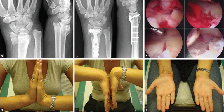
(a) Radiographs showing preoperative wrist posteroanterior and lateral views of patient 1 showing an intraarticular C2 type distal radial fracture, (b) Postoperative radiograph of wrist showing posteroanterior and lateral views of the same patient, following stable fixation of distal radial fracture with a volar fixed angle locking plate, (c) Photograph showing wrist arthroscopic views. A shaver is being used for clearing the hematoma and debris in the joint, while the articular reduction is confirmed, (d-f) Clinical photographs showing range of motion at the final followup
Figure 2.
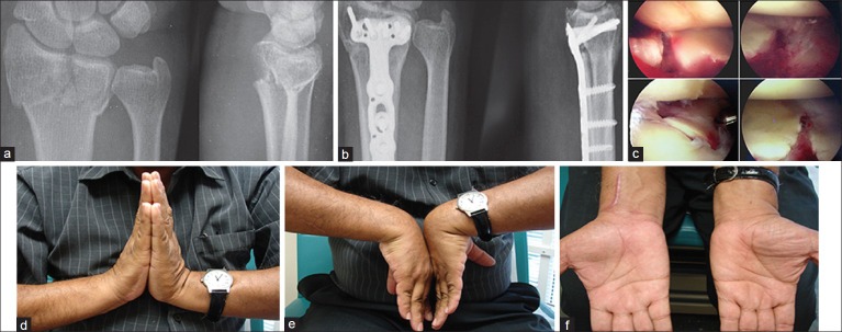
(a) Preoperative radiographs of wrist showing posteroanterior and lateral views of patient 2 with an intraarticular distal radial fracture. (b) Postoperative radiographs showing posteroanterior and lateral views of wrist of patient 2, following stable fixation of distal radial fracture with a volar fixed angle locking plate. (c) Photograph of arthroscopic view of wrist showing joint debridement in progress and assessment of the articular reduction in patient 2. (d-f) Clinical photographs of patient 2 showing range of motion of wrist at the final followup
Operative procedure
The patient was placed in the supine position and a shoulder support was secured to the surgical table on the ipsilateral side of the injured wrist. A regional block, using the three nerve blocking technique at the elbow level was preferred in order to prevent complications caused by axillary block.21,22 Once anesthetized, wrist was held in supination and a nonsterile tourniquet was applied to the upper arm, along with a strap to provide countertraction. The upper extremity was prepared, draped, and then exsanguinated with an esmarch bandage and the tourniquet was inflated to 250 mmHg. Intravenous sedation is used for tourniquet pain. As a part of our surgical protocol, endoscopic carpal tunnel release using the single portal technique [Figure 3] (Microaire, carpal tunnel release system, Charlottesville, VA) was performed routinely at this time.23,24 However, if the displacement and deformity were severe, the carpal tunnel was released after the fracture reduction, to facilitate placement of endoscope in the canal. The extended flexor carpi radial approach25 was used for plate fixation. Once the fracture was preliminarily fixed, longitudinal wrist traction was given by placing finger traps on the index and middle fingers along with 10 pound of weight suspended through a pulley system, which was secured to the shoulder holder. Traction tower was not used to facilitate the use of fluoroscopy throughout the procedure. An 18-gauge needle was then used to identify the radio-carpal joint, because Lister's tubercle is usually displaced and hence cannot be used as an anatomic landmark [Figure 4a and b]. Fluid ingress was obtained by gravity and an esmarch bandage was used to avoid soft tissue infiltration. A 2.7 mm arthroscope was introduced through the 3-4 portal. A 2.9 mm full radius shaver placed through the 4-5 or 6R portal was used to remove blood clots and small intraarticular debris [Figure 5a].
Figure 3.
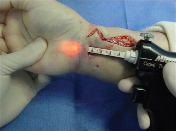
Photograph showing endoscopic carpal tunnel release
Figure 4.
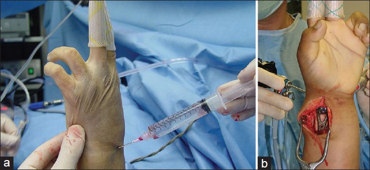
(a) Photograph showing insufflation of radiocarpal joint with saline after putting the wrist on longitudinal traction, (b) Photograph showing arthroscopic evaluation of wrist after stable fixation of the distal radial fracture, with wrist in longitudinal traction
Figure 5.

(a) Photograph showing arthroscopic debridement of the radiocarpal joint, (b) Photograph showing preliminary fixation using the Kirschner wire under a fluoroscopic view, (c) Fluoroscopic images showing lunate fossa reduction under arthroscopic guidance
We first obtained fluoroscopic postero-anterior and lateral wrist views to evaluate fracture reduction and implant positioning [Figures 5b and c]. Then, a systematic careful arthroscopic inspection of the radio-carpal joint including the articular surface of the distal radius and the intraarticular soft tissue structures was performed, in order to judge and rectify if required, the reduction observed by fluoroscopy and not to miss any ligamentous lesion. A 2.9 mm full radius shaver was used for removing the blood clots and debris [Figures 1c and 2c] and a small probe was used to palpate the joint surface in search for articular gaps and step-offs and to test the indemnity of the carpal ligaments and the TFCC [Figure 6a]. Thorough evaluation of the midcarpal joint was also carried out to identify scapho-lunate [Figure 6b], luno-triquetral injuries, and other midcarpal instabilities. Appropriate midcarpal assessment helps in classifying the injuries as per the Geissler17 classification. Partial tears or stretching of the carpal interosseous ligaments was treated with debridement. Severe ligament injuries detected through midcarpal portal have to be treated by aggressive debridement and fluoroscopic assisted pinning of the involved bones.
Figure 6.
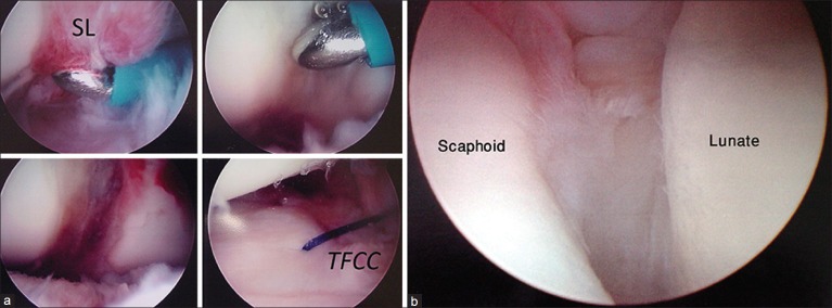
Arthroscopic views showing (a) contused scapholunate ligament being treated with thermal ligamentoplasty and repair of peripheral tear of TFCC using PDS suture, (b) (mid-carpal view) showing scapholunate diastasis
Type IB tears26 of the TFCC were usually seen in significant fracture displacement. Small ones could be managed with debridement while larger tears with loss of the trampoline effect require percutaneous suture repair by suture welding technique.27 To obtain maximum distal radio-ulnar joint (DRUJ) congruency and ensure proper rehabilitation, tears are repaired while holding the wrist in supination.27 Thorough irrigation of the joint through the scope was carried out once all concomitant lesions have been identified and treated. After arthroscopic confirmation of the articular congruency, the volar plate positioning was again checked under fluoroscopy. The volar incision was closed with absorbable stitches. The portal wounds were sealed with steri-strips. Any Kirschner wire was cut and left underneath the skin. Immediate finger motion was started and then wrist rehabilitation protocol was tailored according to the type of fixation. A below elbow cast was given for 4 to 8 weeks depending upon the fracture pattern and associated TFCC and ligamentous injuries. In the absence of significant associated injuries, 4 weeks of cast was given following which active range of motion exercises and strengthening exercises were started and continued for further 4 to 8 weeks. For associated TFCC injury, immediately after surgery, a sugar tong splint was given with wrist in supination for initial 1 week, which was followed by 5 weeks of immobilization in a muenster type cast in order to permit some elbow flexion and extension, while restricting supination and pronation at wrist. Cast removal was followed by 4 to 8 weeks of physical therapy with active range of motion and strengthening exercises. For associated intercarpal ligament injuries, arthroscopic debridement and Kirschner wire fixation was done and wrist maintained in a thumb spica cast for 8 weeks. At 8 weeks, the pins were removed and active range of motion and strengthening exercises were continued for further 6 to 8 weeks. All the patients were followedup for a minimum period of 24 months (average followup being 26 months) and final clinical evaluation was done using the modified Mayo wrist scoring system.28
RESULTS
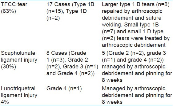
Five patients needed modification of the reduction and fixation after performing wrist arthroscopy for joint evaluation. In two patients who received a volar plate for fracture fixation, it was necessary to remove one of the distal pegs to correct a step off of about 2 mm, which was detected by arthroscopy. This peg was placed again in both the cases after reduction was achieved. In three other patients, arthroscopy showed significant depression of small fragments, especially on the volar-ulnar aspect of the distal radius. These fragments needed to be elevated and finally held in place by supplementary unthreaded 1.5 mm Kirchner wires. Ligamentous lesions and TFCC injuries were found during wrist arthroscopy in 26 patients [Table 1]. Five patients had combined injuries i.e. TFCC tear, scapholunate ligament injury and/or lunotriquetral ligament injury. One associated scaphoid fracture which was diagnosed preoperatively by radiograph was treated by percutaneous Acutrak screw under fluoroscopic and arthroscopic guidance. The average tourniquet time was 59 minutes (range 38-92 minutes). No bone graft was used in any patient.
Table 1.
Associated injuries detected during wrist arthroscopy
Radiographic parameters evaluated at the average followup of 26 months (range 24-52 months) showed 21° of radial inclination (range 18°-22°), 2° of palmar tilt (range 0°-10°) and 0.7 mm of ulnar variance (range 0-1.5 mm). There were 20 excellent [Figures 1d–f and 2d–f], 3 good, and 4 fair results as per the modified Mayo wrist scoring system.28
The complications encountered in our series included two patients with pin tract infection and two patients with extensor carpi ulnaris tendonitis (which resolved with a local cortisone infiltration). One patient who was treated with 2 Kirschner wires developed a complex regional pain syndrome (type I), which resolved due to timely, aggressive treatment based on pain medication and occupational therapy without any stellate ganglion blocks.
DISCUSSION
Several studies have shown that restoration of articular congruency is the most important factor in determining the functional outcome of intraarticular distal radius fractures1,2,3,5,12 Intraarticular step-offs of less than 1 mm reduce the possibility of developing posttraumatic osteoarthritis.1,2,3,12 Arthroscopic evaluation is a minimally invasive procedure that proves to be the best modality to assess the joint surface and residual step-offs once reduction and fixation have been obtained, when compared to radiographs and fluoroscopy.3,5,6,7,12,13,14 Wrist arthroscopy for distal radius fractures is indicated in young adults and high demand middle-aged patients, especially with intraarticular involvement secondary to high energy trauma, where there is a suspicion of associated soft tissue injuries.4,7,8,9,10,11,19 The classic Colle's fracture, in an elderly and low-demand patient with osteopenic bone, does not require joint evaluation.9
Compartment syndrome, open fractures, and irreducible carpal dislocations are relative contraindications for performing wrist arthroscopy for distal radius fractures.9,19 It has also been recommended to wait for 48 hours after the injury to avoid bleeding from the fracture site; however, fragment manipulation can be difficult after 7 days of the fracture.4
Edward et al.,6 reported 15 intraarticular distal radius fractures treated with closed reduction and percutaneous fixation. The reduction was assessed sequentially by fluoroscopy, radiographs, and wrist arthroscopy. They found that in 5 (33%) patients, the ideal reduction achieved with fluoroscopy and radiographs had an articular step of more than 1 mm detected by adjuvant arthroscopy. Wrist motion, grip strength, and final radiographic parameters have been shown to be superior with arthroscopic-assisted reduction as compared to open reduction.12,13 Ruch et al.,29 in a prospective cohort study showed that the patients who underwent assisted arthroscopic procedures had a greater degree of supination, flexion, and extension than the patients undergoing only fluoroscopic-assisted surgery. Wrist arthroscopy improves the diagnosis and treatment of ligament injuries and TFCC injuries that are so frequently associated with intraarticular distal radius fractures.7,8,9,10,11,12,18,19 Arthroscopy has also been demonstrated to be more reliable for the diagnosis of these injuries even when compared to cine-arthrography30 and MRI;31 furthermore, these injuries can be treated in the same surgical sitting with minimal disturbance of the soft tissues.4,9,10,11,12,32,33 Lindau et al.,32 in a prospective case series of 51 patients with displaced distal radius fractures assessed with arthroscopic evaluation found complete or partial TFCC tears in 43 patients (24 peripheral tears, 10 central perforations, and 9 combined tears). At 1 year followup, 10 patients with complete peripheral TFCC tears and 7 with partial tears had DRUJ instability. Shih et al.,33 reported their results using arthroscopy to treat 33 patients of distal radius fractures with associated soft tissue injuries. They found TFCC tears in 18 patients and 6 patients had scapholunate ligament tear with instability. All the peripheral TFCC tears were repaired and scapholunate instabilities were treated by arthroscopic debridement and transfixation of the scapholunate joint with Kirschner wires. At the final followup, 11 patients achieved excellent results. Osterman and Vanduzer34 in their series of 56 patients treated with arthroscopic-assisted distal radial fixations reported restoration of 95% of rotational arc at 5 years of followup. Varitimidis et al.,12 reported a better clinical result with arthroscopic assisted distal radial fixation as compared to fluoroscopy alone. Furthermore, they reported TFCC injury in 60% patients, scapholunate ligament tear in 45% and luno-triquetral tear in 20% of patients in the arthroscopically treated group.
In our series, arthroscopy as an adjunct not only provided useful information regarding the intraarticular pathologies and management of these soft tissue injuries, but also ensured accurate reduction of the fracture fragments. TFCC injuries were seen in 63% of our patients, whereas scapholunate ligament disruption and luno-triquetral ligament disruption were seen in 30% and 4% of patients respectively in our series. Patient satisfaction and objective scores in our series are also similar to those mentioned in the literature. Hence, we conclude that radiocarpal and midcarpal arthroscopy is a useful adjunct to stable fixation of distal radial fractures in young adults and high-demand older population.
Footnotes
Source of Support: Nil
Conflict of Interest: None
REFERENCES
- 1.Knirk JL, Jupiter JB. Intraarticular fractures of the distal end of the radius in young adults. J Bone Joint Surg Am. 1986;68:647–59. [PubMed] [Google Scholar]
- 2.Trumble TE, Schmitt SR, Vedder NB. Factors affecting functional outcome of displaced intraarticular distal radius fractures. J Hand Surg Am. 1994;19:325–40. doi: 10.1016/0363-5023(94)90028-0. [DOI] [PubMed] [Google Scholar]
- 3.Ono H, Katyam T, Furuta K, Suzuki D, Fujitani R, Akahane M. Distal radius fracture arthroscopic intraarticular gap and step-off measurement after open reduction and internal fixation with a volar locked plate. J Orthop Sci. 2012:17443–9. doi: 10.1007/s00776-012-0226-8. [DOI] [PubMed] [Google Scholar]
- 4.Abboudi J, Culp RW. Treating fractures of the distal radius with arthroscopic assistance. Orthop Clin North Am. 2001;32:307–15. doi: 10.1016/s0030-5898(05)70251-0. [DOI] [PubMed] [Google Scholar]
- 5.Chen AC, Chan YS, Yuan LJ, Ye WL, Lee MS, Chao EK. Arthroscopically assisted osteosynthesis of complex intraarticular fractures of the distal radius. J Trauma. 2002;53:354–9. doi: 10.1097/00005373-200208000-00028. [DOI] [PubMed] [Google Scholar]
- 6.Edwards CC, Haraszti CJ, McGillivary GR, Gutlow AP. Intraarticular distal radius fractures: Arthroscopic assessment of radiographically assisted reduction. J Hand Surg Am. 2001;26:1036–41. doi: 10.1053/jhsu.2001.28760. [DOI] [PubMed] [Google Scholar]
- 7.Kuzma GR, Kuzma KR. Distal radius fractures arthroscopic assited fixation. Oper Tech Sports Med. 2010;18:189–96. [Google Scholar]
- 8.Dantuliri PK, Gillon T. Current treatment of distal radiais fractures: Arthroscopic assisted fracture reduction of distal radius fractures. Oper Tech Orthop. 2009;19:88–95. [Google Scholar]
- 9.Badia A, Khanchandani P. Volar plate fixation. In: Slutsky DJ, Osterman AL, editors. Distal radial fractures and carpal injuries: The cutting edge. 1st ed. Philadelphia: Saunders, Elsevier; 2008. pp. 149–56. [Google Scholar]
- 10.Lutsky K, Boyer MI, Steffen JA, Goldfarb CA. Arthroscopic assessment of intraarticular fractures after open reduction and internal reduction and internal fixation from a volar approach. J Hand Surg. 2008;33A:476–84. doi: 10.1016/j.jhsa.2007.12.009. [DOI] [PubMed] [Google Scholar]
- 11.Slutsky DJ. Current innovations in wrist arthroscopy. J Hand Surg. 2012;37A:1932–41. doi: 10.1016/j.jhsa.2012.06.028. [DOI] [PubMed] [Google Scholar]
- 12.Varitimidis SE, Basdekis GK, Dailiana ZH, Hanted ME, Bargiotas K, Malizos K. Treatment of intraarticular fractures of the distal radius: Fluoroscopic or arthroscopic reduction? J Bone Joint Surg. 2008;90B:778–85. doi: 10.1302/0301-620X.90B6.19809. [DOI] [PubMed] [Google Scholar]
- 13.Doi K, Hattori Y, Otsuka K, Abe Y, Yammamoto H. Intraarticular fractures of the distal aspect of the radius: arthroscopically assisted reduction compared with open reduction and internal fixation. J Bone Joint Surg Am. 1999;81:1093–110. doi: 10.2106/00004623-199908000-00005. [DOI] [PubMed] [Google Scholar]
- 14.Kordasiewicz B, Podgórski A, Klich M, Michalik D, Chaberek S, Pomianowski S. Arthroscopic assessment of intraarticular distal radius fractures-results of minimally invasive fixation. Ortop Traumatol Rehabil. 2011;13:369–86. doi: 10.5604/15093492.955727. [DOI] [PubMed] [Google Scholar]
- 15.Whipple TL. The role of arthroscopy in the treatment of intraarticular wrist fractures. Hand Clin. 1995;11:13–8. [PubMed] [Google Scholar]
- 16.Del Piñal F. Treatment of explosion-type distal radius fractures. In: Del Pinal F, Mathoulin C, Luchetti C, editors. Arthroscopic management of distal radius fractures. Berlin: Springer Verlag; 2010. pp. 41–65. [Google Scholar]
- 17.Geissler WB, Freeland AE, Savoie FH, McIntyre LW, Whipple TL. Intracarpal soft-tissue lesions associated with an intraarticular fracture of the distal end of the radius. J Bone Joint Surg Am. 1996;78:357–65. doi: 10.2106/00004623-199603000-00006. [DOI] [PubMed] [Google Scholar]
- 18.Forward DP, Lindau TR, Melsom DS. Intercarpal ligament injuries associated with fractures of the distal part of the radius. J Bone Joint Surg. 2007;89A:2334–340. doi: 10.2106/JBJS.F.01537. [DOI] [PubMed] [Google Scholar]
- 19.Geissler WB. Arthroscopically assisted reduction of intraarticular fractures of the distal radius. Hand Clin. 1995;11:19–29. [PubMed] [Google Scholar]
- 20.Muller ME, Allgower M, Schneider R, Willenegger H. 3rd ed. New York: Springer; 1990. Manual of internal fixation: Techniques recommended by the AO group; pp. 118–50. [Google Scholar]
- 21.Stark RH. Neurologic injury from axillary block anesthesia. J Hand Surg Am. 1996;21:319–26. doi: 10.1016/S0363-5023(96)80350-9. [DOI] [PubMed] [Google Scholar]
- 22.Bouaziz H, Narchi P, Mercier FJ, Khoury A, Poirier T, Benhamou D. The use of a selective axillary nerve block for outpatient hand surgery. Anesth Analg. 1998;86:746–8. doi: 10.1097/00000539-199804000-00013. [DOI] [PubMed] [Google Scholar]
- 23.Agee JM, McCarroll HR, Jr, Tortosa RD, Berry DA, Szabo RM, Peimer CA. Endoscopic release of the carpal tunnel: A randomized prospective multicenter study. J Hand Surg Am. 1992;17:987–95. doi: 10.1016/s0363-5023(09)91044-9. [DOI] [PubMed] [Google Scholar]
- 24.Badia A. Median Nerve Compression Secondary to Fractures of Distal Radius. In: Luchetti R, Amadeo P, editors. Carpal Tunnel Syndrome. Berlin: Springer; 2006. pp. 247–52. [Google Scholar]
- 25.Orbay JL, Badia A, Indriago IR, Infante A, Khouri KR, Gonzalez E, et al. The extended flexor carpi radialis approach: A new perspective for the distal radius fracture. Tech Hand Upper Extrem Surg. 2001;5:204–11. doi: 10.1097/00130911-200112000-00004. [DOI] [PubMed] [Google Scholar]
- 26.Palmer AK. Triangular fibrocartilage complex lesions: A classification. J Hand Surg Am. 1989;14:594–606. doi: 10.1016/0363-5023(89)90174-3. [DOI] [PubMed] [Google Scholar]
- 27.Badia A, Khanchandani P. Suture welding for arthroscopic repair of Peripheral triangular fibrocartilage complex tear. Tech Hand Upper Extrem Surg. 2007;11:45–50. doi: 10.1097/bth.0b013e3180336cec. [DOI] [PubMed] [Google Scholar]
- 28.Amadio PC, Berquist TH, Smith DK, Ilstrup DM, Cooney WP, Linscheid RL. Scaphoid malunion. J Hand Surg Am. 1989;14:679–87. doi: 10.1016/0363-5023(89)90191-3. [DOI] [PubMed] [Google Scholar]
- 29.Ruch DS, Valle J, Poehling GG, Smith BP, Kuzma GR. Arthroscopic reduction versus fluoroscopic reduction in the management of intraarticular distal radius fractures. Arthroscopy. 2004;20:225–30. doi: 10.1016/j.arthro.2004.01.010. [DOI] [PubMed] [Google Scholar]
- 30.Weiss AP, Akelman E, Lainbiase R. Comparison of the findings of triple injection cinearthrography of the wrist with those of arthroscopy. J Bone Joint Surg Am. 1996;78:348–56. doi: 10.2106/00004623-199603000-00005. [DOI] [PubMed] [Google Scholar]
- 31.Rominger MB, Bernreuter WK, Kenney PJ, Lee DH. MR imaging of anatomy and tears of wrist ligaments. Radiographics. 1993;13:1233–48. doi: 10.1148/radiographics.13.6.8290721. [DOI] [PubMed] [Google Scholar]
- 32.Lindau T, Adlercreutz C, Aspenberg P. Peripheral tears of the triangular fibrocartilage complex cause distal radioulnar joint instability after distal radius fractures. J Hand Surg Am. 2000;25:464–8. doi: 10.1053/jhsu.2000.6467. [DOI] [PubMed] [Google Scholar]
- 33.Shih JT, Lee HM, Hou YT, Tan CM. Arthroscopically-assisted reduction of intraarticular fractures and soft tissue management of distal radius. Hand Surg. 2001;6:127–35. doi: 10.1142/s021881040100059x. [DOI] [PubMed] [Google Scholar]
- 34.Osterman AL, Vanduzer ST. Arthroscopy in the treatment of distal radial fractures with assessment and treatment of associated injuries. Atlas Hand Clin. 2006;11:231–41. [Google Scholar]


