Abstract
We established a plasmid-based system for generating infectious Ebola virus-like particles (VLPs), which contain an Ebola virus-like minigenome consisting of a negative-sense copy of the green fluorescent protein gene. This system produced nearly 103 infectious particles per ml of supernatant, equivalent to the titer of Ebola virus generated by a reverse genetics system. Interestingly, infectious Ebola VLPs were generated, even without expression of VP24. Transmission and scanning electron microscopic analyses showed that the morphology of the Ebola VLPs was indistinguishable from that of authentic Ebola virus. Thus, this system allows us to study Ebola virus entry, replication, and assembly without biosafety level 4 containment. Furthermore, it may be useful in vaccine production against this highly pathogenic agent.
Ebola and Marburg viruses are filamentous, enveloped, nonsegmented, and negative-stranded RNA viruses of the family Filoviridae in the order Mononegavirales (15). These viruses cause severe hemorrhagic fever in humans and other primates with high mortality rates (15). The genus Ebolavirus consists of four species, Zaire, Sudan, Ivory Coast, and Reston Ebolavirus, with genomes of approximately 19 kb in length, encoding seven structural proteins and one nonstructural protein (15). Four of these viral proteins—nucleoprotein (NP), VP35, VP30, and the RNA-dependent RNA polymerase (L) —are necessary for replication and transcription of viral RNA (vRNA) (10), while three others —glycoprotein (GP), VP40, and VP24—are membrane-associated virion proteins (5, 15). GP is the surface glycoprotein that forms spikes on virions and is responsible for receptor binding and membrane fusion, supporting viral penetration into cells (8, 20, 25). VP40, the most abundant membrane viral protein, is responsible for formation of filamentous virus particles (9, 12, 22). VP24 is a minor viral protein whose functions remain elusive, although a recent study suggests its involvement in nucleocapsid formation and in virus assembly and budding (5, 7). Although the properties of some of these viral proteins have been investigated (3, 5-7, 9, 10, 12, 14, 16-18, 21, 27), their functions during viral replication, especially the nature of their protein-protein and protein-vRNA interactions, are largely unknown.
Recently, Volchkov et al. (24) and members of our laboratory (11) established reverse genetics systems that allow one to generate infectious Ebola viruses entirely from cloned cDNAs. Although use of these reverse genetics systems has helped to elucidate the roles of the nonstructural viral protein, sGP, and/or GP in Ebola virus replication (11, 24), the requirement for biosafety level 4 (BL4) containment facilities has hampered further progress in Ebola virus research. In an effort to devise alternative strategies that would allow investigators to study Ebola virus under non-BL4 conditions, we established a plasmid-driven system to generate infectious Ebola virus-like particles (VLPs) containing an Ebola virus minigenome. As described here, the features of this new system are conducive to the study of Ebola virus assembly, receptor binding, and fusion under non-BL4 conditions.
Transcription and replication of an artificial Ebola virus minigenome.
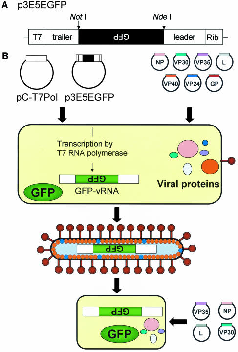
The first step in establishing a system for the production of VLPs containing an Ebola virus minigenome was to generate a plasmid containing the green fluorescent protein (GFP) gene in antisense orientation between the leader and trailer sequences of the Ebola virus genome, flanked by the T7 RNA polymerase promoter and a ribozyme, using plasmid 3E-5E as a template (10). The resulting plasmid was designated p3E5EGFP (Fig. 1A). Transfection of this plasmid together with a plasmid expressing T7 RNA polymerase into eukaryotic cells should result in transcription of the GFP reporter gene, thereby generating Ebola virus-like GFP-vRNA. Four nucleocapsid proteins—L, NP, VP35, and VP30—are sufficient to support the transcription and replication of artificial Ebola vRNAs (10); hence, we next tested whether expression of L, NP, VP35, and VP30 leads to replication and transcription of the artificial GFP-vRNA. Using the amounts of plasmid DNA described in a previous report (10) as a guide, we first transfected 106 human embryonic kidney (293T) cells with 1 μg of p3E5EGFP, 1 μg of pCEZ-L, 0.5 μg of pCEZ-NP, 0.5 μg of pCEZ-VP35, and 0.1 μg of pCEZ-VP30, for the production of the Ebola virus Zaire L, NP, VP35, and VP30 proteins, respectively. pC-T7Pol (11) was also cotransfected into cells to provide T7 RNA polymerase. Forty-eight hours later, more than 50% of the transfected cells expressed GFP (data not shown), confirming that the four nucleocapsid proteins (L, NP, VP35, and VP30) are sufficient for replication and transcription of T7 RNA polymerase-derived GFP-vRNA. In addition, like Mühlberger et al. (10), we found that VP30 expression increased the number of GFP-expressing cells (data not shown).
FIG. 1.
Production of Ebola VLPs from plasmids. (A) Schematic diagram of the construct for production of the Ebola virus minigenome, p3E5EGFP. This construct was made from plasmid 3E-5E, which contains the chloramphenicol acetyltransferase (CAT) gene in antisense orientation between the leader and trailer sequences of the Ebola virus genome, flanked by the T7 RNA polymerase promoter (T7) and a ribozyme (Rib) (10). The CAT gene was replaced with the GFP gene using NotI and NdeI restriction enzyme sites; thus, the GFP gene is also cloned in the antisense orientation, as indicated by the inverted letters. (B) Schematic diagram of a system for Ebola VLP generation. 293T cells were transfected with plasmids for the expression of the Ebola virus NP, L, VP35, VP30, GP, VP40, and VP24 proteins and with p3E5EGFP. pC-T7Pol was also cotransfected into cells to provide T7 RNA polymerase. Culture supernatants of 293T cells were then harvested 72 h after transfection and were incubated with 293T cells which had been transfected with plasmids for expressing L, NP, VP35, and VP30 proteins 24 h prior to inoculation with the culture supernatant.
To determine the optimal amounts of plasmid DNA required for the most efficient GFP expression, we tested various amounts of the L, NP, VP35, and VP30 expression plasmids. Changing the amounts of plasmids expressing NP and VP35, from 0.5 to 1.0 μg, did not affect GFP expression in transfected cells (data not shown). Altering the amounts of the L and VP30 expression plasmids, however, appreciably affected the number of GFP-expressing cells; among different amounts of plasmids tested, 4.0 μg of pCEZ-L and 0.3 μg of pCEZ-VP30 were the most efficient in producing GFP-expressing cells (Table 1). Thus, 4.0 μg of pCEZ-L, 0.5 μg of pCEZ-NP and pCEZ-VP35, and 0.3 μg of pCEZ-VP30 were used in all subsequent experiments.
TABLE 1.
Optimal amounts of plasmid DNA required for the transcription and replication of an artificial Ebola virus minigenomea
| Amt of Plasmid DNA (μg) expressing:
|
Relative efficiency of GFP expressionb | |||||
|---|---|---|---|---|---|---|
| L | NP | VP35 | VP30 | T7 Pol | GFP-vRNA | |
| 1.0 | 0.5 | 0.5 | 1.0 | 1.0 | 1.0 | 1.0 |
| 1.0 | 0.5 | 0.5 | 0.3 | 1.0 | 1.0 | 2.5 |
| 1.0 | 0.5 | 0.5 | 0.1 | 1.0 | 1.0 | 3.2 |
| 2.0 | 0.5 | 0.5 | 1.0 | 1.0 | 1.0 | 1.5 |
| 2.0 | 0.5 | 0.5 | 0.3 | 1.0 | 1.0 | 7.5 |
| 2.0 | 0.5 | 0.5 | 0.1 | 1.0 | 1.0 | 3.7 |
| 3.0 | 0.5 | 0.5 | 1.0 | 1.0 | 1.0 | 2.9 |
| 3.0 | 0.5 | 0.5 | 0.3 | 1.0 | 1.0 | 8.3 |
| 3.0 | 0.5 | 0.5 | 0.1 | 1.0 | 1.0 | 7.2 |
| 4.0 | 0.5 | 0.5 | 1.0 | 1.0 | 1.0 | 0.0 |
| 4.0 | 0.5 | 0.5 | 0.3 | 1.0 | 1.0 | 13.9 |
| 4.0 | 0.5 | 0.5 | 0.1 | 1.0 | 1.0 | 2.5 |
293T cells were transfected with plasmids for the expression of the Ebola virus Zaire L, NP, VP35, and VP30 proteins and with p3E5EGFP and pC-T7Pol. Forty-eight hours later, the number of GFP-expressing cells was determined with a fluorescence microscope.
Determined by counting the number of GFP-expressing cells in five microscopic fields. The sample containing 1 μg of plasmids expressing L, VP30, p3E5EGFP, and pC-T7Pol and 0.5 μg of plasmids expressing NP and VP35 was chosen as the reference (value of 1); approximately 500 GFP-expressing cells were observed with these amounts of plasmid DNA.
Formation of Ebola VLPs entirely from cloned cDNAs.
To generate Ebola VLPs, we transfected 106 293T cells with plasmids for the expression of GP (pCEboZGP; 1 μg [8]), VP40 (pCEboZVP40; 1 μg [9]), VP24 (pCEboZVP24; 1 μg) in addition to the optimal amounts of plasmids for the expression of proteins required for transcription and replication of the artificial Ebola virus minigenome (listed above) together with 1 μg of p3E5EGFP and pC-T7Pol. Interestingly, the numbers of GFP-expressing cells were more than 10-fold lower than those found when cells were transfected with plasmids for the expression of proteins required for transcription and replication of the artificial Ebola virus minigenome only (i.e., pCEZ-L, pCEZ-NP, pCEZ-VP35, pCEZ-VP30, p3E5EGFP, and pC-T7Pol). This reduction in GFP-expressing cells is likely due to the cytotoxicity of membrane-associated proteins GP, VP40, and VP24 (1, 2, 18, 21, 30; L. Jasenosky, S. Watanabe, and Y. Kawaoka, unpublished data). Culture supernatants of 293T cells were harvested 72 h after transfection and incubated with other 293T cells which had been transfected with plasmids for expressing L, NP, VP35, and VP30 proteins (required for replication and transcription of GFP-vRNA) 24 h prior to inoculation (Fig. 1B). Seventy-two hours later, we detected GFP-expressing cells corresponding to 5.8 × 10² VLPs/ml of supernatant (data not shown). We also confirmed that no GFP-expressing cells were detected when supernatants were incubated with 293T cells without cotransfection of plasmids encoding L, NP, VP35, and VP30 proteins (data not shown).
We next investigated whether the efficiency of VLP formation would increase if the amounts of certain expression plasmids were modified. Because VP24 is a minor protein, we thought its expression level in the initial experiment might be too high. Reduction of the VP24 expression plasmid from 1 to 0.03 μg resulted in a 10-fold increase in GFP-expressing cells (approximately 103 GFP-positive cells/106 plasmid-transfected cells). When supernatants of 293T cells collected at 72 h posttransfection were used to infect 293T cells expressing L, NP, VP35, and VP30, we found a slight (∼1.6-fold) increase in the number of GFP-positive cells, corresponding to the number of VLPs (9.2 × 10² infectious particles per ml of supernatant at 72 h postinfection) (Table 2). Interestingly, even without the addition of the VP24-expressing plasmid, infectious VLPs were produced, indicating that this protein is not essential for either the formation or entry of Ebola VLPs into cells. By contrast, without GP or VP40, infectious Ebola VLPs were not produced.
TABLE 2.
Optimal amounts of plasmid DNA required for the production of infectious VLPsa
| Amt of Plasmid DNA (μg) expressing: | Relative efficiency of VLP formationb | ||||||||
|---|---|---|---|---|---|---|---|---|---|
| L | NP | VP35 | VP30 | GP | VP40 | VP24 | T7 Pol | GFP-vRNA | |
| 4.0 | 0.5 | 0.5 | 0.3 | 1.0 | 1.0 | 1.0 | 1.0 | 1.0 | 1.0 |
| 4.0 | 0.5 | 0.5 | 0.3 | 1.0 | 1.0 | 0.3 | 1.0 | 1.0 | 1.3 |
| 4.0 | 0.5 | 0.5 | 0.3 | 1.0 | 1.0 | 0.1 | 1.0 | 1.0 | 1.2 |
| 4.0 | 0.5 | 0.5 | 0.3 | 1.0 | 1.0 | 0.03 | 1.0 | 1.0 | 1.6 |
| 4.0 | 0.5 | 0.5 | 0.3 | 1.0 | 1.0 | 0 | 1.0 | 1.0 | 1.2 |
| 4.0 | 0.5 | 0.5 | 0.3 | 1.0 | 0 | 0.03 | 1.0 | 1.0 | 0 |
| 4.0 | 0.5 | 0.5 | 0.3 | 0 | 1.0 | 0.03 | 1.0 | 1.0 | 0 |
293T cells were transfected with plasmids for the expression of seven Ebola Zaire virus structural proteins and with p3E5EGFP and pC-T7Pol. Seventy-two hours later, the supernatants containing Ebola VLPs were harvested and incubated with fresh 293T cells transfected with plasmids expressing L, NP, VP35, and VP30 proteins. Seventy-two hours after infection, the number of GFP-expressing cells (corresponding to the number of infectious VLPs) was determined with a fluorescence microscope.
Determined by counting the number of GFP-expressing cells in all microscopic fields. The sample containing 4.0 μg of plasmid expressing L, 0.5 μg of plasmids expressing NP and VP35, 0.3 μg of plasmid expressing VP30, and 1 μg of plasmids expressing GP, VP40, VP24, p3E5EGFP, and pC-T7Pol (which produced ∼600 infectious VLPs/ml of supernatant) was chosen as the reference (value of 1).
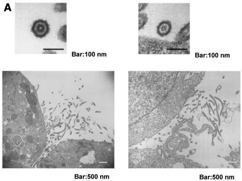
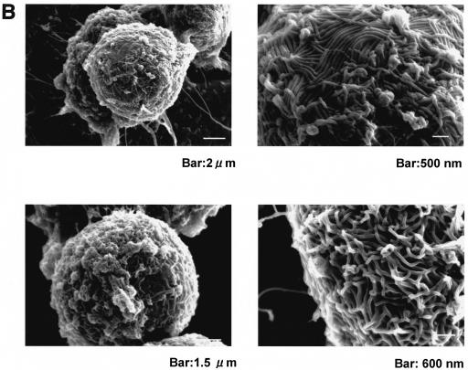
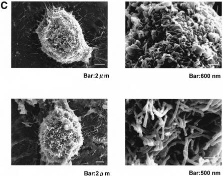
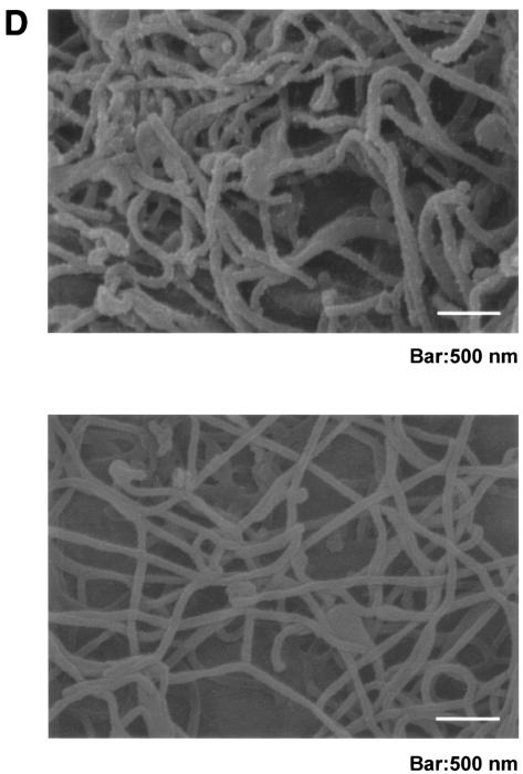
To determine the morphology of Ebola VLPs generated entirely from plasmids, we performed transmission and scanning electron microscopy (TEM and SEM, respectively) on COS-7 cells transfected with all plasmids required for VLP production. In ultrathin sections of these cells, filamentous particle-like structures budding from the plasma membrane and containing electron-dense structures such as viral nucleocapsid were observed by TEM (Fig. 2A, right). Under the SEM, we also observed many filamentous structures on the plasmid-transfected cell surface (Fig. 2C). These structures were indistinguishable from those observed in cells infected with authentic Ebola virus (Fig. 2A, left, and B) (4, 15). To confirm whether these filamentous structures were Ebola VLPs, we performed immuno-SEM using mouse anti-Ebola virus Zaire GP monoclonal antibodies (MAb) (12, 19). Under the SEM, we detected filaments with rough surfaces due to decoration with anti-Ebola virus Zaire GP MAb (Fig. 2D, top). In contrast, we could not detect rough-surfaced filaments using anti-hemagglutinin (HA; influenza A virus A/WSN/33) polyclonal antibodies (Fig. 2D, bottom). These results demonstrated that GP was expressed on the surface, indicating that these filamentous structures were Ebola VLPs.
FIG. 2.
Morphology of Ebola VLPs generated entirely from cDNAs. (A) TEM of COS-7 cells transfected with plasmids (VLP, right) or Vero E6 cells infected with Ebola virus (left). Cells were fixed with 2.5% glutaraldehyde in 0.1 M cacodylate buffer (pH 7.4) and postfixed with 2% osmium tetroxide in the same buffer. Samples were stained en bloc with 1% aqueous uranyl acetate and then processed as described previously (12). (B and C) SEM of Vero E6 cells infected with Ebola virus (B) or COS-7 cells transfected with plasmids (VLP) (C). Cells were fixed as described in the legend for panel A. The fixed samples were dehydrated with a series of ethanol gradients, substituted with t-butanol, and dried in a Hitachi ES-2030 freeze dryer. Dried specimens were coated with osmium tetroxide by using an HPC-1S osmium coater (Vacuum Device Inc.)and examined with a Hitachi S-4200 microscope. (D) Immuno-SEM of COS-7 cells transfected with plasmids. Cells were fixed with 3% paraformaldehyde and 0.1% glutaraldehyde in phosphate-buffered saline (PBS). After incubation with PBS containing 1% bovine serum albumin (PBS-BSA), the samples were treated with a mixture of mouse anti-GP monoclonal antibodies (1:100 dilution in PBS-BSA) (top image) or rabbit anti-HA (influenza A virus A/WSN/33; 1:250 dilution) (bottom image) polyclonal antibodies. After washing with PBS, samples were incubated with a goat anti-mouse immunoglobulin conjugated to 15-nm gold particles (1:50 dilution) or anti-rabbit immunoglobulin with 5-nm gold particles (1:50 dilution). After washing with PBS, the specimens were processed as described in the legend for panels B and C for SEM.
Thus, the expression of all viral structural proteins leads to the formation of infectious Ebola VLPs containing an artificial Ebola virus minigenome (GFP-vRNA) that can be delivered to subsequent cells.
Authenticity of Ebola VLPs containing an Ebola virus-like GFP-vRNA.
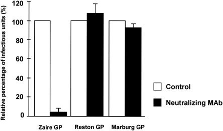
To validate that the GP-containing particles initiate infection in the same manner as authentic Ebola virus, we first determined whether a neutralizing MAb to the Ebola virus Zaire GP could inhibit Ebola VLP infection. Culture supernatants containing Ebola VLPs were mixed with a MAb to the Zaire GP known to neutralize Ebola Zaire virus and a recombinant vesicular stomatitis virus pseudotyped with the Zaire GP (19). Thirty minutes after incubation at 4°C, the mixture was used to infect 293T cells expressing L, NP, VP35, and VP30. Incubation of VLPs with the MAb abolished GFP-positive (VLP-infected) cells completely, indicating that the antibody neutralized the VLPs (Fig. 3). Normal mouse ascites did not abolish GFP expression in VLP-incubated cells.
FIG. 3.
Neutralization of Ebola VLPs by a MAb to the Zaire GP. Culture supernatants containing Ebola VLPs possessing the Zaire, Reston, or Marburg GP were mixed with a MAb to the Zaire GP known to neutralize the Ebola Zaire virus (19). Thirty minutes after incubation at 4°C, the mixture was used to infect 293T cells expressing L, NP, VP35, and VP30. Normal mouse ascites was used as a negative control. Experiments were performed three times. The percentage of infectious units indicates VLP infectivities relative to those in the presence of normal mouse ascites, which were assigned a value of 100%.
We previously identified amino acid substitutions in the Ebola virus Zaire GP that inhibit the ability of this protein to support the infectivity of a mutant vesicular stomatitis virus pseudotyped with Ebola virus Zaire GP (25). Since one of the GP mutants, I623A (Ile-to-Ala substitution at position 623), reduced the infectivity of the pseudotyped virus, we attempted to generate Ebola VLPs containing this mutant protein. As a positive control, we also generated Ebola VLPs containing GP with a Leu-to-Ala substitution at position 585 (L585A), which does not impair GP function (25). The numbers of GFP-positive 293T cells transfected with plasmids for production of VLPs possessing mutant GP, I623A or L585A, did not differ (data not shown), but the infectivity of Ebola VLPs possessing I623A GP was 1.5 log lower than that of VLPs possessing L585A GP (Table 3). These results confirmed the GP-mediated attachment and entry of VLPs into cells, suggesting that such VLPs generated from plasmids can be used to study the mechanisms of Ebola virus entry into cells.
TABLE 3.
Infectivities of Ebola VLPs possessing GPs of a different Ebola subtype, Marburg virus, or mutant GPsa
| GP | Infectious units (log10/ml) |
|---|---|
| Ebola Zaire GP mutants | |
| GP I623A (mutant GP) | 0.8 ± 0.3 |
| GP L585A (mutant GP) | 2.3 ± 0.4 |
| Ebola virus | |
| Zaire | 2.3 ± 0.5 |
| Reston | 2.7 ± 0.4 |
| Sudan | 1.9 ± 0.2 |
| Ivory Coast | 2.3 ± 0.3 |
| Marburg virus | 2.7 ± 0.5 |
293T cells were transfected with plasmids for the expression of seven Ebola Zaire virus structural proteins and with p3E5EGFP and pC-T7Pol. Seventy-two hours later, the supernatant containing Ebola VLPs was harvested and incubated with fresh 293T cells transfected with plasmids expressing L, NP, VP35, and VP30. Seventy-two hours after infection, the number of GFP-expressing cells (corresponding to the number of infectious VLPs) was determined with a fluorescence microscope. Results are means ± standard deviations.
GPs of other subtypes of Ebola and Marburg viruses can be incorporated into Ebola virus Zaire VLPs.
Viral protein-protein interactions are important in virus assembly. It was recently reported that coexpression of GP and VP40 resulted in formation of filamentous particles with GP spikes, suggesting their interaction (12). To investigate whether a type- or subtype-specific GP is needed for infectious virion formation, we attempted to generate Ebola VLPs possessing the GP of other Ebola subtypes (Sudan, Ivory Coast, or Reston) or of Marburg virus instead of the Zaire GP. Substitution of these GPs did not appreciably affect the infectivity of Ebola VLPs, with the exception of the Sudan GP (Table 3). In fact, replacement of the Zaire GP with the Reston or Marburg protein resulted in a 2.5-fold increase in VLP infectivity. Figure 3 confirms that the infectivity of these VLPs was indeed mediated by GP. That is, infectivity of VLPs possessing the Reston or Marburg GP was not affected upon incubation with anti-Zaire GP MAbs with neutralizing abilities (19), in contrast to those possessing the Zaire GP. Thus, the GPs of other Ebola virus subtypes can also be incorporated into Ebola VLPs, where they function to mediate virus entry into cells.
Here we describe a plasmid-driven system for the generation of infectious Ebola VLPs containing an artificial Ebola vRNA. 293T cells transfected with plasmids for Ebola VLP formation produced nearly 103 infectious particles per ml, equivalent to the titer of Ebola virus generated by a reverse genetics system (11). Huang et al. (7) reported that NP, VP35, and VP24 proteins are minimally required for nucleocapsid formation and that the structures produced were similar to the authentic Ebola virus nucleocapsids. Our results suggest that VP24 is not essential for nucleocapsid formation or for the formation of and entry of Ebola VLPs into cells. Since the protein components of actual Ebola virus are identical to those of Ebola VLPs, the only difference between authentic virus and these artificial VLPs lies in the length of their genomic RNAs. The minigenome RNA used in this study is approximately 2 kb long, or approximately 10 times shorter than the full-length genome. Thus, the lack of a requirement for VP24 in nucleocapsid formation in our system may be related to the length of the genomic RNA. Despite this difference, the Ebola VLPs function in an authentic manner. Therefore, we suggest that infectious Ebola VLPs containing an Ebola virus minigenome could be used productively, under non-BL4 conditions, to identify the structure-function relationships among Ebola virus components that contribute to the extreme pathogenicity of this agent.
Effective anti-Ebola virus vaccines for primates remain to be developed (13, 23, 28, 29). It was recently demonstrated that replication-incompetent influenza VLPs protected mice against challenge with lethal doses of influenza virus (26). By analogy, combining our systems for generating Ebola virus (11) and Ebola VLPs (this report) from plasmids should enable us to produce replication-incompetent Ebola VLPs that would infect cells and express protective viral antigens (unlike inactivated vaccines) without generating progeny particles in infected cells, thus constituting a safe protective vaccine. Studies to develop such vaccines are under way.
Acknowledgments
We thank Stephan Becker and Elke Mühlberger for providing the 3E-5E plasmid. We also thank Krisna Wells and Martha McGregor for excellent technical assistance and John Gilbert for editing the manuscript.
This work was supported by a National Institute of Allergy and Infectious Diseases Public Health Service research grant, by CREST (Japan Science and Technology Corporation), and by Grants-in-Aid by the Ministry of Education, Culture, Sports, Science and Technology and by the Ministry of Health, Labor and Welfare, Japan.
REFERENCES
- 1.Chan, S. Y., M. C. Ma, and M. A. Goldsmith. 2000. Differential induction of cellular detachment by envelope glycoproteins of Marburg and Ebola (Zaire) viruses. J. Gen. Virol. 81:2155-2159. [DOI] [PubMed] [Google Scholar]
- 2.Chan, S. Y., R. F. Speck, M. C. Ma, and M. A. Goldsmith. 2000. Distinct mechanisms of entry by envelope glycoproteins of Marburg and Ebola (Zaire) viruses. J. Virol. 74:4933-4937. [DOI] [PMC free article] [PubMed] [Google Scholar]
- 3.Dessen, A., V. Volchkov, O. Dolnik, H. D. Klenk, and W. Weissenhorn. 2000. Crystal structure of the matrix protein VP40 from Ebola virus. EMBO J. 19:4228-4236. [DOI] [PMC free article] [PubMed] [Google Scholar]
- 4.Feldmann, H., and H. D. Klenk. 1996. Marburg and Ebola viruses. Adv. Virus Res. 47:1-52. [DOI] [PubMed] [Google Scholar]
- 5.Han, Z., H. Boshra, J. O. Sunyer, S. H. Zwiers, J. Paragas, and R. N. Harty. 2003. Biochemical and functional characterization of the Ebola virus VP24 protein: implications for a role in virus assembly and budding. J. Virol. 77:1793-1800. [DOI] [PMC free article] [PubMed] [Google Scholar]
- 6.Harty, R. N., M. E. Brown, G. Wang, J. Huibregtse, and F. P. Hayes. 2000. A PPxY motif within the VP40 protein of Ebola virus interacts physically and functionally with a ubiquitin ligase: implications for filovirus budding. Proc. Natl. Acad. Sci. USA 97:13871-13876. [DOI] [PMC free article] [PubMed] [Google Scholar]
- 7.Huang, Y., L. Xu, Y. Sun, and G. J. Nabel. 2002. The assembly of Ebola virus nucleocapsid requires virion-associated proteins 35 and 24 and posttranslational modification of nucleoprotein. Mol. Cell 10:307-316. [DOI] [PubMed] [Google Scholar]
- 8.Ito, H., S. Watanabe, A. Sanchez, M. A. Whitt, and Y. Kawaoka. 1999. Mutational analysis of the putative fusion domain of Ebola virus glycoprotein. J. Virol. 73:8907-8912. [DOI] [PMC free article] [PubMed] [Google Scholar]
- 9.Jasenosky, L. D., G. Neumann, I. Lukashevich, and Y. Kawaoka. 2001. Ebola virus VP40-induced particle formation and association with the lipid bilayer. J. Virol. 75:5205-5214. [DOI] [PMC free article] [PubMed] [Google Scholar]
- 10.Mühlberger, E., M. Weik, V. E. Volchkov, H. D. Klenk, and S. Becker. 1999. Comparison of the transcription and replication strategies of Marburg virus and Ebola virus by using artificial replication systems. J. Virol. 73:2333-2342. [DOI] [PMC free article] [PubMed] [Google Scholar]
- 11.Neumann, G., H. Feldmann, S. Watanabe, I. Lukashevich, and Y. Kawaoka. 2002. Reverse genetics demonstrates that proteolytic processing of the Ebola virus glycoprotein is not essential for replication in cell culture. J. Virol. 76:406-410. [DOI] [PMC free article] [PubMed] [Google Scholar]
- 12.Noda, T., H. Sagara, E. Suzuki, A. Takada, H. Kida, and Y. Kawaoka. 2002. Ebola virus VP40 drives the formation of virus-like filamentous particles along with GP. J. Virol. 76:4855-4865. [DOI] [PMC free article] [PubMed] [Google Scholar]
- 13.Pushko, P., J. Geisbert, M. Parker, P. Jahrling, and J. Smith. 2001. Individual and bivalent vaccines based on alphavirus replicons protect guinea pigs against infection with Lassa and Ebola viruses. J. Virol. 75:11677-11685. [DOI] [PMC free article] [PubMed] [Google Scholar]
- 14.Ruigrok, R. W., G. Schoehn, A. Dessen, E. Forest, V. Volchkov, O. Dolnik, H. D. Klenk, and W. Weissenhorn. 2000. Structural characterization and membrane binding properties of the matrix protein VP40 of Ebola virus. J. Mol. Biol. 300:103-112. [DOI] [PubMed] [Google Scholar]
- 15.Sanchez, A., A. S. Khan, S. R. Zaki, G. J. Nabel, T. G. Ksiazek, and C. J. Peters. 2001. Filoviridae: Marburg and Ebola viruses, p. 1279-1304. In D. M. Knipe and P. M. Howley (ed.), Fields virology, 4th ed. Lippincott-Raven Publishers, Philadelphia, Pa.
- 16.Sanchez, A., Z. Y. Yang, L. Xu, G. J. Nabel, T. Crews, and C. J. Peters. 1998. Biochemical analysis of the secreted and virion glycoproteins of Ebola virus. J. Virol. 72:6442-6447. [DOI] [PMC free article] [PubMed] [Google Scholar]
- 17.Scianimanico, S., G. Schoehn, J. Timmins, R. H. Ruigrok, H. D. Klenk, and W. Weissenhorn. 2000. Membrane association induces a conformational change in the Ebola virus matrix protein. EMBO J. 19:6732-6741. [DOI] [PMC free article] [PubMed] [Google Scholar]
- 18.Simmons, G., R. J. Wool-Lewis, F. Baribaud, R. C. Netter, and P. Bates. 2002. Ebola virus glycoproteins induce global surface protein down-modulation and loss of cell adherence. J. Virol. 76:2518-2528. [DOI] [PMC free article] [PubMed] [Google Scholar]
- 19.Takada, A., H. Feldmann, U. Stroeher, M. Bray, S. Watanabe, H. Ito, M. McGregor, and Y. Kawaoka. 2003. Identification of protective epitopes on Ebola virus glycoprotein at the single amino acid level by using recombinant vesicular stomatitis viruses. J. Virol. 77:1069-1074. [DOI] [PMC free article] [PubMed] [Google Scholar]
- 20.Takada, A., C. Robison, H. Goto, A. Sanchez, K. G. Murti, M. A. Whitt, and Y. Kawaoka. 1997. A system for functional analysis of Ebola virus glycoprotein. Proc. Natl. Acad. Sci. USA 94:14764-14769. [DOI] [PMC free article] [PubMed] [Google Scholar]
- 21.Takada, A., S. Watanabe, H. Ito, K. Okazaki, H. Kida, and Y. Kawaoka. 2000. Downregulation of β1 integrins by Ebola virus glycoprotein: implication for virus entry. Virology 278:20-26. [DOI] [PubMed] [Google Scholar]
- 22.Timmins, J., S. Scianimanico, G. Schoehn, and W. Weissenhorn. 2001. Vesicular release of Ebola virus matrix protein VP40. Virology 283:1-6. [DOI] [PubMed] [Google Scholar]
- 23.Vanderzanden, L., M. Bray, D. Fuller, T. Roberts, D. Custer, K. Spik, P. Jahrling, J. Huggins, A. Schmaljohn, and C. Schmaljohn. 1998. DNA vaccines expressing either the GP or NP genes of Ebola virus protect mice from lethal challenge. Virology 246:134-144. [DOI] [PubMed] [Google Scholar]
- 24.Volchkov, V. E., V. A. Volchkova, E. Mühlberger, L. V. Kolesnikova, M. Weik, O. Dolnik, and H. D. Klenk. 2001. Recovery of infectious Ebola virus from complementary DNA: RNA editing of the GP gene and viral cytotoxicity. Science 291:1965-1969. [DOI] [PubMed] [Google Scholar]
- 25.Watanabe, S., A. Takada, T. Watanabe, H. Ito, H. Kida, and Y. Kawaoka. 2000. Functional importance of the coiled-coil of the Ebola virus glycoprotein. J. Virol. 74:10194-10201. [DOI] [PMC free article] [PubMed] [Google Scholar]
- 26.Watanabe, T., S. Watanabe, G. Neumann, H. Kida, and Y. Kawaoka. 2002. Immunogenicity and protective efficacy of replication-incompetent influenza virus-like particles. J. Virol. 76:767-773. [DOI] [PMC free article] [PubMed] [Google Scholar]
- 27.Weissenhorn, W., A. Carfi, K. H. Lee, J. J. Skehel, and D. C. Wiley. 1998. Crystal structure of the Ebola virus membrane fusion subunit, GP2, from the envelope glycoprotein ectodomain. Mol. Cell 2:605-616. [DOI] [PubMed] [Google Scholar]
- 28.Wilson, J. A., M. Bray, R. Bakken, and M. K. Hart. 2001. Vaccine potential of Ebola virus VP24, VP30, VP35, and VP40 proteins. Virology 286:384-390. [DOI] [PubMed] [Google Scholar]
- 29.Wilson, J. A., and M. K. Hart. 2001. Protection from Ebola virus mediated by cytotoxic T lymphocytes specific for the viral nucleoprotein. J. Virol. 75:2660-2664. [DOI] [PMC free article] [PubMed] [Google Scholar]
- 30.Yang, Z. Y., H. J. Duckers, N. J. Sullivan, A. Sanchez, E. G. Nabel, and G. J. Nabel. 2000. Identification of the Ebola virus glycoprotein as the main viral determinant of vascular cell cytotoxicity and injury. Nat. Med. 6:886-889. [DOI] [PubMed] [Google Scholar]


