Abstract
Although the papillomavirus structural proteins, L1 and L2, can spontaneously coassemble to form virus-like particles, currently available methods for production of L1/L2 particles capable of transducing reporter plasmids into mammalian cells are technically demanding and relatively low-yield. In this report, we describe a simple 293 cell transfection method for efficient intracellular production of papillomaviral-based gene transfer vectors carrying reporter plasmids. Using bovine papillomavirus type 1 (BPV1) and human papillomavirus type 16 as model papillomaviruses, we have developed a system for producing papillomaviral vector stocks with titers of several billion transducing units per milliliter. Production of these vectors requires both L1 and L2, and transduction can be prevented by papillomavirus-neutralizing antibodies. The stocks can be purified by an iodixanol (OptiPrep) gradient centrifugation procedure that is substantially more effective than standard cesium chloride gradient purification. Although earlier data had suggested a potential role for the viral early protein E2, we found that E2 protein expression did not enhance the intracellular production of BPV1 vectors. It was also possible to encapsidate reporter plasmids devoid of BPV1 DNA sequences. BPV1 vector production efficiency was significantly influenced by the size of the target plasmid being packaged. Use of 6-kb target plasmids resulted in BPV1 vector yields that were higher than those with target plasmids closer to the native 7.9-kb size of papillomavirus genomes. The results suggest that the intracellular assembly of papillomavirus structural proteins around heterologous reporter plasmids is surprisingly promiscuous and may be driven primarily by a size discrimination mechanism.
Papillomaviruses are small nonenveloped viruses with double-stranded circular DNA genomes. They replicate in stratified squamous epithelial tissues, such as the skin or mucosa. The outermost layers of these tissues are thought to be relatively secluded from immunological surveillance. Papillomaviruses exploit this weakness by restricting virion production to the outer, terminally differentiated layers of the epithelium (36). A consequence of the extensive regulation of the late phase of the papillomavirus life cycle is that recapitulating the assembly of papillomaviruses in cultured cells has posed a substantial challenge. A variety of systems have been developed for in vitro production of infectious papillomaviruses and papillomavirus-based gene transfer vectors (which are also known as papillomavirus pseudoviruses) (5, 6, 16, 25, 28, 35, 41, 47, 49). However, currently available systems are technically demanding and relatively low-yield.
Many details of the assembly of papillomaviruses remain unclear. Previous work using recombinant Semliki Forest virus (SFV) and vaccinia virus expression systems has shown that the papillomavirus minor virion protein, L2, can induce relocalization of the major virion protein, L1, to subnuclear domains known as promyelocytic oncogenic domains (PODs) or nuclear domain 10 bodies (9). L2 can also relocalize the early protein E2 to PODs (9), possibly through a direct interaction between the two proteins (15). E2 binds with high affinity to specific recognition sites within papillomavirus genomes and functions as a transcriptional regulator (reviewed in reference 14). Taken together, the data suggested a model in which L2 serves to promote virion assembly by concentrating L1, E2, and the viral genome at PODs. Subsequent work using a vaccinia virus expression system has shown that expression of bovine papillomavirus type 1 (BPV1) E2 can augment the intracellular production of BPV1-based gene transfer vectors (46), although the basis for this increase is not known.
In this report, we describe a simple plasmid transfection method for efficient intracellular production of papillomavirus-based gene transfer vectors utilizing the viral L1 and L2 proteins. Contrary to our expectations, the coexpression of the BPV1 E2 protein within transfected cells did not significantly affect vector production efficiency. The availability of high-titer papillomavirus vector stocks should facilitate future studies of papillomavirus replication and tropism. The vectors may also have future utility as vaccine or gene therapy vehicles.
MATERIALS AND METHODS
Plasmid construction.
Nucleotide maps of the plasmids described in this work are available at the website at http://ccr.cancer.gov/staff/links.asp?staffID=443.
A BPV1 L2 expression vector, pZ-L2P, was created by transferring the partially codon-modified L2 open reading frame (ORF) from the construct pCDNA/HBL2 (8) into the backbone of the plasmid expression vector pCI-PRE (4). Inspection of the partially codon-modified L2 ORF reported by Zhou and colleagues (48) revealed an inadvertent incorporation of a proline→arginine mutation at codon 31. We used PCR to convert codon 31 to CCC (proline).
A synthetic ORF encoding the BPV1 L1 protein was designed according to the system presented at the website at http://genome.nci.nih.gov/publications/papilloma_ADAP.html. A/T-rich tracts, sequences resembling splice donors, and potential polyadenylation signals were silently removed from the converted ORF. The synthetic ORF was constructed out of polyacrylamide gel electrophoresis-purified oligonucleotides (Invitrogen), each approximately 75 bases in length with overlaps of at least 23 bp. The primers were annealed and extended using Pwo DNA polymerase (Roche).
All the target plasmids shown express enhanced green fluorescent protein (GFP) (Clontech) under control of the human EF1α promoter and 5′ untranslated region (UTR) derived from pEFP-2 (1). Target plasmids named with the letter “U” carry an NlaIII fragment of the BPV1 genome (bases 7,276 to 89) encompassing the viral upstream regulatory region (URR). Stuffer fragments encompassing the lacZ gene derived from pSC11MCS1 (13) and/or fragments of pVgRXR (Invitrogen) encoding the insect ecdysone receptor were ligated 5′ of the URR in the plasmid pU-5864 to generate larger versions of the plasmid. The target plasmid pSU-5697 was generated by replacing the ampicillin resistance gene of pU-5864 with an SV40 Ori/blasticidin S deaminase cassette derived from pUb6-V5-HisA (Invitrogen). pUP-7665, which carries a putative packaging signal identified by Zhao and colleagues (45, 47), was generated by ligating a SmaI-AvrII fragment of the BPV1 genome (bases 948 to 2770) in sense orientation upstream of the URR in the plasmid pU-5864.
The BPV1 E1, E2, or E1-IRES-E2 expression plasmids were all engineered to contain the intron and PRE cassette from pCI-PRE. In some instances we used an E2 mutant, M162I, that is defective for expression of the 27-kDa internally initiated repressor isoform of E2 (24). An episomally maintained heavy metal-inducible E2 expression construct, pMEP-E2, and CV-1 lines stably carrying the construct were provided by Alison McBride (27). Plasmids encoding the cDNA of SV40 T antigen were derived from pRSV-Tex, which was provided by Mary Loeken (23). A constitutive T-antigen expression plasmid, pTIH, was generated by transferring the T-antigen cDNA into pIRES1-Hyg (Clontech).
Further details of plasmid construction are available from the authors.
Cell lines and transfection.
All cell lines were maintained in Dulbecco's modified Eagle medium (DMEM) (Invitrogen) supplemented with 10% fetal calf serum (HyClone), Glutamax I (Invitrogen), and modified Eagle medium nonessential amino acids (Invitrogen) (DMEM-10). The adenovirus-transformed human embryonic kidney cell line 293H was purchased from Invitrogen. Ecdysone-inducible cell lines were generated using EcR-293 cells (Invitrogen) subjected to selection in 400 μg (each) of Zeocin and Geneticin (Invitrogen)/ml. 293T, a human embryonic kidney cell line carrying an integrated copy of the SV40 genome, was acquired from Xuefei Shen (Scripps Research Institute, La Jolla, Calif.). 293T lines have previously been found to express very low levels of SV40 T antigen due to a splicing bias in favor of the SV40 t antigen mRNA (8, 11). The 293TT cell line was generated by transfection of 293T cells with linearized pTIH plasmid, followed by selection in 400 μg of hygromycin (Roche)/ml. Expression of T antigen was verified by fluorescence-activated cell sorter (FACS) analysis (Becton Dickinson) of cells subjected to permeabilization (Cytofix/Cytoperm kit; BD Pharmingen) and staining with anti-T-antigen monoclonal antibody PAb101 (BD Pharmingen).
Plasmid DNA used for transfection was prepared using a HiSpeed Maxiprep kit (Qiagen). CV-1 and COS-7 cells were transfected using Lipofectamine Plus (Invitrogen) according to the manufacturer's instructions. 293 cells were transfected using Lipofectamine2000 (Invitrogen), according to the manufacturer's instructions, with minor modifications. Cells were preplated 18 h in advance at 1.1 × 10⁵ cells per cm2 of culture area. The cells were then transfected with 0.5 μg of DNA (total) and 1.1 μl of Lipofectamine2000 reagent per cm2 of culture area. The lipid-DNA mixture was added to the cell cultures for 5 h and then removed and replaced with fresh medium. Transfected cells were trypsinized and replated in a vessel with three-fold-greater culture area 20 h after transfection.
Western blotting.
Quantitation of BPV1 L1 expression was accomplished by comparison to BPV1 L1 virus-like particle (VLP) standards (19) in Western blots using monoclonal antibody 1H8 (Chemicon). Anti-BPV1 L2 Western blots used monoclonal C6 (22). Anti-BPV1 E2 Western blots used monoclonal B201 (provided by Elliot Androphy [Tufts University School of Medicine, Boston, Mass.]). The signal was detected by chemiluminescence using Western Lightning reagents (Perkin-Elmer) and subjected to densitometric analysis using a ChemiImager 4400 system (Alpha Innotech).
Harvest of papillomaviral vector stocks.
293 producer cells were harvested 44 h after transfection. In initial experiments (Fig. 1 and 2), the cells were suspended in Dulbecco's phosphate-buffered saline containing calcium and magnesium (DPBS) (Invitrogen) and then sonicated three times (45 s each) at 200 W using a Misonix 3000 sonicator fitted with a 3-in. cup attachment. The sonicated lysate was partially clarified by centrifugation at 100 × g for 2 min.
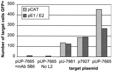
FIG. 1.
Expression of E1 and E2 proteins fails to enhance BPV1 vector production efficiency. 293H target cells were incubated for 48 h with various types of producer cell lysates. Ten thousand target cells were analyzed by FACS to detect GFP expression resulting from BPV1 vector transduction. Producer cells were generated by cotransfection of 293H cells with an L1/L2 expression plasmid (pSheLL) and the target plasmid shown, together with either control plasmid pCAT (striped bars) or pE1/E2 (solid bars). Target plasmid names denote the presence or absence of the BPV1 URR (U) or of a putative packaging signal (P) and the plasmid size (in base pairs). Two negative controls for BPV1 vector transduction are shown at left.
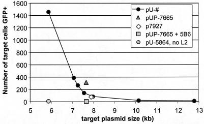
FIG. 2.
Target plasmid size influences BPV1 vector production efficiency. The graph shows the number of 293H target cells expressing GFP after treatment with cell lysates generated by cotransfection of pSheLL together with GFP-expressing plasmids of various sizes. See the legend to Fig. 1 for a description of target plasmid nomenclature.
For the experiments shown in Fig. 3 and 4, 293TT producer cells were lysed using the nonionic detergent Brij 58 (Sigma) at a final concentration of 0.5% in DPBS supplemented with an additional 9.5 mM MgCl2. Lysates were digested at 37° for 1 h with 1,500 U of Serratia marcescens nuclease (Benzonase) (Sigma)/ml and 20 U of Plasmid Safe ATP-dependent DNase (Epicentre)/ml. Although Epicentre declined to identify Plasmid Safe, polyacrylamide gel electrophoresis analysis suggested that it is exonuclease V. The nuclease-digested lysate was chilled on ice, mixed with a 0.17 volume of 5 M NaCl, and then clarified by centrifugation at 1500 × g for 10 min. The resulting supernatant was titrated on 293H cells (see below).
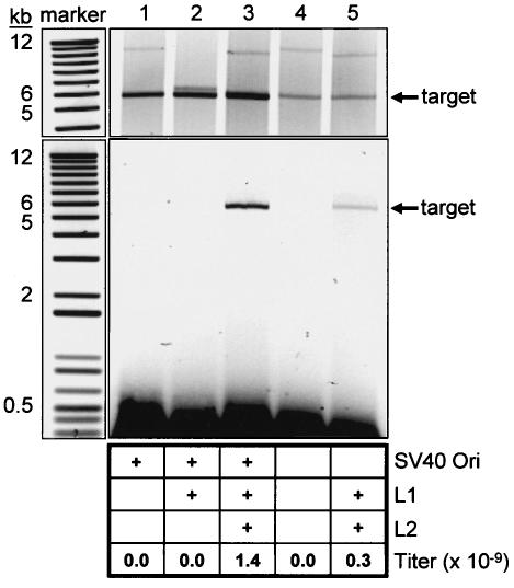
FIG. 3.
Quantitation of low-molecular-weight and nuclease-resistant DNA in transiently transfected 293TT cells. The top image shows a SYBR Green I-stained agarose gel loaded with low-molecular-weight DNA extracted from 293TT cells transfected with various plasmids. Cells were cotransfected with the following: lane 1, pU-10151 and pSU-5697; lane 2, pADAP-L1P and pSU-5697; lane 3, pSheLL and pSU-5697; lane 4, pU-10151 and pU-5864; lane 5, pSheLL and pU-5864. In the top image, either pSheLL or a control plasmid (pADAP-L1P or pU-10151) is visible above the band marked “target.” Each lane in the top image was loaded with 100,000 cell equivalents of low-molecular-weight DNA linearized by digestion with the restriction enzyme BsrGI. The lower image shows nuclease-resistant DNA isolated from same transfectants. The nuclease-resistant DNA was linearized by digestion with BsrGI and visualized by SYBR Green I staining of an agarose gel. Each lane of the gel in the bottom image was loaded with 1.5 million cell equivalents of extracted DNA. The chart at the bottom of the figure shows the presence or absence of various components within the producer cells and the BPV1 vector titer of the cell lysate.
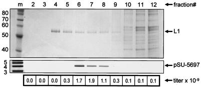
FIG. 4.
Purification of a BPV1 vector stock produced by transfection of 293TT cells. The top image depicts a protein-stained polyacrylamide gel loaded with samples from various fractions of an OptiPrep gradient. Western blotting of the fractions using an anti-L1 antibody showed a pattern identical to the band marked as L1 and showed negligible amounts of L1 above fraction 11 (data not shown). Lane m contains protein standards marked in kilodaltons (top image) or DNA standards marked in kilobases (lower image). The lower image shows nuclease-resistant DNA precipitated from each OptiPrep fraction and visualized by agarose gel electrophoresis followed by SYBR Green I staining. The target plasmid DNA migrated at the same rate as the supercoiled target plasmid standard. The BPV1 vector titer of each cell lysate (in transducing units per milliliter) is given in the chart at the bottom of the figure.
Further details concerning the handling of papillomaviral vectors are available from the authors upon request.
Titration of papillomavirus vector stocks.
293H cells were used as targets for determining the transducing capacity of clarified producer-cell lysates or OptiPrep (Axis-Shield) fractions through titration (see below). 293H target cells were plated 18 h in advance in 0.5 ml of DMEM at 105 cells per well in 24-well plates. One microliter of papillomaviral vector stock or, in some cases, 1 μl of stock diluted 1:10 or 1:100 in DPBS-800 mM NaCl was then added directly to the culture medium. Forty-eight hours after inoculation, the target cells were trypsinized and subjected to FACS analysis using a FACSCalibur machine (Becton Dickinson). For FACS analysis (CellQuest Pro v4.0.1 [Becton Dickinson]), 10,000 live cell events were collected, and a marker region was adjusted such that <0.1% of a population of mock-transduced 293H cells registered as FL1+. Only negligible amounts of fluorescence were detectable in target cells 24 h after inoculation (data not shown). Because the target reporter plasmids used in these studies have no mechanism for replicating in transduced 293H target cells, we reasoned that at a low multiplicity of infection each fluorescent cell observed 48 h after inoculation could be regarded as a single transduction event. Thus, the titer (in transducing units per milliter) was calculated using the formula (% of cells FL1+ at 48 h) × (number of cells harvested) × (1,000 μl/ml) × (dilution factor). The titer was calculated based on dilutions resulting in fluorescence of 1 to 25% of target cells. Within this range we observed a linear relationship between stock dilution and apparent titer.
Antibody neutralization of BPV1- or human papillomavirus type 16 (HPV16)-based vectors was performed by preincubating the vector stock on ice for 1 h with ascites stocks of monoclonal antibody 5B6 (29) or V5 (7) diluted 1:1,000.
DNA analysis.
Low-molecular-weight DNA was extracted from one million cells using a previously reported neutral lysis method (3). For the top panel of Fig. 3, low-molecular-weight DNA was extracted from cells and then linearized by digestion with BsrGI (NEB). One hundred thousand cell equivalents of digested DNA were loaded into a Tris-acetate-EDTA-0.9% agarose gel (Owl, Inc.). After electrophoresis, the gel was stained with SYBR Green I (Sigma) in 25 mM Tris (pH 8) for 30 min. Quantitation was performed by comparison to a BsrGI-linearized pSU-5697 plasmid subjected to twofold serial dilution (data not shown). Size was assessed by comparison to a 1-kb Plus marker ladder (Invitrogen) loaded such that the 5-kb band contained 6 ng of DNA.
To analyze encapsidated DNA, cell lysates were nuclease digested, salt treated, and clarified as described above. The clarified lysate (or OptiPrep fractionated material) was then digested with proteinase K (Qiagen) and subjected to phenol-chloroform extraction followed by ethanol precipitation. The purified DNA was quantitated by SYBR Green I staining of agarose gels as described above.
OptiPrep purification of papillomaviral vector stocks.
OptiPrep solutions were adjusted to 1× DPBS-800 mM NaCl; 27-33-39% OptiPrep gradients were cast by underlayering and then allowed to diffuse for 3 to 4 h at room temperature. Clarified Brij 58-salt lysate (see above) of 25 million cells in a 0.5-ml volume was layered on top of the gradient. Tubes were then centrifuged in an SW50.1 rotor (Beckman) at 234,000 × g for 3.5 h at 16°C. After centrifugation, fractions were collected by bottom puncture of the tubes. Each of the fractions shown in Fig. 4 was approximately 0.2 ml. A 10-μl sample of each fraction was separated on a 10% NuPage gel (Invitrogen) and then stained with Microwave Blue protein stain (Protiga). Protein molecular weight was determined by comparison to a Benchmark (Invitrogen) unstained protein ladder. L1 was quantitated by densitometric comparison to bovine serum albumin standards (Bio-Rad) subjected to twofold serial dilution.
Nucleotide sequence accession number.
The sequence of the codon-modified ORF encoding the BPV1 L1 protein was submitted to GenBank (accession number AY312992).
RESULTS
Plasmid-based expression of the BPV1 L1 and L2 proteins.
Papillomavirus L1 and L2 genes are generally expressed at low levels in the context of standard plasmid-based mammalian cell expression systems. This restriction is thought to be due to the presence of rare codons that have correspondingly low levels of cognate tRNAs and to cis-acting elements that inhibit RNA production, processing, and translation (12, 18, 34, 38, 39), reviewed in references 33 and 31. To overcome this theoretical limitation on papillomavirus structural gene expression, Zhou and colleagues used a PCR-based mutagenesis strategy to silently mutate clusters of rare codons in the BPV1 L1 and L2 genes (48). We observed high levels of L2 expression in 293T cells transfected with the pCDNA/HBL2 construct described by Zhou and colleagues (data not shown). In contrast, expression from the codon-modified L1 construct, pCDNA/HBL1, was near the lower limit of detection by Western blotting (data not shown).
To more completely overcome the expression inhibition of the wild-type BPV1 L1 ORF, we changed every codon that could be silently mutated. When transfected into 293T cells, expression plasmids carrying the resulting “as different as possible” L1 ORF drove L1 protein expression at levels at least 100-fold higher than those of Zhou and colleagues' pCDNA/HBL1 (data not shown).
To maximize the coexpression of BPV1 L1 and L2, we constructed a bicistronic L1/L2 expression plasmid using the internal ribosome entry site of murine encephalomyocarditis virus. To reduce encapsidation of the plasmid (pSheLL), we inserted stuffer DNA into the plasmid backbone in order to increase its size to 10.8 kb.
Expression of E2 is not required for intracellular production of BPV1 vectors.
We generated a series of GFP-expressing target plasmids that might be suitable for packaging within L1/L2 particles. Each of the initial panel of plasmids was similar in size to the wild-type BPV1 genome (7.9 kb). Some target plasmids also included the BPV1 URR and a putative packaging signal contained within the BPV1 E1 ORF (45). The target plasmids p7927, pU-7981, and pUP-7665 were named according to their size (in base pairs) and the presence or absence of the URR (U) or putative packaging signal (P). p7927 does not contain any BPV1 DNA sequences.
To test the efficiency of BPV1 vector production using these target plasmids, 293H cells were cotransfected with the L1/L2-expressing plasmid pSheLL, a target plasmid, and either control plasmid pCAT or pE1/E2, which expresses BPV1 E1 and E2. pE1/E2 was found to drive expression of the E2 protein at levels appropriate for supporting its transcription activation function and its role in E1-mediated replication of the viral genome (data not shown). Two days after transfection, the transfected producer cells were sonicated, and the resulting lysates were titered for their capacity to transduce fresh 293H target cells with GFP. Figure 1 shows the number of target 293H cells fluorescing after treatment with lysates of producer cells transfected with the plasmids shown. The results show that cotransfection of producer cells with pE1/E2 did not increase their BPV1 vector yield regardless of which target plasmid was used. Similar results were observed in three independent experiments. E2 also failed to increase vector yield when it was expressed in the absence of E1 or at different levels using either a previously characterized E2 expression plasmid, pMEP-E2 (27), or using an ecdysone-inducible expression system (Invitrogen) (data not shown). E2 expression also failed to enhance BPV1 vector production in CV-1 and COS-7 cells (data not shown). In the course of performing these experiments we never observed an apparent enhancing effect attributable to E2 expression.
Pretreatment of lysates with anti-L1 neutralizing monoclonal antibody 5B6 (29) resulted in a >30-fold reduction in the number of fluorescent target cells (Fig. 1), demonstrating that GFP expression was due to BPV1 vector transduction. Substitution of pSheLL with a plasmid expressing only L1 (pADAP-L1P) resulted in very low levels of GFP transduction. This result is consistent with past observations showing that expression of L2 is required for efficient intracellular production of infectious papillomavirus virions (49), pseudotypes (28), and reporter vectors (41).
Smaller target plasmids improve BPV1 vector yield.
In some viral vector systems, target nucleic acids smaller than the native viral genome may be preferred substrates for packaging (37, 44, 47). We therefore examined the relationship between target plasmid size and BPV1 vector yield. Figure 2 shows the number of target cells fluorescing after treatment with lysates of cells cotransfected with pSheLL and target plasmids of various sizes. Except for p7927, each of the target plasmids carries the BPV1 URR. Each plasmid also contains a GFP expression cassette and stuffer DNA fragments of various sizes. BPV1 vector production was as much as 10-fold more efficient in cells transfected with the 5.9-kb plasmid pU-5864 than in cells transfected with plasmids closer to the 7.9-kb size of the wild-type BPV1 genome. Use of the target plasmid pUP-7665 resulted in a ∼2-fold improvement in vector yield compared to similar-sized target plasmids. As expected, BPV1 vector production in cells transfected with target plasmids substantially larger than 8 kb was very inefficient. Similar results were observed in five independent experiments.
Production and characterization of crude BPV1 vector stocks.
Although it is possible to produce BPV1 vectors in transfected 293H cells, we reasoned that intracellular replication of target plasmids might improve the BPV1 vector yield. We therefore generated a panel of stable 293 cell lines expressing various levels of either SV40 T antigen or BPV1 E1 and E2. These lines supported intracellular replication of a target plasmid, pSU-5697, that carries both the SV40 and BPV1 origins of replication (Oris) (data not shown). In a series of BPV1 vector production experiments, we observed a linear relationship between the number of copies of replicated plasmid present in the cells and the titer of BPV1 vector recovered, regardless of whether the replication was mediated by E1 and E2 or T antigen (data not shown).
One stable 293 cell line that was engineered to overexpress T antigen supported high levels of BPV1 vector production. We used this cell line, named 293TT, to assess the suitability of a variety of methods for harvesting BPV1 vectors. We found that lysis of cells using the nonionic detergent Brij 58 at a final concentration of 0.5% gave better vector yields than lysis by sonication or freeze-thaw cycles or lysis with other detergents, such as Triton X-100, Tween 80, NP 40, or sodium deoxycholate. Recovery of the vector from Brij 58-lysed cells was substantially enhanced by nuclease digestion (data not shown).
Attempts to clarify crude lysates by centrifugation showed that the majority of the vector present in sonicated or Brij 58-lysed 293TT cells could be pelleted at forces as low as 16,000 × g (data not shown). This unexpected finding suggests that the transduction-competent virions in such lysates are associated with larger particles, such as cell debris or multivirion aggregates (42, 50). Increasing the sodium chloride concentration of sonicated or Brij 58-lysed producer cells to 0.8 M increased the apparent titer of the lysates and allowed clarification by centrifugation at 16,000 × g.
Using the Brij 58 and high-salt extraction method, we examined target plasmid encapsidation in 293TT cells transiently cotransfected with pSheLL (or control plasmids) and target plasmids with or without the SV40 Ori (pSU-5697 or pU-5864, respectively). Forty-four hours after transfection, low-molecular-weight DNA was isolated from the transfected cells. Plasmid DNA from these extracts was readily detectable by direct staining of agarose gels using the nucleic acid stain SYBR Green I (Fig. 3, upper image). A portion of the transfected cell population was subjected to Brij 58 lysis and nuclease digestion, followed by phenol-chloroform extraction and ethanol precipitation of nuclease-resistant DNA. Cells expressing L1 and L2 contained substantial amounts of nuclease-resistant full-length target plasmid DNA (Fig. 3, lower image). In contrast, very little of the 10.8-kb pSheLL plasmid was rendered nuclease-resistant, suggesting it is too large to serve as an effective substrate for packaging. The appearance of nuclease-resistant DNA was strictly dependent on the expression of L2, indicating that L2 plays a critical role in encapsidation in this system.
Table 1 summarizes quantitation of the target plasmid, L1 protein, and BPV1 vector yield for the cell lysates depicted in Fig. 3. Values similar to those shown in Table 1 were observed in eight independent experiments. The data show that the BPV1 vector yield was roughly proportional to the number of copies of the target plasmid within the cells. The findings suggest that target plasmid content is the primary limiting factor for papillomavirus vector production in this system. The results are consistent with the conclusion that transfected bacterially derived target plasmid can be packaged about as efficiently as plasmids subject to endogenous replication by T antigen.
TABLE 1.
BPV1 vector yielda
| Parameter | Value for plasmid
|
Ratio (SV40 Ori +/−) | |
|---|---|---|---|
| pSU-5697 | pU-5864 | ||
| No. of L1 VLP equivalents/cellb | 52,000 | 37,000 | |
| Total no. of plasmids/cell | 12,000 | 1,700 | 7 |
| No. of protected plasmids/cell | 600 | 100 | 6 |
| No. of GFP-transducing units/cell | 75 | 15 | 5 |
| % of plasmid protected | 5 | 6 | |
| Protected plasmid/infectivity ratio | 8 | 7 | |
Stocks were derived from the same cell lysates analyzed in Fig. 3 (lane 3 for pSU5697 and lane 5 for pU-5864).
Number of VLPs present in cell lysate assuming that the measured amount of L1 protein was entirely assembled into VLPs.
Comparison of the number of copies of nuclease-resistant plasmid to the BPV1 vector titer of the lysates suggested a DNA-containing “full” particle-to-infectivity ratio of about 8 (Table 1). The actual particle-to-infectivity ratio may be lower than 8, since our titration method was optimized for speed and reproducibility rather than for maximum sensitivity.
Purification of BPV1 vector stocks.
Cesium chloride (CsCl) gradients are often used for purification of infectious papillomavirus particles. However, experiments using CsCl gradients to purify BPV1 vectors out of Brij 58-salt lysates (see above) resulted in a 99% loss of apparent titer (data not shown). The ultracentrifugation medium iodixanol (trade name OptiPrep) can be a suitable alternative for purifying virions and viral vectors while still preserving their infectivity (50). OptiPrep is an iodinated dihexanol compound originally developed for use as an injectable X-ray contrast agent. It is nonionic, has relatively low osmotic content, and is nontoxic to cultured cells at concentrations of up to 30% (wt/vol) (2).
Because OptiPrep is effective both as a velocity gradient medium and as a buoyant density gradient medium, we reasoned that it might be possible to perform both types of separation in a single gradient. Using a single partially diffused 27-33-39% OptiPrep gradient prepared in DPBS with 0.8 M NaCl, L1 protein could be purified away from most of the cellular protein in lysates of 293TT cells cotransfected with pSheLL and pSU-5697 (Fig. 4, upper image). The OptiPrep gradient also achieved substantial separation between DNA-containing full particles and empty VLPs. Although fractions 4 and 5 of the gradient contained the majority of the L1 protein, the packaged target plasmid and BPV1 vector titer were found primarily in fractions 6, 7, and 8 (Fig. 4). Electron microscopic analysis of fractions 4 and 7 showed that both fractions contained particles resembling papillomavirus virions (data not shown). The data indicate that empty VLPs migrated to higher-density fractions of the gradient than full particles. This was surprising, since empty papillomavirus VLPs are known to exhibit lower density than full particles in CsCl gradients. However, many types of particles exhibit greater levels of hydration in OptiPrep, thus altering their apparent density relative to values observed with CsCl gradients (50). A separate set of experiments involving equilibrium centrifugation of 293TT lysates in self-forming OptiPrep density gradients showed a peak in L1 content at fractions weighing 1.25 g/ml, while the peak titer fraction weighed 1.20 g/ml (data not shown). These values are lower than the apparent densities of empty and full particles in CsCl (1.28 and 1.32, respectively).
The ratio of VLP equivalents to infectivity for OptiPrep fraction 7 was 88, compared to 680 for the original crude lysate (data not shown). Thus, the OptiPrep fractionation resulted in an eightfold increase in the overall particle-to-infectivity ratio. In contrast, the ratio of target plasmid content to titer was essentially unchanged by OptiPrep purification, suggesting that the procedure does not significantly compromise virion infectivity. The overall recovery of titer after OptiPrep fractionation typically varied from 50 to 70%. We suspect that the losses may have been due to nonspecific adsorption of L1 particles to plastic surfaces (42), since a comparable fraction of L1 protein was lost during the purification process (data not shown).
Generation and antibody neutralization of an HPV16-based vector.
One potential application for papillomaviral vectors is the detection of antipapillomavirus neutralizing antibodies elicited during natural infection with HPV or following vaccination against HPV. Leder and colleagues have recently achieved high-level expression of the HPV16 L1 and L2 genes using expression plasmids carrying codon-modified versions of the genes (20). We found that these constructs can be used to generate HPV16-based vector stocks with titers comparable to those of BPV1 vector stocks. In both instances, 5 × 10⁹ GFP-transducing units can be obtained from a single 75-cm2 flask of 293TT cells cotransfected with pSU-5697 and either pSheLL or Leder and colleagues' pL1h and pL2h constructs. The initial handling steps used for generating crude BPV1 vector stocks are also suitable for harvesting HPV16 vectors.
To determine whether HPV16 vectors could be neutralized by HPV16-specific antibodies, we tested the transducing capacity of an HPV16 vector stock after preincubation with serial dilutions of the anti-HPV16 L1 neutralizing monoclonal antibody, V5 (7). At dilutions lower than one to a million, there was a >95% reduction in HPV16 vector transduction relative to vector stock that was untreated or pretreated with control BPV1-neutralizing monoclonal antibody 5B6 (data not shown). Because currently available methods for assaying sera for HPV16-neutralizing capacity are arduous, the availability of efficient methods for production of HPV16 reporter vectors should greatly facilitate the study of antibody responses to the virus.
DISCUSSION
We have used conventional mammalian cell transfection methods to generate high-titer BPV1 and HPV16 gene transfer vectors. This novel method, which makes use of codon-modified versions of the papillomavirus structural genes, is 10 million-fold more efficient than our previous SFV-based methods. The new production system is straightforward and can be used to quickly generate papillomavirus vector stocks with titers of several billion transducing units per milliliter.
The intracellular encapsidation of recombinant reporter plasmids into papillomaviral L1/L2 particles is an unexpectedly promiscuous process. Our laboratory had previously suggested a model in which the papillomavirus early protein E2 might lend specificity to the process of virion morphogenesis (9). Zhao and colleagues have reported data consistent with this theory, finding that E2 expression modestly enhanced the production of BPV1 vectors using vaccinia virus and baculovirus overexpression systems (46). This finding partly conflicts with those in prior studies by Unckell and colleagues (41) and Stauffer and colleagues (35) showing, respectively, that HPV33 and HPV18 vectors can be generated in the absence of E2 expression. Although Stauffer and colleagues did not examine the effect of expressing E2, the work of Unckell and colleagues showed that vaccinia virus-based overexpression of E2 had no effect on papillomavirus vector yield. However, the system reported by Unckell and colleagues made use of a plasmid carrying the SV40 Ori and cells expressing SV40 T antigen. Our lab (9) and Stauffer and colleagues (35) have suggested that the interaction of T antigen with the SV40 Ori might functionally replace the E2/papillomavirus Ori interaction proposed to facilitate assembly. The data in the present report clearly demonstrate that in several types of mammalian cell lines, expression of E2 fails to enhance encapsidation of plasmid DNA carrying multiple E2 binding sites, even in the absence of SV40 Ori or T antigen. Although our results do not rule out the proposed role for E2 in the morphogenesis of authentic virions in terminally differentiated epithelia, the data do support the possibility of an E2-independent assembly mechanism for papillomaviruses in vivo.
Although it is generally believed that L2 is required for intracellular encapsidation of papillomavirus genomes (35, 49), purified L1 protein can be used to generate papillomavirus vectors in the absence of L2 using cell-free production systems (17, 26, 40, 43). L2 has also been shown to be dispensable for intracellular encapsidation of target plasmids in Unckell and colleagues' system for production of HPV33 vectors (41). Because the encapsidation of target plasmids in our system is strictly dependent on the intracellular expression of L2 (Fig. 3), it may serve as a relevant method for studying the potential role of L2 in encapsidation.
Cotransfection of small target plasmids together with a BPV1 L1 and L2 expression vector resulted in conversion of approximately 5% of cell-associated target plasmid into an encapsidated, nuclease-resistant form (Table 1). However, it is unlikely that there is complete overlap between the populations of transfected cells with abundant nucleus-localized copies of target plasmid and cells with appropriate amounts of L1 and L2 protein. Thus, 5% should be considered a minimum estimate of the actual encapsidation efficiency.
Given the apparently sequence-independent nature of the packaging process, it seems surprising that a substantial portion of the transfected target plasmid can become encapsidated despite the large excess of cellular DNA in the nuclear environment. It is possible, however, to envision a size-dependent assembly model in which the papillomavirus structural proteins sample the nuclear environment for appropriately small DNA molecules. In this model, association of the papillomavirus structural proteins with cellular DNA might result in a reversible, partially assembled state. In contrast, assembly of L1 and L2 around appropriate-size DNA species (such as a papillomavirus genome or the target plasmids we have studied) could proceed to an irreversibly assembled state. Since SFV, vaccinia viruses, and baculoviruses can induce fragmentation of cellular DNA, the existence of a size-dependent, sequence-nonspecific assembly mechanism could contribute to the low yield of papillomaviral vectors produced using these systems. This concept (41) is supported by the observation that L1 and L2 can nonspecifically encapsidate cellular DNA fragments smaller than 8 kb during long-term culture of baculovirus-infected insect cells (10). Although a size-dependent assembly model might explain our observations using transfected 293 cells, the fact that as much as 95% of the target plasmid in these cells remains unencapsidated at the time of harvest, despite an apparent excess of L1, leaves room for the possibility that additional specificity mechanisms facilitate papillomavirus assembly in vivo.
The availability of a rapidly titerable papillomaviral vector production system allowed us to empirically examine a wide range of factors involved in the harvest and handling of the vectors. The revised purification methods described in this work are substantially simpler and more efficient than previous methods. Using a hybrid velocity-density OptiPrep ultracentrifugation method, we have been able to enrich DNA-containing particles away from empty VLPs and cell debris in a single purification step. In contrast to CsCl equilibrium centrifugation, the OptiPrep centrifugation method did not substantially alter the particle-to-infectivity ratio, suggesting that OptiPrep purification is physically gentler than CsCl purification.
High-titer papillomaviral vector stocks should facilitate future investigation of a variety of topics. We have shown here that an HPV16 vector expressing GFP can be used to detect papillomavirus-neutralizing antibodies. Minor modifications of the technology have allowed us to develop a high-throughput method for titration of papillomavirus-neutralizing antibodies (D. V. Pastrana, C. B. Buck, Y. Y. S. Pang, et al., submitted for publication). The vectors may also be applied to genetic screens that might identify host cell proteins involved in the entry phase of the viral life cycle. The production methods described here are sufficiently robust to consider using papillomavirus vectors as vaccine vehicles. Papillomavirus vectors may be potent inducers of mucosal and systemic immunity (32), at least in part because L1 capsids can trigger innate immune responses in certain types of professional antigen-presenting cells, including dendritic cells (21, 30).
REFERENCES
- 1.Anborgh, P. H., X. Qian, A. G. Papageorge, W. C. Vass, J. E. DeClue, and D. R. Lowy. 1999. Ras-specific exchange factor GRF: oligomerization through its Dbl homology domain and calcium-dependent activation of Raf. Mol. Cell. Biol. 19:4611-4622. [DOI] [PMC free article] [PubMed] [Google Scholar]
- 2.Andersen, K. J., H. Vik, H. P. Eikesdal, and E. I. Christensen. 1995. Effects of contrast media on renal epithelial cells in culture. Acta Radiol. Suppl. 399:213-218. [DOI] [PubMed] [Google Scholar]
- 3.Arad, U. 1998. Modified Hirt procedure for rapid purification of extrachromosomal DNA from mammalian cells. BioTechniques 24:760-762. [DOI] [PubMed] [Google Scholar]
- 4.Buck, C. B., X. Shen, M. A. Egan, T. C. Pierson, C. M. Walker, and R. F. Siliciano. 2001. The human immunodeficiency virus type 1 gag gene encodes an internal ribosome entry site. J. Virol. 75:181-191. [DOI] [PMC free article] [PubMed] [Google Scholar]
- 5.Campo, M. S. 2002. Animal models of papillomavirus pathogenesis. Virus Res. 89:249-261. [DOI] [PubMed] [Google Scholar]
- 6.Chow, L. T., and T. R. Broker. 1997. In vitro experimental systems for HPV: epithelial raft cultures for investigations of viral reproduction and pathogenesis and for genetic analyses of viral proteins and regulatory sequences. Clin. Dermatol. 15:217-227. [DOI] [PubMed] [Google Scholar]
- 7.Christensen, N. D., J. Dillner, C. Eklund, J. J. Carter, G. C. Wipf, C. A. Reed, N. M. Cladel, and D. A. Galloway. 1996. Surface conformational and linear epitopes on HPV-16 and HPV-18 L1 virus-like particles as defined by monoclonal antibodies. Virology 223:174-184. [DOI] [PubMed] [Google Scholar]
- 8.Cole, C. N., and S. D. Conzen. 2001. Polyomaviridae: the viruses and their replication, p. 2141-2174. In D. M. Knipe and P. M. Howley (ed.), Fields virology, 4th ed., vol. 2. Lippincott Williams & Wilkins, Philadelphia, Pa.
- 9.Day, P. M., R. B. Roden, D. R. Lowy, and J. T. Schiller. 1998. The papillomavirus minor capsid protein, L2, induces localization of the major capsid protein, L1, and the viral transcription/replication protein, E2, to PML oncogenic domains. J. Virol. 72:142-150. [DOI] [PMC free article] [PubMed] [Google Scholar]
- 10.Fligge, C., F. Schafer, H. C. Selinka, C. Sapp, and M. Sapp. 2001. DNA-induced structural changes in the papillomavirus capsid. J. Virol. 75:7727-7731. [DOI] [PMC free article] [PubMed] [Google Scholar]
- 11.Fu, X. Y., and J. L. Manley. 1987. Factors influencing alternative splice site utilization in vivo. Mol. Cell. Biol. 7:738-748. [DOI] [PMC free article] [PubMed] [Google Scholar]
- 12.Furth, P. A., W. T. Choe, J. H. Rex, J. C. Byrne, and C. C. Baker. 1994. Sequences homologous to 5′ splice sites are required for the inhibitory activity of papillomavirus late 3′ untranslated regions. Mol. Cell. Biol. 14:5278-5289. [DOI] [PMC free article] [PubMed] [Google Scholar]
- 13.Hammond, S. A., R. P. Johnson, S. A. Kalams, B. D. Walker, M. Takiguchi, J. T. Safrit, R. A. Koup, and R. F. Siliciano. 1995. An epitope-selective, transporter associated with antigen presentation (TAP)-1/2-independent pathway and a more general TAP-1/2-dependent antigen-processing pathway allow recognition of the HIV-1 envelope glycoprotein by CD8+ CTL. J. Immunol. 154:6140-6156. [PubMed] [Google Scholar]
- 14.Hegde, R. S. 2002. The papillomavirus E2 proteins: structure, function, and biology. Annu. Rev. Biophys. Biomol. Struct. 31:343-360. [DOI] [PubMed] [Google Scholar]
- 15.Heino, P., J. Zhou, and P. F. Lambert. 2000. Interaction of the papillomavirus transcription/replication factor, E2, and the viral capsid protein, L2. Virology 276:304-314. [DOI] [PubMed] [Google Scholar]
- 16.Howett, M. K., N. D. Christensen, and J. W. Kreider. 1997. Tissue xenografts as a model system for study of the pathogenesis of papillomaviruses. Clin. Dermatol. 15:229-236. [DOI] [PubMed] [Google Scholar]
- 17.Kawana, K., H. Yoshikawa, Y. Taketani, K. Yoshiike, and T. Kanda. 1998. In vitro construction of pseudovirions of human papillomavirus type 16: incorporation of plasmid DNA into reassembled L1/L2 capsids. J. Virol. 72:10298-10300. [DOI] [PMC free article] [PubMed] [Google Scholar]
- 18.Kennedy, I. M., J. K. Haddow, and J. B. Clements. 1991. A negative regulatory element in the human papillomavirus type 16 genome acts at the level of late mRNA stability. J. Virol. 65:2093-2097. [DOI] [PMC free article] [PubMed] [Google Scholar]
- 19.Kirnbauer, R., J. Taub, H. Greenstone, R. Roden, M. Durst, L. Gissmann, D. R. Lowy, and J. T. Schiller. 1993. Efficient self-assembly of human papillomavirus type 16 L1 and L1-L2 into virus-like particles. J. Virol. 67:6929-6936. [DOI] [PMC free article] [PubMed] [Google Scholar]
- 20.Leder, C., J. A. Kleinschmidt, C. Wiethe, and M. Muller. 2001. Enhancement of capsid gene expression: preparing the human papillomavirus type 16 major structural gene L1 for DNA vaccination purposes. J. Virol. 75:9201-9209. [DOI] [PMC free article] [PubMed] [Google Scholar]
- 21.Lenz, P., P. M. Day, Y. Y. Pang, S. A. Frye, P. N. Jensen, D. R. Lowy, and J. T. Schiller. 2001. Papillomavirus-like particles induce acute activation of dendritic cells. J. Immunol. 166:5346-5355. [DOI] [PubMed] [Google Scholar]
- 22.Liu, W. J., L. Gissmann, X. Y. Sun, A. Kanjanahaluethai, M. Muller, J. Doorbar, and J. Zhou. 1997. Sequence close to the N-terminus of L2 protein is displayed on the surface of bovine papillomavirus type 1 virions. Virology 227:474-483. [DOI] [PubMed] [Google Scholar]
- 23.Loeken, M., I. Bikel, D. M. Livingston, and J. Brady. 1988. trans-activation of RNA polymerase II and III promoters by SV40 small t antigen. Cell 55:1171-1177. [DOI] [PubMed] [Google Scholar]
- 24.McBride, A. A., J. B. Bolen, and P. M. Howley. 1989. Phosphorylation sites of the E2 transcriptional regulatory proteins of bovine papillomavirus type 1. J. Virol. 63:5076-5085. [DOI] [PMC free article] [PubMed] [Google Scholar]
- 25.Meyers, C., and L. A. Laimins. 1994. In vitro systems for the study and propagation of human papillomaviruses. Curr. Top. Microbiol. Immunol. 186:199-215. [DOI] [PubMed] [Google Scholar]
- 26.Muller, M., L. Gissmann, R. J. Cristiano, X. Y. Sun, I. H. Frazer, A. B. Jenson, A. Alonso, H. Zentgraf, and J. Zhou. 1995. Papillomavirus capsid binding and uptake by cells from different tissues and species. J. Virol. 69:948-954. [DOI] [PMC free article] [PubMed] [Google Scholar]
- 27.Penrose, K. J., and A. A. McBride. 2000. Proteasome-mediated degradation of the papillomavirus E2-TA protein is regulated by phosphorylation and can modulate viral genome copy number. J. Virol. 74:6031-6038. [DOI] [PMC free article] [PubMed] [Google Scholar]
- 28.Roden, R. B., H. L. Greenstone, R. Kirnbauer, F. P. Booy, J. Jessie, D. R. Lowy, and J. T. Schiller. 1996. In vitro generation and type-specific neutralization of a human papillomavirus type 16 virion pseudotype. J. Virol. 70:5875-5883. [DOI] [PMC free article] [PubMed] [Google Scholar]
- 29.Roden, R. B., E. M. Weissinger, D. W. Henderson, F. Booy, R. Kirnbauer, J. F. Mushinski, D. R. Lowy, and J. T. Schiller. 1994. Neutralization of bovine papillomavirus by antibodies to L1 and L2 capsid proteins. J. Virol. 68:7570-7574. [DOI] [PMC free article] [PubMed] [Google Scholar]
- 30.Rudolf, M. P., S. C. Fausch, D. M. Da Silva, and W. M. Kast. 2001. Human dendritic cells are activated by chimeric human papillomavirus type-16 virus-like particles and induce epitope-specific human T cell responses in vitro. J. Immunol. 166:5917-5924. [DOI] [PubMed] [Google Scholar]
- 31.Schwartz, S. 2000. Regulation of human papillomavirus late gene expression. Upsala J. Med. Sci. 105:171-192. [PubMed] [Google Scholar]
- 32.Shi, W., J. Liu, Y. Huang, and L. Qiao. 2001. Papillomavirus pseudovirus: a novel vaccine to induce mucosal and systemic cytotoxic T-lymphocyte responses. J. Virol. 75:10139-10148. [DOI] [PMC free article] [PubMed] [Google Scholar]
- 33.Smith, D. W. 1996. Problems of translating heterologous genes in expression systems: the role of tRNA. Biotechnol. Prog. 12:417-422. [DOI] [PubMed] [Google Scholar]
- 34.Sokolowski, M., W. Tan, M. Jellne, and S. Schwartz. 1998. mRNA instability elements in the human papillomavirus type 16 L2 coding region. J. Virol. 72:1504-1515. [DOI] [PMC free article] [PubMed] [Google Scholar]
- 35.Stauffer, Y., K. Raj, K. Masternak, and P. Beard. 1998. Infectious human papillomavirus type 18 pseudovirions. J. Mol. Biol. 283:529-536. [DOI] [PubMed] [Google Scholar]
- 36.Stubenrauch, F., and L. A. Laimins. 1999. Human papillomavirus life cycle: active and latent phases. Semin. Cancer Biol. 9:379-386. [DOI] [PubMed] [Google Scholar]
- 37.Tai, H. T., C. A. Smith, P. A. Sharp, and J. Vinograd. 1972. Sequence heterogeneity in closed simian virus 40 deoxyribonucleic acid. J. Virol. 9:317-325. [DOI] [PMC free article] [PubMed] [Google Scholar]
- 38.Tan, W., B. K. Felber, A. S. Zolotukhin, G. N. Pavlakis, and S. Schwartz. 1995. Efficient expression of the human papillomavirus type 16 L1 protein in epithelial cells by using Rev and the Rev-responsive element of human immunodeficiency virus or the cis-acting transactivation element of simian retrovirus type 1. J. Virol. 69:5607-5620. [DOI] [PMC free article] [PubMed] [Google Scholar]
- 39.Tan, W., and S. Schwartz. 1995. The Rev protein of human immunodeficiency virus type 1 counteracts the effect of an AU-rich negative element in the human papillomavirus type 1 late 3′ untranslated region. J. Virol. 69:2932-2945. [DOI] [PMC free article] [PubMed] [Google Scholar]
- 40.Touze, A., and P. Coursaget. 1998. In vitro gene transfer using human papillomavirus-like particles. Nucleic Acids Res. 26:1317-1323. [DOI] [PMC free article] [PubMed] [Google Scholar]
- 41.Unckell, F., R. E. Streeck, and M. Sapp. 1997. Generation and neutralization of pseudovirions of human papillomavirus type 33. J. Virol. 71:2934-2939. [DOI] [PMC free article] [PubMed] [Google Scholar]
- 42.Volkin, D. B., L. Shi, and G. Sanyal. March. 2002. Stabilized human papillomavirus formulations. U.S. patent 6,358,744 B1.
- 43.Yeager, M. D., M. Aste-Amezaga, D. R. Brown, M. M. Martin, M. J. Shah, J. C. Cook, N. D. Christensen, C. Ackerson, R. S. Lowe, J. F. Smith, P. Keller, and K. U. Jansen. 2000. Neutralization of human papillomavirus (HPV) pseudovirions: a novel and efficient approach to detect and characterize HPV neutralizing antibodies. Virology 278:570-577. [DOI] [PubMed] [Google Scholar]
- 44.Yoshiike, K. 1968. Studies on DNA from low-density particles of SV40. I. Heterogeneous defective virions produced by successive undiluted passages. Virology 34:391-401. [DOI] [PubMed] [Google Scholar]
- 45.Zhao, K. N., I. H. Frazer, W. Jun Liu, M. Williams, and J. Zhou. 1999. Nucleotides 1506-1625 of bovine papillomavirus type 1 genome can enhance DNA packaging by L1/L2 capsids. Virology 259:211-218. [DOI] [PubMed] [Google Scholar]
- 46.Zhao, K. N., K. Hengst, W. J. Liu, Y. H. Liu, X. S. Liu, N. A. McMillan, and I. H. Frazer. 2000. BPV1 E2 protein enhances packaging of full-length plasmid DNA in BPV1 pseudovirions. Virology 272:382-393. [DOI] [PubMed] [Google Scholar]
- 47.Zhao, K. N., X. Y. Sun, I. H. Frazer, and J. Zhou. 1998. DNA packaging by L1 and L2 capsid proteins of bovine papillomavirus type 1. Virology 243:482-491. [DOI] [PubMed] [Google Scholar]
- 48.Zhou, J., W. J. Liu, S. W. Peng, X. Y. Sun, and I. Frazer. 1999. Papillomavirus capsid protein expression level depends on the match between codon usage and tRNA availability. J. Virol. 73:4972-4982. [DOI] [PMC free article] [PubMed] [Google Scholar]
- 49.Zhou, J., D. J. Stenzel, X. Y. Sun, and I. H. Frazer. 1993. Synthesis and assembly of infectious bovine papillomavirus particles in vitro. J. Gen. Virol. 74:763-768. [DOI] [PubMed] [Google Scholar]
- 50.Zolotukhin, S., B. J. Byrne, E. Mason, I. Zolotukhin, M. Potter, K. Chesnut, C. Summerford, R. J. Samulski, and N. Muzyczka. 1999. Recombinant adeno-associated virus purification using novel methods improves infectious titer and yield. Gene Ther. 6:973-985. [DOI] [PubMed] [Google Scholar]


