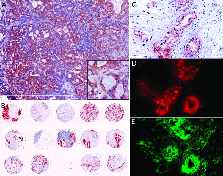
Figure 6.
Histology of human biopsy samples. (A) Human tissue tumor microarray. (B) Human PDAC metathesis in the liver (fine needle aspirate). (C) Anti-CTSE immunohistochemistry (adjacent slice as D and E). (D) Anti-CTSE immunofluorescence. (E) RIT-TMB staining, same slice as D (20 µM RIT-TMB for 1 hour).

