

Abstract
There is no doubt that the success of minimally invasive parathyroidectomy (MIP) has changed the whole treatment of patients with primary hyperparathyroidism, especially the approach towards traditional bilateral neck exploration. A single adenoma is the most common cause of primary hyperparathyroidism and its removal results in cure. Hence, it is worth the effort to localise and excise the single adenoma using modern technologies such as high-quality sestamibi scans and to confirm complete excision using rapid intra operative parathormone (IOPTH) assays. The objective of the study was to evaluate the feasibility of rapid IOPTH assay in successfully facilitating minimally invasive parathyroid excision. This research involved the retrospective study of seven patients, who underwent MIP at Sagar Hospital in Bengaluru, India, for parathyroid adenoma. All patients with evidence of unifocal disease on sestamibi scanning and cervical ultrasonography, underwent MIP via 2–3 cm lateral incision. Blood samples for measurement of IOPTH were taken at the time of induction of anaesthesia and 10 min after the adenoma excision. Reduction of parathormone (PTH) levels of more than 50 % in the postexcision sample was taken as evidence for complete extirpation of parathyroid adenoma. A solitary adenoma was identified in all the seven patients. After MIP, IOPTH levels fell in six of the seven patients. Following the surgery, all the cases were followed up for a period of 1 month. During this time, except for one patient, six patients remained asymptomatic and blood tests revealed normal serum calcium levels. A histopathological examination confirmed the diagnosis of parathyroid adenoma in six of the seven patients. After accurate preoperative localisation of the adenoma in patients with primary hyperparathyroidism, MIP with IOPTH measurement offers a safe and successful outcome.
Keywords: Hyperparathyroidism, Parathyroidectomy, Minimally invasive, Parathormone, Intraoperative parathormone
Introduction
Nowadays, there is an increasing trend towards minimally invasive surgery (MIS) for parathyroid disease in many medical institutions worldwide, because hyperparathyroidism results from the enlargement of a single gland or parathyroid adenoma in 85 % of cases [1]. MIS is an open form of surgery performed using a transverse skin incision of 2.5 cm on the side of the affected gland [2]. This offers the dual advantage of being less painful and cosmetically more acceptable. Conventional surgery using a long incision is required only for multiple adenomata or hyperplasia, which occurs in 15–20 % of patients [3]. Thus, a large majority of patients with primary hyperparathyroidism can be offered MIS. The recommended localisation procedures before the initial operation are sestamibi scanning and ultrasonography. Research has shown that the sensitivity of ultrasonography and sestamibi localisation individually are 65 % and 80 %, respectively. Similarly, the sensitivity and accuracy of combined parathyroid imaging are 78–96 % and 88 % [4–6]. However, the sensitivity, specificity and accuracy rate of intraoperative parathormone assay (IOPTH) are 97 %, 100 % and 97 %, respectively, which is very high [7]. Also, in a substantial number of patients, no definitive localisation was made, as preoperative localisation studies were concordant only 50–60 % of the time [1, 5, 8]. But in 13–19 % of patients, monitoring the IOPTH levels during parathyroidectomy directly aids the surgeon’s operative approach [7, 9]. Finally, to ensure that complete extirpation of the disease has taken place with excision of a solitary pathological parathyroid gland, rapid intraoperative estimation of PTH (IOPTH) has been introduced as an adjunct to preoperative localising imaging studies. A substantial fall in the parathormone (PTH) levels after excision of the gland will offer confirmation of the fact that surgery has been completely successful.
Aim
To evaluate the feasibility of rapid parathormone assay to enable minimally invasive parathyroid excision.
Patients and Methods
The present study is a retrospective observational study of seven patients undergoing minimally invasive parathyroid surgery at Sagar Hospital between August 2007 and September 2010. The symptoms of these patients were noted and all the seven patients underwent sestamibi scans and cervical ultrasonography (CUS) preoperatively. A nuclear medicine physician interpreted all the sestamibi scans. The CUS results were read by various radiologists.
Once the glands were localised, the patients underwent parathyroidectomy under general anaesthesia. Our technique uses a 2–3 cm transverse incision situated on the side of the abnormal gland, medial to the medial margin of the sternocleidomastoid muscle. The platysma is transected and the sternocleidomastoid muscle is retracted laterally to expose the strap muscles. These are retracted medially, exposing a space bounded anteromedially by the thyroid gland, laterally by the carotid sheath and the prevertebral fascia posteriorly. All the glands were sought and detected within this space (Fig. 1). Parathyroidectomy was performed by careful dissection close to the gland with visualisation and preservation of the recurrent laryngeal nerve (Fig. 2).
Fig. 1.
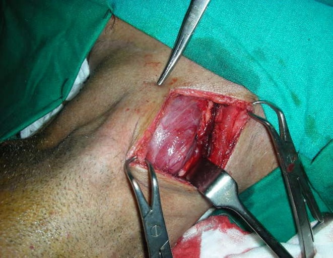
Dissection of parathyroid adenoma
Fig. 2.
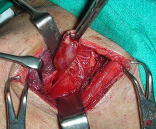
Excision of parathyroid adenoma
Rapid PTH Assay
Blood samples were collected into ethylenediaminetetraacetic acid tubes from a peripheral vein immediately after induction of anaesthesia, prior to excision of abnormal gland and 10 min after excision of the adenoma to measure PTH levels using IMMULITE 1000 Turbo intact PTH (chemiluminescent immunometric) assay. After confirmation of a reduction in PTH concentration of more than 50 % from the highest baseline value, the operation was concluded without visualising the remaining parathyroid glands.
All the patients were discharged either on the day of surgery or the following morning if surgery was performed in the afternoon. Serum calcium levels were measured on the first postoperative day and a month after the operation. Patients returned to the outpatient department a month later for a follow-up visit and were assessed for symptoms and complications along with a review of the histological findings.
Results
A total of seven patients, four men, a boy and two women with a mean age of 45 (range 9–74) years, referred with various symptoms of hyperparathyroidism, ranging from subjective symptoms such as general malaise or depressed mood to objective signs and symptoms, such as decreased bone mineral density or nephrolithiasis, underwent minimally invasive parathyroid surgery. Mean preoperative calcium and PTH levels were 13.7 (range 11.9–17.4 mg/dl) and 571 (range 92.1–1500 pg/ml), respectively. Preoperative localisation identified a solitary lesion with sestamibi scan and CUS in all the patients.
All operations were completed using the MIS approach. A solitary adenoma was found in all the patients. Circulating PTH levels had fallen by more than 50 % from baseline levels, 10 min after gland excision in six of the seven patients. In all the patients except one, the boy, parathyroidectomy was curative as determined by normal postoperative serum calcium levels (Table 1).
Table 1.
Patient demographics, biochemical values, intraoperative and postoperative data
| SL No | Age & Gender | PTH (pre-operative) | Ca++ (Pre) | Ph | PTH at Induction | PTH after Excision | Ca++ (Post) | Follow-up Symptoms | Ca++ after 1m |
|---|---|---|---|---|---|---|---|---|---|
| 1. | 60 years Male | 1235 | 13.6 | NA | 1000 | 200 | 10.5 | None | Not Available |
| 2. | 62 years Female | 313 | 14.4 | 2.5 | 466 | 35.7 | 11.4 | None | Not Available |
| 3. | 45 years Female | 257.6 | 12.9 | Low | 206 | 44.4 | 10.5 | None | 9.7 |
| 4 | 74 years Male | 1500 | 17.4 | 5.5 | 2435 | 104 | 8.0 | None | 8.6 |
| 5. | 9 years Male | 92.1 | 12.7 | 4.3 | 91.6 | 118(1st) 151(2nd) | 13.2 | Not Available | Not Available |
| 6. | 33 years Male | 127.3 | 11.9 | NA | 503 | 125 | 8.5 | None | 8.5 |
| 7. | 37 years Male | 458.4 | 13.5 | NA | 1524 | 190 | 11 | None | 8.55 |
PTH parathormone, Ca++ serum calcium, Ph serum phosphorus, Pre preoperative, Post postoperative, m month, NA not available
Failure of IOPTH in One Patient
In the 9-year-old male, despite the removal of a typical large adenoma, based on preoperative localisation studies, the IOPTH level did not fall. The time taken to get the report of this patient’s PTH value from the laboratory was more than 90 min, as against the normal period of 15 min. On enquiry, it was found that a new kit was used and there was some ambiguity about the report. The credibility of the report was questionable, and hence it was decided not to explore other parathyroid glands in the 9-year-old boy. This decision was conveyed to the patient’s family soon after surgery. Postoperatively, high PTH levels and hypercalcaemia persisted with this patient. The need for further studies and re-exploration was explained to the family, but they got the patient discharged against medical advice. The operative specimen was reported as thymus on histology.
Discussion
The first description of the use of a rapid assay for measurement of intact parathyroid hormone in patients with hyperparathyroidism undergoing parathyroidectomy was reported in the late 1980s. Since then, its use has been enhanced by improved efficiency of analytical performance and has led to its increased clinical utility in the surgical management of hyperparathyroidism. Later, in 1994, Irvin proposed the combination of Tc-sestamibi (MIBI) scintigraphy, a preoperative tumour localisation method with rapid intraoperative parathyroid monitoring, which was considered as a notable breakthrough in the management of hyperparathyroidism. Since then, compared with the traditional bilateral neck exploration of all glands, this combination method simplified identification of the affected gland and prevented unnecessary bilateral neck exploration [10–12].
Surgery for primary hyperparathyroidism remains curative and is the treatment of choice for the majority of patients. Advances in technology, such as improvement in preoperative localisation and IOPTH measurement, have facilitated the introduction of minimally invasive parathyroid excision as an effective mode of treatment. Thus, for a surgeon, the area of exploration identified by sestamibi scan, combined with the adequacy of resection confirmed by a satisfactory decrease in levels of intraoperative PTH, provides certainty of success in surgery [13]. Minimally invasive parathyroidectomy not only shortens the operative time but has added advantages such as a cosmetically better scar compared with the traditional bilateral neck exploration, reduction of patient/consumer expenses with shorter hospitalisation, and restoration of normal secretory activity of the gland by preventing unnecessary dissection of the healthy part of the gland [6, 14–16]. Also, when the operation is in progress, based on the variations in the plasma PTH, further exploration of the neck can be conducted [17]. However, when removing the localised single gland, the surgeon must be sure that the disease is extirpated completely. It has been proved that parathyroid glands are the only endocrine glands in the body to secrete parathormone and this 84-residue parathormone molecule has a half-life of <5 min and usually its secretion is suppressed by properly functioning parathyroid glands. Therefore, blood concentrations of intact PTH should decrease immediately within a short period of time subsequent to the removal of all hypersecreting parathyroid glands [18].
In the setting of primary hyperparathyroidism, numerous studies have shown that a rapid intraoperative PTH assay was accurate in predicting surgical success and reported cure rates of 96–98 % [7, 19]. Meanwhile, studies also revealed the recurrence rate after limited parathyroidectomy to be 1.5 %. This is comparable to the rates reported after conventional bilateral neck exploration [20]. The parathyroid imaging studies reported 96 % sensitivity and 88 % accuracy [5, 6]. The limited accuracy of parathyroid imaging precludes the surgeon from completely depending on the preoperative localisation studies. Hence, adjuncts such as intraoperative isotope scanning and IOPTH measurement have been utilised. However, a study reported that preoperative sestamibi imaging (81 %) was more accurate than intraoperative gamma probe (50 %) detection in localising abnormal parathyroid glands [21]. Similarly, based on their study, Perrier et al. [22] came to the conclusion that they need no longer use the gamma-probe, even though the gamma-probe is an effective investigation to preoperative sestamibi scanning in identifying abnormal glands, because this technique has contributed very little to the preoperative radio nucleotide study results and prolonged the operation time [22]. Often, imaging studies such as CUS and MIBI scans cannot differentiate parathyroid glands correctly from other structures, such as lymph node or thyroid nodules, preoperatively. The additional benefit of IOPTH can be used in these situations to confirm the findings on MIBI and CUS scans. Likewise, rapid IOPTH measurement changed the operative management in 29 % of concordant (where the CUS and sestamibi scans agreed completely on the site of the lesion) and 66 % of discordant (where the CUS and sestamibi scans did not agree completely on the site of the lesion) cases [9]. In addition, in secondary and tertiary hyperparathyroidism cases, the rapid PTH assay has been shown to be useful, and the success rate in reoperative cases for failed surgery or recurrent disease improved from 76 % to 94 % [23, 24]. Another study reported that the assay has also been able to predict severe postoperative hypocalcaemia in reoperative patients with multiglandular disease [25]. However, in a small number of reports, it has been suggested that intraoperative PTH monitoring does not significantly benefit the overall success rate of traditional surgery with bilateral neck exploration when it does not accurately detect the presence of double adenomas [26, 27]. This assay is not intended to replace, but to complement and provide guidance for the judgment and experience of the surgeon to determine surgical cure. Perrier et al. mentioned that, often with IOPTH results, good surgical judgement is still essential to prevent unnecessarily prolonged operations. They also reported that poor surgical judgement may result in needless exploration of abnormal glands, even after identifying all of them [22]. In our Sagar Hospital study, localisation was inaccurate in one of the seven patients. IOPTH did not show a postexcision reduction in this case. But, for various reasons outlined, formal complete exploration of the neck was not undertaken. This resulted in failure to extirpate the disease.
In the contemporary world, patients are concerned about their cosmetic appearance, and with the MIS approach, patients are satisfied more than ever before by the improved cosmetic results and reduced postoperative pain. Even Beyer and colleagues [12] agreed that this technique minimises unnecessary costs associated with operating room time, length of hospital stay and related services fees[23]. In addition, this technique significantly decreased the number of reoperations and it allowed the use of local anaesthesia for parathyroidectomy [19]. Udelsman and colleagues [13] reported that when compared with traditional methods of parathyoidectomy, MIP resulted in 50 % cost saving through the avoidance of an overnight hospital stay and use of local anaesthesia instead of general anaesthesia. They also reported that the mean hospital charge was reduced to 40 % of the previous charges [13]. Cosmesis and costing have not been formally assessed in this study.
The average operative time for parathyroidectomy with PTH monitoring and localisation was shortened to 36 min from 90 min compared with patients having parathyroidectomy without localisation and PTH assay [10]. Finally, the conventional assay for parathormone takes a significantly longer time to perform; in contrast, the rapid PTH assay takes less time and yields clinically similar results [17].
In our study, in six of the seven cases, the location of the abnormal parathyroid gland was predicted accurately by preoperative imaging (sestamibi and CUS), and IOPTH levels fell by more than 50 % from baseline level within 10 min of abnormal parathyroid gland excision. In some patients, PTH might fall much more slowly than others, and hence Perrier et al. [22] repeated the PTH assay later in patients whose PTH levels did not fall by more than 50 % in 10 min.
Several PTH criteria can be used to confirm complete excision are as follows:
>50 % PTH drop from preincision level only (disregarding pre-excision level) 10 min after parathyroid gland excision [17, 28, 29].
>50 % PTH drop from the highest preincision or pre-excision level 10 min after parathyroid excision, with the requirement that the IOPTH levels returns to normal range [6, 28, 30].
>50 % PTH drop from the highest preincision or pre-excision level 10 min after parathyroid excision and falling below the preincision IOPTH level [24].
>50 % PTH drop from the highest either preincision or pre-excision level 5 min after parathyroid excision [10, 13].
>50 % PTH drop from pre-excision level no more than 10 min after gland excision [31].
Communication between the laboratory and the surgeon is essential for the proper planning and execution of intraoperative PTH tests [32]. In the one case that failed in our Sagar Hospital series, the delay and lack of communication already mentioned resulted in the decision not to explore the neck further and this caused the disease to persist.
Failure of PTH serum levels to fall promptly after removal of an abnormal parathyroid gland indicates that either the presence of residual hypersecreting parathyroid tissue was missed by preoperative localising studies, or the tissue removed was not the abnormally functioning parathyroid gland. Additionally, a failure to fall can occur during adenoma mobilisation [30]. This, however, is rare but can mislead the surgeon.
Our study has small numbers; it is a retrospective study and the follow-up is short. It can therefore be considered a pilot study that establishes the feasibility of the use of IOPTH assay intraoperatively. Larger prospective studies and a comparison of IOPTH assay with intraoperative scintigraphic localisation, with particular attention to the complexity and cost of the two procedures, will help resolve the contentious issue of confirmation of complete extirpation of parathyroid disease with the minimally invasive approach.
Conclusion
To conclude, even with the minimal number of cases evaluated in our study, rapid PTH has been a clear success in six of the seven patients. Through imaging studies and rapid PTH assay, it is feasible to successfully localise the uniglandular disease of hyperparathyroidism and parathyroidectomy can be performed through a more limited dissection. The whole procedure is more cost effective than traditional surgery and results in earlier discharge of patients, improved cosmetic appearance, and reduced postoperative pain. These are significant benefits for the patient and healthcare service alike.
Acknowledgment
The author thanks Mr K. S. Jagadish, Dr Kamala, Dr John Fielder, Mr Clay Patrick Evans, and last but not the least Dr C. J. Muddukiran for their excellent assistance with the preparation of this article.
References
- 1.Mihai R, Palazzo FF, Gleeson FV, Sadler GP. Minimally invasive parathyroidectomy without intra-operative parathyroid hormone monitoring in patients with primary hyperparathyroidism. Br J Surg. 2007;94:42–47. doi: 10.1002/bjs.5574. [DOI] [PubMed] [Google Scholar]
- 2.Gurnell EM, Thomas SK, McFarlane I, Munday I, Balan KK, Berman L, et al. Focused parathyroid surgery with intraoperative parathyroid hormone measurement as a day-case procedure. Br J Surg. 2004;91:78–82. doi: 10.1002/bjs.4463. [DOI] [PMC free article] [PubMed] [Google Scholar]
- 3.Kaplan EL, Yashiro T, Salti G. Primary hyperparathyroidism in the 1990s. Ann Surg. 1992;215:300–317. doi: 10.1097/00000658-199204000-00002. [DOI] [PMC free article] [PubMed] [Google Scholar]
- 4.Purcell GP, Dirbas FM, Jeffrey BR, et al. Parathyroid localisation with high-resolution ultrasound and technetium Tc 99 m sestamibi. Arch Surg. 1999;134:824–830. doi: 10.1001/archsurg.134.8.824. [DOI] [PubMed] [Google Scholar]
- 5.Arici C, Cheah WK, Ituarte PHG, et al. Can localization studies be used to direct focused parathyroid operations? Surgery. 2001;129:720–729. doi: 10.1067/msy.2001.114556. [DOI] [PubMed] [Google Scholar]
- 6.Burkey SH, Snyder WH, III, Nwariaku F, Watumull L, Mathews D. Directed parathyroidectomy: feasibility and performance in 100 consecutive patients with primary hyperparathyroidism. Arch Surg. 2003;138:604–609. doi: 10.1001/archsurg.138.6.604. [DOI] [PubMed] [Google Scholar]
- 7.Boggs JE, Irwin GL, III, Molinari AS, Deriso GT. Intraoperative parathyroid hormone monitoring as an adjunct to parathyroidectomy. Surgery. 1996;124:954–958. doi: 10.1016/S0039-6060(96)80040-7. [DOI] [PubMed] [Google Scholar]
- 8.Gawande AA, Monachik JM, Abbruzzese TA, Iannuccilli JD, Ibrahim SI, Moore FD. Reassessment of parathyroid hormone monitoring during parathyroidectomy for primary hyperparathyroidism after 2 preoperative localization studies. Arch Surg. 2006;141:381–384. doi: 10.1001/archsurg.141.4.381. [DOI] [PubMed] [Google Scholar]
- 9.Lew JI, Solorzano CC, Montano RE, Carneiro DM, Irvin GL., III Role of intra-operative parathormone monitoring during parathyroidectomy in patients with discordant localization studies. Surgery. 2008;144:299–306. doi: 10.1016/j.surg.2008.03.039. [DOI] [PubMed] [Google Scholar]
- 10.Irvin GL, III, Prudhomme DL, Deriso GT, Sfakianakis G, Chandarlapaty SKC. A new approach to parathyroidectomy. Ann Surg. 1994;219:574–581. doi: 10.1097/00000658-199405000-00015. [DOI] [PMC free article] [PubMed] [Google Scholar]
- 11.Chen H. Surgery for primary hyperparathyroidism: what is the best approach? Ann Surg. 2002;236:552–553. doi: 10.1097/00000658-200211000-00002. [DOI] [PMC free article] [PubMed] [Google Scholar]
- 12.Beyer TD, Chen E, Ata A, DeCresce R, Prinz RA, Solorzano CC. A prospective evaluation of the effect of sample collection site on intra-operative parathormone monitoring during parathyroidectomy. Surgery. 2008;144:504–510. doi: 10.1016/j.surg.2008.07.004. [DOI] [PubMed] [Google Scholar]
- 13.Udelsman R, Donovan PI, Sokoll LJ. One hundred consecutive minimally invasive parathyroid explorations. Ann Surg. 2000;232:331–339. doi: 10.1097/00000658-200009000-00005. [DOI] [PMC free article] [PubMed] [Google Scholar]
- 14.Beyer TD, Solorzano CC, Starr F, Nilubol N, Prinz RA. Parathyroidectomy outcomes according to operative approach. Am J Surg. 2007;193:368–373. doi: 10.1016/j.amjsurg.2006.09.023. [DOI] [PubMed] [Google Scholar]
- 15.Bergenfelz A, Lindblom P, Tibblin S, et al. Unilateral versus bilateral neck exploration for primary hyperparathyroidism. A prospective randomized controlled trial. Ann Surg. 2002;236:543–551. doi: 10.1097/00000658-200211000-00001. [DOI] [PMC free article] [PubMed] [Google Scholar]
- 16.Udelsman R. Six hundred fifty-six consecutive explorations for primary hyperparathyroidism. Ann Surg. 2002;235:665–672. doi: 10.1097/00000658-200205000-00008. [DOI] [PMC free article] [PubMed] [Google Scholar]
- 17.Libutti SK, Alexander HR, Bartlett DL, et al. Kinetic analysis of the rapid intraoperative parathyroid hormone assay in patients during operation for hyperparathyroidism. Surgery. 1999;126:1145–1151. doi: 10.1067/msy.2099.101835. [DOI] [PubMed] [Google Scholar]
- 18.Irvin GL, III, Deriso GT., III A new, practical intraoperative parathyroid hormone assay. Am J Surg. 1994;168:466–468. doi: 10.1016/S0002-9610(05)80101-1. [DOI] [PubMed] [Google Scholar]
- 19.Garner SC, Leight GS., Jr Initial experience with intraoperative PTH determinations in the surgical management of 130 consecutive cases of primary hyperparathyroidism. Surgery. 1999;126:1132–1138. doi: 10.1067/msy.2099.101429. [DOI] [PubMed] [Google Scholar]
- 20.Carneiro DM, Solorzano CC, Irvin GL., III Recurrent disease after limited parathyroidectomy for sporadic primary hyperparathyroidism. J Am Coll Surg. 2004;199:849–853. doi: 10.1016/j.jamcollsurg.2004.08.013. [DOI] [PubMed] [Google Scholar]
- 21.Saaristo RA, Salmi JJO, Koobi T, Turjanmaa V, Sand JA, Nordback IH. Intra-operative localization of parathyroid glands with gamma counter probe in primary hyperparathyroidism: a prospective study. J Am Coll Surg. 2002;195:19–22. doi: 10.1016/S1072-7515(02)01178-X. [DOI] [PubMed] [Google Scholar]
- 22.Perrier ND, Ituarte P, Morita E, et al. Parathyroid surgery: separating promise from reality. J Clin Endocrinol Metab. 2002;87:1024–1029. doi: 10.1210/jcem.87.3.8310. [DOI] [PubMed] [Google Scholar]
- 23.Sokoll LJ, Drew H, Udelsman R. Intraoperative parathyroid hormone analysis: a study of 200 consecutive cases. Clin Chem. 2000;46:1662–1668. [PubMed] [Google Scholar]
- 24.Irvin GL, III, Molinari AS, Figueroa C, Carneiro DM. Improved success rate in reoperative parathyroidectomy with intra-operative PTH assay. Ann Surg. 1999;229:874–879. doi: 10.1097/00000658-199906000-00015. [DOI] [PMC free article] [PubMed] [Google Scholar]
- 25.Elaraj DM, Remaley AT, Simonds WF, et al. Utility of rapid intraoperative parathyroid hormone assay to predict severe postoperative hypocalcemia after reoperation for hyperparathyroidism. Surgery. 2002;132:1028–1034. doi: 10.1067/msy.2002.128480. [DOI] [PubMed] [Google Scholar]
- 26.Starr FL, DeCresce R, Prinz RA. Use of intraoperative parathyroid hormone measurement does not improve success of bilateral neck exploration for hyperparathyroidism. Arch Surg. 2001;136:536–542. doi: 10.1001/archsurg.136.5.536. [DOI] [PubMed] [Google Scholar]
- 27.Gauger PG, Agarwal G, England BG, Delbridge LW, Matz KA, Wilkinson M, et al. Intraoperative parathyroid hormone monitoring fails to detect double parathyroid adenomas: a 2-institution experience. Surgery. 2001;130:1005–1010. doi: 10.1067/msy.2001.118385. [DOI] [PubMed] [Google Scholar]
- 28.Agarwal G, Barakate MS, Robinson B, Wilkinson M, Barraclough B, Reeve TS, Delbridge LW. Intraoperative quick parathyroid hormone versus same-day parathyroid hormone testing for minimally invasive parathyroidectomy: a cost-effectiveness study. Surgery. 2001;130:963–970. doi: 10.1067/msy.2001.118376. [DOI] [PubMed] [Google Scholar]
- 29.Yang GP, Levine S, Weigel RJ. A spike in parathyroid hormone during neck exploration may cause a false-negative intraoperative assay resul. Arch Surg. 2001;136:945–949. doi: 10.1001/archsurg.136.8.945. [DOI] [PubMed] [Google Scholar]
- 30.Weber KJ, Misra S, Lee JK, Wilhelm SW, DeCresce R, Prinz RA. Intra-operative PTH monitoring in parathyroid hyperplasia requires stricter criteria for success. Surgery. 2004;136:1154–1159. doi: 10.1016/j.surg.2004.05.060. [DOI] [PubMed] [Google Scholar]
- 31.Carter AB, Howanitz PJ. Intra-operative testing for parathyroid hormone: a comprehensive review of the use of the assay and the relevant literature. Arch Pathol Lab Med. 2003;127:1424–1442. doi: 10.5858/2003-127-1424-ITFPHA. [DOI] [PubMed] [Google Scholar]