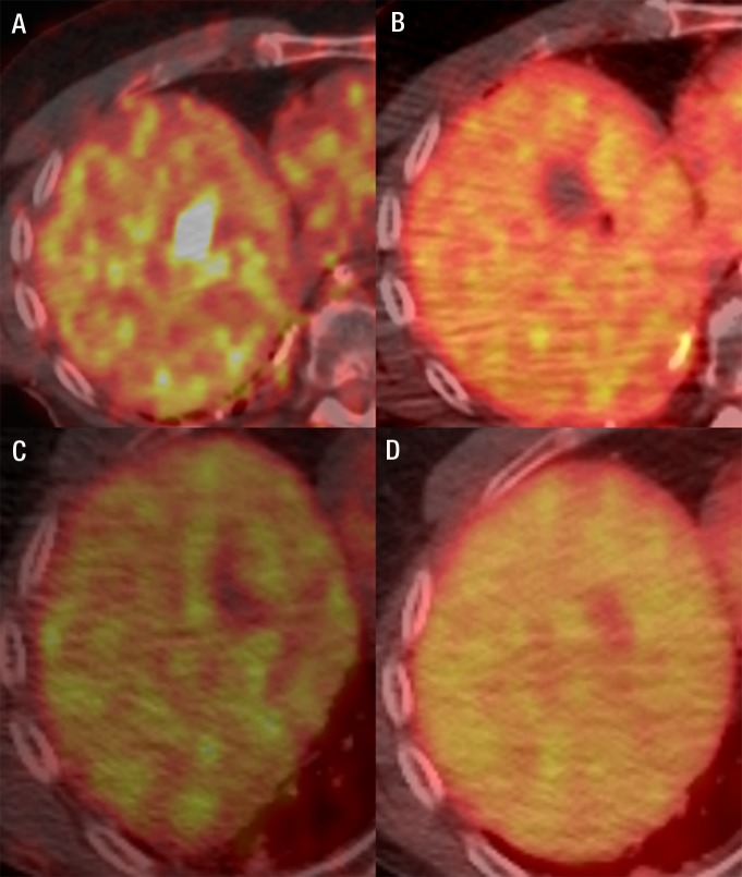
Figure 2:
Fused FDG PET/CT images in a 60-year-old man with metastatic melanoma. A, FDG-avid liver metastasis in the right lobe (standardized uptake value, 18.2). B, Image immediately after microwave ablation shows the photopenic ablation defect. C, Image at 4-week follow-up shows good ablation result. D, Image 5 months after abaltion shows a further decrease of the ablation zone size and no metabolic activity.

