

Abstract
We report a rare case of polymelia in a 6-month-old female child who presented with developed lower limbs and an additional underdeveloped left lower limb.
Keywords: Anatomy, infant, polymelia, radiology
INTRODUCTION

Polymelia is a congenital anomaly, which is defined as the presence of accessory limbs attached to various body regions and could be classified as cephalomelia (extra-limb attached to the head), notomelia (extra-limb attached to the back bone), thoracomelia (extra-limb attached to the thorax), and pyromelia (extra-limb attached to the pelvis). These anomalies are usually associated with genetic factors including tamogenes, chromosomes, and environmental agents. Here, we report a rare case of pyromelia, a form of polymelia.
CASE REPORT
A 6-month-old female presented to us with an extra-underdeveloped lower limb [Figure 1]. She also had Grade 3 weakness in the left lower limb. There was no bladder or bowel incontinence. Plain X-ray demonstrated a normal left hip joint with an accessory left underdeveloped limb. The accessory limb had rudimentary femur falsely attached to normal acetabulam of the left hip joint. Clinically left hip joint movements were normal. Both rudimentary femur and the tibia formed a false knee joint. This joint had no flexion or extension movement. Distal most end of the accessory limb had a single false digit with a single curved false metatarsal [Figure 2]. There was no history of congenital anomalies in the family members. There was no history of teratogenic drug intake during pregnancy by the mother. The patient was lost to follow-up as the family relocated to another city.
Figure 1.
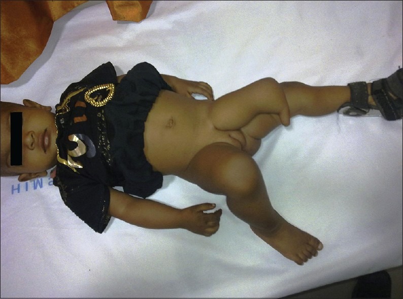
Patient's photograph shows left lower limb polymelia.
Figure 2.
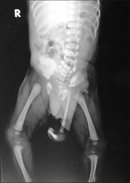
Radiograph of the patient reveals a normal left hip joint with an accessory left underdeveloped limb. The accessory limb has rudimentary femur falsely attaching to normal acetabulam of left hip joint. Both rudimentary femur and the tibia form a false knee joint. Distal most end of the accessory limb shows a single false digit with a single curved false metatarsal.
DISCUSSION
Isolated limb duplication is a rare congenital condition and only a few cases have been documented. Various adverse embryogenic influences are responsible for this kind of anomaly.[1,2,3,4]
Limb differentiation occurs roughly between the 4th and 5th weeks of embryonic development,[5] and it follows a dorsal to ventral and proximal to distal pattern, with many factors involved in the process.[3,4]
Initially, two pairs of limb buds – anterior and posterior – protrude from both sides of the embryo, and comprise cells of ectoderm and mesoderm. Their interaction is responsible for cell positioning and limb differentiation. The covering ectodermal layer of the limb bud is termed the apical ectodermal ridge (AER). The zone of proliferating activity (ZPA), another group of cells is located subjacent to the AER. Both are necessary for limb development. Mesodermal cells in the ZPA stimulate AER formation and the AER maintain the ZPA.[6,7]
The level and manifestation of limb deformity can thus be used to determine the approximate timing of the teratogenic event that occurred during limb development. As the AER grows more distal, the induced mesoderm cells, comprising rudimentary parts of the limb, can continue to grow without any developmental interference even if the AER is transplanted to the adjacent region. This leads to an assumption that duplication of the limb arises from the influence of the AER with abnormal splitting creating two sets of limbs.[8,9]
Other deformities in our patient such as semivertebrae, spina bifida, scoliosis, and hip dislocation were absent. We found only a left club foot.
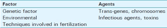
Several factors lead to this anomaly [Table 1]. Over 80% of children born to mothers who took thalidomide had limb defects. These defects ranged from absence of the limb (amelia) or proximal limb elements (phocomelia) to loss of the thumb or digit tip. Women taking hormone (progestrone etc.) during pregnanacy have chances of the structural anomalies like polydactyly, polymelia, and other congenital defects. Antenatal screening with ultrasonography can be a useful tool to diagnose such conditions in utero.
Table 1.
Factors leading to polymelia
Treatment for such a case like ours requires surgery to detach the soft tissue between false limb and the true pelvic region.
CONCLUSION
Radiologist plays a significant role in patients with polymelia to assess for additional congenital anamolies before surgical intervention.
Footnotes
Available FREE in open access from: http://www.clinicalimagingscience.org/text.asp?2013/3/1/18/111235
Source of Support: Nil
Conflict of Interest: None declared.
REFERENCES
- 1.Griffet J, Bastiani-Griffet F, Jund S, Moreigne M, Zabjek KF. Duplication of the leg - Renal agenesis: Congenital malformation syndrome. J Pediatr Orthop B. 2000;9:306–8. doi: 10.1097/01202412-200010000-00015. [DOI] [PubMed] [Google Scholar]
- 2.La Torre R, Fusaro P, Anceschi MM, Montanino-Oliva M, Modesto S, Cosmi EV. Unusual case of caudal duplication (dipygus) J Clin Ultrasound. 1998;26:163–5. doi: 10.1002/(sici)1097-0096(199803/04)26:3<163::aid-jcu10>3.0.co;2-c. [DOI] [PubMed] [Google Scholar]
- 3.Kher AS, Gahankari DR, Tambwekar SR, Doraiswamy A, Iyer S, Bharucha BA, et al. Supernumerary limbs: A case report of a rare congenital anomaly. Ann Plast Surg. 1996;37:549–52. doi: 10.1097/00000637-199611000-00016. [DOI] [PubMed] [Google Scholar]
- 4.Kojima T, Hirakawa M, Hirase Y, Hwang KH. Complete congenital duplication with incomplete separation of a lower extremity. Plast Reconstr Surg. 1993;91:926–9. doi: 10.1097/00006534-199304001-00032. [DOI] [PubMed] [Google Scholar]
- 5.O’Rahilly R, Muller F. Developmental Stages in Human Embryos. Washington: Carnegie Institution; 1987. Publication No 637. [Google Scholar]
- 6.Roberts DJ, Tabin C. The genetics of human limb development. Am J Hum Genet. 1994;55:1–6. [PMC free article] [PubMed] [Google Scholar]
- 7.Scherz PJ, Harfe BD, McMahon AP, Tabin CJ. The limb bud Shh-Fgf feedback loop is terminated by expansion of former ZPA cells. Science. 2004;305:396–9. doi: 10.1126/science.1096966. [DOI] [PubMed] [Google Scholar]
- 8.Packard DS Jr, Levinsohn EM, Hootnick DR. Extent of duplication in lower-limb malformations suggests the time of the teratogenic insult. Pediatrics. 1993;91:411–3. [PubMed] [Google Scholar]
- 9.Zhao L, Li MQ, Sun XT, Ma ZS, Guo G, Huang YT. Congenital lumbosacral limb duplication: A case report. J Orthop Surg (Hong Kong) 2006;14:187–91. doi: 10.1177/230949900601400216. [DOI] [PubMed] [Google Scholar]