Abstract
Purpose
The aim of this study was to investigate eccentric torque production capacity of the ankle, knee and hip muscle groups in patients with unilateral chronic ankle instability (CAI) as compared to healthy matched controls.
Methods
In this case-control study, 40 participants (20 with CAI and 20 controls) were recruited based on convenient non-probability sampling. The average peak torque to body weight (APT/BW) ratio of reciprocal eccentric contraction of ankle dorsi flexor/plantar flexor, ankle evertor/invertor, knee flexor/extensor, hip flexor/extensor and hip abductor/adductor was determined using an isokinetic dynamometer. All subjects participated in two separate sessions with a rest interval of 48 to 72 hours. In each testing session, the torque production capacity of the ankle, knee, and hip muscle groups of only one lower limb was measured. At first, 3 repetitions of maximal eccentric-eccentric contraction were performed for the reciprocal muscles of a joint in a given movement direction. Then, the same procedure of practice and testing trials was repeated for the next randomly-ordered muscle group or joint of the same limb.
Results
There was no significant interaction of group (CAI and healthy controls) by limb (injured and non-injured) for any muscle groups. Main effect of limb was not significant. Main effect of group was only significant for eccentric torque production capacity of ankle dorsi flexor and hip flexor muscle groups. The APT/BW ratio of these muscles was significantly lower in the CAI group than the healthy controls (P<0.05).
Conclusion
CAI is associated with eccentric strength deficit of ankle dorsi flexor and hip flexor muscles as indicated by reduction in torque production capacity of these muscles compared to healthy controls. This strength deficit appeared to exist in both the injured and non-injured limbs of the patients.
Keywords: Eccentric Strength, Lower Limb, Chronic Ankle Instability
INTRODUCTION
Lateral ankle sprain is the most common type of ankle injury among both general and athlete populations [1]. Ankle injuries account for 10-30% of all sports injuries and it has been reported that approximately 80% of ankle sprains lead to injury recurrence and instability [1, 2]. Ankle injuries have caused more participation restriction than any other single sport injuries [2].
Chronic ankle instability (CAI) is attributed to the contribution of mechanical and functional instability in the development of residual pain, giving way, and disability of patients suffering from the initial acute ankle sprain [3–6]. The high prevalence [7] and rate of recurrence and instability followed by ankle sprain [5] has motivated the researchers and clinicians to identify some relevant factors associated with CAI. Factors such as ligamentous laxity, proprioceptive impairment, balance instability, and strength deficit (especially ankle evertors) have been hypothesized as potential causes of CAI [6, 8–13], among them ankle muscle weakness is the most questionable factor [11]. Identifying the potential contribution of these factors is important for the patients’ return to pre-injury daily/sports activity and also to prevent recurrence and instability associated with the CAI [5].
Muscle strength evaluation and training are important parts of rehabilitation of individuals with musculoskeletal disorders [6, 8, 11]. Ankle muscle strength has been studied extensively in patients suffering from CAI. Most of these studies have focused on the strength of evertor and invertor muscle groups [7, 10–14] and some have tested strength of dorsi flexors and plantar flexors [5, 9, 15]. However the results are inconclusive: while some studies reported isokinetic strength deficit for the evertor [7, 10, 13], invertor [7, 10], dorsi flexor [9, 15], and plantar flexor [5] in CAI; others failed to find any significant muscle weakness between the two study groups [11, 12, 14].
In addition to the above case-control studies in which healthy participants served as a control group, other studies that used the non-injured ankles as the control [16–19] have also reported inconclusive results. The reason for these conflicting results is not clear. Variations in contraction mode (concentric vs. eccentric), velocity of the tests (slow vs. fast), and methodological limitations (e.g. small sample size) have been addressed as possible reasons [8].
Optimal function of the lower limb during weight bearing and closed chain activities requires both concentric and eccentric contraction of the involved muscles in order to minimize ground reaction forces imposed on the ankle-foot complex [6, 11]. For example, lateral ankle sprain occurs when the ankle is forced into plantar flexion/inversion; therefore eccentric activity of ankle dorsiflexor and evertor muscle groups is necessary to prevent this injury [10, 12–14]. For this reason, clinicians and researchers should focus on evaluation and training of eccentric as well as concentric contractions [7, 9] in musculoskeletal disorders including CAI.
Moreover, synergistic muscle activity of the hip, knee and ankle joints is necessary for appropriate gait mechanics and foot positions during heel strike [20, 21]. Therefore, from a biomechanical perspective, dysfunction of a single joint could influence the others in the kinetic chain [6, 22, 23]. Despite this relationship, to our knowledge, there are only two studies investigating the strength of the muscles acting on knee and hip joints in patients with CAI [5, 21]. In these studies, only isometric strength of the hip abductor/extensor [21] and concentric strength of the knee and hip flexor/extensor [5] were investigated using hand-held and isokinetic dynamometers, respectively. Observation of isometric strength weakness of hip abductor and concentric strength deficit of knee flexor/extensor in the CAI could support the notion of inter-joint interaction of the lower limb kinetic chain secondary to a single joint injury.
Given the conflicting results on the ankle strength deficit and limited research on muscle weakness of the knee and hip muscles in CAI, it is necessary to conduct a more comprehensive study to evaluate torque production capacity of all muscle groups acting on hip, knee and ankle. Moreover, eccentric torque of the knee and hip muscle groups has not yet been investigated in the CAI. Therefore, the purpose of this study was to investigate eccentric torque production capacity of the ankle, knee, and hip muscle groups in patients suffering from CAI as compared to healthy matched controls. Research on this topic may provide important insight on designing an appropriate rehabilitative intervention to restore functional performance and prevent injury and instability followed by lateral ankle sprain.
METHODS AND SUBJECTS
Subjects
The sample consisted of 40 participants (20 with CAI and 20 controls) between the age of 18 and 35. All participants provided a written informed consent prior for participation in this investigation.
Sample size determination in the current research was done based on the approach suggested by Campbell et al for repeated measure designs [24]. For this purpose, we focused on the findings of Gribble and Robinson for concentric strength data of the ankle, knee and hip muscle groups in patients with CAI [5]. A sample of 33 participants (16.5 per group) was required to provide 90% power (β = 0.10) with a significance level of 0.05 for detecting effect size equal to 1.1 for each variable based on a repeated measure design with one between-group factor. The planned total study size was finally considered as 40 subjects (20 per group).
Subject recruitment from orthopedic and physiotherapy clinics was based on the medical records of patients who had been seeking medical care for “acute ankle sprain” in the past year. Inclusion criteria were: (1) experience of at least one unilateral acute ankle sprain without fracture which resulted in pain, swelling, and temporary use of crutches (but not in the past 3 months) [5, 21]; (2) at least two episodes of the ankle “giving way” in the past 6 months [5, 11]; (3) no participation in the rehabilitation program at the time of study [5, 7, 11, 21]; and (4) no evidence of mechanical instability [25] as evaluated by an orthopaedist using anterior drawer test [11] or talar tilt test [3]. All patients were pain free and were able to perform full weight bearing on both legs at the time of assessment [7, 11, 14, 21]. Patients were also excluded if they reported history of lower limb injury other than the unilateral CAI.
To evaluate functional limitation, all patients completed the foot and ankle ability measure (FAAM) questionnaire [26]. The FAAM is a 29-item self-reported instrument with two main subscales: activity of daily living (ADL) with 21 items and sport with 8 items. Total scores of ADL and sports were transformed to percentage for which higher scores represent higher level of functional status. An Iranian version of FAAM has been validated for use in our country [26]. To match the activity level of two study groups, the ankle activity score (AAS) [27] was completed by all participants. The AAS is an adapted version of Tegner activity rating scale [28] which has been originally developed for knee ligament injuries. Similar to the Tegner activity rating scale, AAS has 11 items with a scoring range of 0-10; higher scores indicating higher level of physical activity. The Persian version of the Tegner has acceptable psychometric properties for use in patients with anterior cruciate ligament injury [28].
Both patients and healthy participants were excluded if they had (self-report) any history of vestibular dysfunction or head injury in the previous 6 months [5], back pain, discopathy, radicular pain, prior surgery in the back or lower limbs [21], recent pregnancy for the female participants and history of neurological disorders [21]. Two groups of CAI and controls were matched according to gender (15 female, 5 male), age, height, body mass index, and activity level.
Procedures
This study was approved by Institutional Review Board (ETH-073) of Ahvaz Jundishapur University of Medical Sciences. The Biodex System IV isokinetic dynamometer and Biodex Software Package (Biodex Inc., Shirley. NY, USA) was used to determine the average peak torque to body weight (APT/BW) ratio of reciprocal eccentric strength of ankle dorsi flexor/plantar flexor, ankle evertor/invertor, knee flexor/extensor, hip flexor/extensor, and hip abductor/adductor muscles. Normalizing APT to BW allows for comparing the participants despite body-weight differences [6, 13]. Reliability of isokinetic variables for assessing muscle strength of ankle dorsi flexor (intra-class correlation coefficient of 0.77 to 0.93) and ankle plantar flexor (intra-class correlation coefficient of 0.78 to 0.95) has been reported in the literature [29]. Furthermore, the recent data available on muscle strength studies of CAI patients reveal that APT/BW ratio is the most commonly used isokinetic parameter [5, 9]. Therefore, to increase the comparability of the current study, the authors decided to report the results of APT/BW.
All subjects participated in two separate sessions with a rest interval of 48 to 72 hours. In each testing session, the torque production capacity of the ankle, knee, and hip muscle groups of only one randomly-ordered lower limb was assessed. The order of joints and their motion direction were also randomized for each participant. The injured limb of the patient group was matched with the corresponding dominant/non- dominant limb of healthy controls [9]. Dominant limb was defined as a limb used to kick a ball.
At first, 3 practice repetitions of sub-maximal and 3 practice trials of maximal contractions were performed to familiarize each participant with the equipment and procedure [11]. After a two minute rest, three repetitions of maximal eccentric-eccentric contraction (reactive eccentric mode) with no rest were performed by the reciprocal muscles of a joint in a given movement direction. A two minute rest was considered between the practice and test trials to avoid potential fatigue [5]. The highest peak torque value was recorded for each of the 3 repetitions and the average of these values was considered as APT [5]. After a five minute rest period, the same procedure was repeated for the next randomly-ordered muscle group or joint of the same limb. Participants were given verbal encouragement throughout the isokinetic muscle testing procedure [7, 11, 13]. All practice and testing trials were performed at the angular velocity of 60o.s-1 [5]. The literature suggests that strength testing must be performed in a velocity between 30o.s-1 and 240o.s-1 [8]. Moreover, choosing a slower velocity within this range would increase the chance of detecting possible differences in torque production capacity [8]. Angular velocity of 60o.s-1 has been used in some literature aiming to assess lower limb strength of patients suffering from CAI [5, 18].
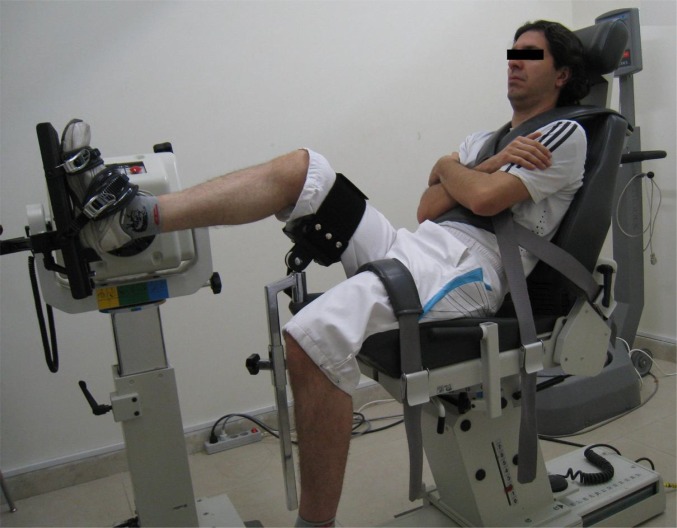
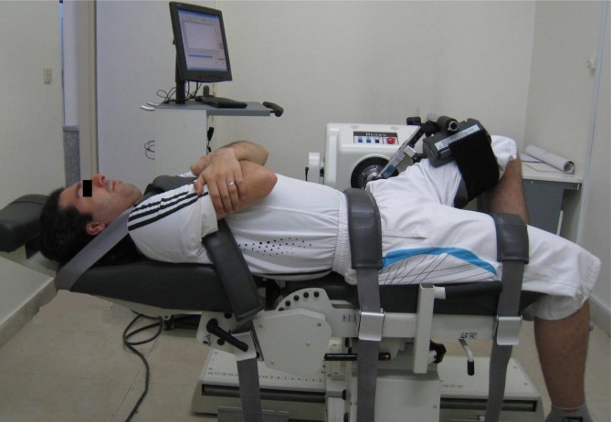
Patients were positioned based on the manufacturer guidelines for assessing strength testing of the hip, knee, and ankle in various directions. Eccentric-eccentric contraction of ankle dorsi flexor/plantar flexor was performed in a sitting position in which the isokinetic chair was in a semi-recumbent position and the knee of the ipsilateral limb was in 200-300 flexion (Fig. 1). Ankle evertor/invertor strength was assessed in a similar way in which the subject was seated in a semi-recumbent position [11, 13] while the knee of the ipsilateral limb was in 300-450 flexion. The movements were performed within the active range of motion, i.e. about 0-30 degree of dorsi flexion, 0-50 degree of plantar flexion, 0-40 degree of eversion and 0-55 degree of inversion. Eccentric strength of knee flexor/extensor was assessed as participants moved their knee from 900 flexion in to near full extension in the sitting position, while the hip joints were in 900 flexion [20]. For testing the hip flexor/extensor and hip abductor/adductor, participants were placed in the supine and side position, respectively (Fig. 2). The test was performed within the available active range of motion from the neutral position of the hip joint, i.e. about 0-120 degrees of flexion and 0-45 degrees of abduction.
Fig. 1.
Isokinetic testing of ankle dorsi flexor-plantar flexor
Fig. 2.
Isokinetic testing of hip flexor-extensor
Statistical Analyses
Data were analyzed using Statistical Package for the Social Sciences (SPSS version 16) and the level of statistical significance was set at p<0.05. For each joint direction, a separate 2 × 2 (2 groups by 2 limbs) mixed model of analysis of variance (ANOVA) test was used to determine main effects and interactions of group and limb factors for each APT/BW ratio.
RESULTS
Table 1 shows that there were no significant differences between the two study groups regarding the age, height, body mass index and activity level. Table 2 shows the mean and standard deviation (SD) for the APT/BW ratio of the ankle, knee and hip muscle groups in the CAI and healthy participants. A summary of ANOVA results is shown in Table 3.
Table 1.
Demographic and functional characteristics of CAI and healthy groups
| Demographic data | CAI group (n = 20) | Healthy group (n = 20) | P. value |
|---|---|---|---|
| Mean (SD) | Mean (SD) | (Mean difference) | |
| Age (yr) | 25.1 (4.52) | 24.7 (4.57) | 0.78 |
| Height (m) | 1.68 (0.75) | 1.68 (0.62) | 0.98 |
| Body mass index(kg/m 2 ) | 22.5 (2.79) | 22.6 (2.12) | 0.91 |
| Time since last lateral ankle sprain (month) | 6.65 (3.43) | not applicable | - |
| Frequency of giving way (in the past 6 month) | 7.50 (5.85) | not applicable | - |
| Ankle activity score * | 7.05 (2.16) | 6.35 (1.95) | 0.29 |
| foot and ankle ability measure † | |||
| Activity of daily living | 91.4 (6.33) | not applicable | - |
| Sports | 80.4 (13.5) | not applicable | - |
Range of scores is from 0-10
Range of scores is from 0-100
CAI: chronic ankle instability; SD: Standard deviation
Table 2.
Mean (SD) of average peak torque to body weight ratio (N.m-1.kg-1) for the ankle, knee and hip muscle groups in both of the CAI and healthy participants
| Parameter | Average Torque to Body Weight Ratio | ||
|---|---|---|---|
| CAI | Healthy | ||
| Isokinetic testing the injured limb (and its matched limb in the control group) | Ankle Dorsi-flexor | 0.51 (0.08) | 0.59 (0.11) |
| Ankle Plantar-flexor | 1.17 (0.39) | 1.40 (0.39) | |
| Ankle Evertor | 0.33 (0.09) | 0.38 (0.11) | |
| Ankle Invertor | 0.36 (0.11) | 0.40 (0.09) | |
| Knee Flexor | 1.23 (0.27) | 1.36 (0.38) | |
| Knee Extensor | 2.40 (0.48) | 2.58 (0.75) | |
| Hip Flexor | 1.41 (0.59) | 1.94 (0.93) | |
| Hip Extensor | 2.24 (0.73) | 2.51 (0.64) | |
| Hip Abductor | 1.21 (0.50) | 1.33 (0.50) | |
| Hip Adductor | 1.78 (0.55) | 1.74 (0.37) | |
| Isokinetic testing the non-injured limb (and its matched limb in the control group) | Ankle Dorsi-flexor | 0.53 (0.15) | 0.61 (0.10) |
| Ankle Plantar-flexor | 1.14 (0.47) | 1.39 (0.40) | |
| Ankle Evertor | 0.33 (0.11) | 0.36 (0.08) | |
| Ankle Invertor | 0.37 (0.13) | 0.40 (0.08) | |
| Knee Flexor | 1.29 (0.34) | 1.33 (0.37) | |
| Knee Extensor | 2.55 (0.65) | 2.53 (0.78) | |
| Hip Flexor | 1.41 (0.54) | 1.96 (0.98) | |
| Hip Extensor | 2.25 (0.69) | 2.49 (0.74) | |
| Hip Abductor | 1.16 (0.44) | 1.28 (0.45) | |
| Hip Adductor | 1.69 (0.52) | 1.76 (0.41) | |
CAI: chronic ankle instability; SD: Standard deviation
Table 3.
Summary of analysis of variance for the average peak torque to body weight ratio of the ankle, knee and hip muscle groups: F-ratios and P-values by variable
| Parameter | Main effect | Interaction | ||||
|---|---|---|---|---|---|---|
| Limb | Group | Limb×Group | ||||
| F | P | F | P | F | P | |
| Ankle dorsi-flexor | 1.16 | 0.3 | 5.46 | 0.02 | 0.00 | 1 |
| Ankle plantar-flexor | 0.18 | 0.7 | 3.61 | 0.06 | 0.10 | 0.7 |
| Ankle evertor | 0.22 | 0.6 | 1.88 | 0.2 | 0.38 | 0.5 |
| Ankle invertor | 0.04 | 0.8 | 1.25 | 0.3 | 0.31 | 0.6 |
| Knee flexor | 0.16 | 0.7 | 0.75 | 0.4 | 0.89 | 0.3 |
| Knee extensor | 0.56 | 0.4 | 0.16 | 0.7 | 2.51 | 0.1 |
| Hip flexor | 0.01 | 0.9 | 5.04 | 0.03 | 0.05 | 0.8 |
| Hip extensor | ≤0.01 | 0.95 | 1.42 | 0.24 | 0.02 | 0.86 |
| Hip abductor | 1.17 | 0.28 | 0.65 | 0.42 | 0.00 | 0.98 |
| Hip adductor | 0.58 | 0.45 | ≤0.01 | 0.93 | 1.22 | 0.27 |
There was no significant interaction of group (CAI and healthy controls) by limb (injured and non-injured) for any muscle groups. Main effect of limb was not significant, meaning that the eccentric APT/BW ratio of the injured limb muscles was not different from that of a non-injured limb. Similar finding was obtained when comparing dominant and non-dominant limbs of the healthy control group.
Main effect of group was only significant for eccentric torque production capacity of ankle dorsi flexor and hip flexor muscle groups. APT/BW ratio of these muscles was significantly lower in the CAI group than the healthy controls. The effect size for this main finding was calculated and based on Cohen's suggestion [30], large and medium effect sizes were detected for the between-group differences of ankle dorsi-flexor (effect size= 0.81) and hip flexor (effect size= 0.67) muscle groups, respectively.
DISCUSSION
The results of this study showed that CAI is associated with eccentric strength deficit of ankle dorsi flexors and hip flexors as indicated by reduction in torque production capacity of these muscle groups in patients with CAI compared with healthy matched controls.
This strength deficit appeared to exist in both the injured and non-injured limbs of the patients. Similar findings were seen in the literature focusing on strength evaluation of CAI when compared with healthy matched controls [9, 14, 31]. This finding could be attributed to “cross-training” or “cross-over” phenomena in which strength change of one limb could affect the strength of the contra-lateral limb [9]. The likelihood of a bilateral deficit of eccentric torque production preceded or followed by the ankle injury addresses an important limitation for the findings of those studies that use the non-injured limb as healthy control limb in their study [14, 31, 32]. This may lead to misinterpretation of the results obtained in these studies.
Eccentric torque of ankle muscle groups
Because there are a few studies that assessed eccentric torque of ankle dorsi flexor/plantar flexor [9], comparability of our findings is limited. Fox et al (2008) in a recent investigation on 20 patients with functional ankle instability found lower eccentric torque value of ankle plantar flexor in the patients as compared to healthy controls [9]. It is necessary to note that they found eccentric torque reduction in ankle “plantar flexion movement” of the injured limb and this would be related to eccentric torque deficit of ankle “dorsi flexor muscles”. However, the authors discussed about the finding of eccentric torque deficit of ankle “plantar flexor muscles”. If we assume eccentric torque deficit of ankle dorsi flexor in the Fox et al study, one may conclude that this deficit is associated with recurrent ankle sprain in the CAI patients. Considering the fact that most ankle sprains occur during the forced movement of ankle plantar flexion/inversion, adequate eccentric strength of ankle dorsi flexor and evertor is necessary to prevent this injury [15]. Therefore, eccentric strength deficit of these muscles is believed to predispose individuals with CAI to injury or recurrent sprains. Alternatively, several mechanisms such as muscle atrophy and muscle reflex inhibition secondary to initial ankle injury [9, 12] could explain the eccentric strength deficit of the ankle dorsi flexor in this patient population.
Based on the injury mechanism described for the ankle sprain, it was expected to observe eccentric strength deficit of ankle evertor in CAI. However, we did not find any significant differences between two study groups. Some investigators have focused on examination of eccentric torque values of ankle evertor/invertor in CAI [7, 9–11, 13, 14]. Similar to the results of the current study, some have found no eccentric strength deficit in CAI [9, 11, 14] while the others have reported a significant decrement in eccentric torque production capacity of patients compared to healthy controls [7, 10, 13]. The reason for these inconsistencies is not clear. However, several factors such as heterogeneity of sample sizes, differences in movement velocity and selection of patients with different disability levels could partially explain these inconsistencies. None of the studies that have considered non-injured ankles as controls for comparison, have found a deficit in eccentric torque production of ankle evertors of the injured compared to non-injured ankles [16–19]. On the other hand, concentric and/or eccentric torque production capacity of ankle inventors was lower in the injured compared to non-injured ankles. Consistent with the later findings, a recent systematic review demonstrated concentric invertor torque deficit in patients with recurrent ankle sprain [33]. These authors acknowledged that this finding was not expected. However, several mechanisms such as selective reflex inhibition of the ankle invertors and deep peroneal nerve stretching secondary to forced inversion movement have been theorized by some investigators to explain this finding [18, 19].
Eccentric torque of knee and hip muscle groups
Only a limited number of researches have focused on the knee and hip torque production capacity in individuals suffering from ankle sprain [5, 21]. Gribble and Robinson (2009) examined the concentric torque of ankle, knee and hip muscle groups in the sagittal plane and found a strength deficit in knee flexor/extensors in addition to ankle plantar flexors [5]. In another study, Friel et al (2006) in an attempt to find a relationship between CAI and hip muscle strength (hip extensor and abductor) using a hand-held dynamometer, reported lower strength in the hip abductor muscle of the injured limb relative to the non-injured limb [21]. They had no healthy control group in their study and comparisons were made between the injured and non-injured limbs of the CAI group [21].
Eccentric torque examination of hip and knee muscle groups in the CAI patients has not been studied yet, so it was not possible for us to compare our results with other studies. However, other functions of proximal muscles such as muscle activation pattern have been investigated in previous studies on patients with inversion ankle sprain [34, 35]. The authors have observed a delay in activation of hip extensor muscles during hip extension in the patients compared to healthy controls. It has been hypothesized that failure in mechanoreceptor afferent inputs (dys-afferentation) from the distal injured joint could affect the efferent motor outputs (dys-efferentation) of the proximal limb muscles [5, 34]. Deficit in eccentric torque production capacity of hip flexor in patients with CAI might be attributed to the role of this muscle during inversion ankle sprain. We hypothesized that the ankle sprain occurs when the lower limb is in the position of slight hip extension and at the same time the ankle forced into the plantar flexion/inversion. Therefore, adequate eccentric strength of hip flexor is necessary to prevent this injury. Based on this hypothesis, we searched for the position of the proximal joints of the lower limb (especially hip position) during ankle sprain and found no study to explain this issue. This issue could be considered as a possible topic to investigate in future projects.
Limitations and recommendations
Due to the cross-sectional design of the present study, it is not clear whether the observed deficit in eccentric torque of ankle dorsi flexor and hip flexor muscle groups is a preexistent and a possible causal factor, or a consequence of the ankle sprain. The design of the present study did not also allow determining the clinimetric properties of isokinetic dynamometry. To overcome the above limitations, future studies should focus on exploring the cause and effect relationship. Furthermore, as the assessment of peak torque by isokinetic system is subjected to human error, future studies should determine the reliability of these measures.
CONCLUSION
The findings showed that CAI is associated with eccentric strength deficit of ankle dorsi flexor and hip flexor muscles as indicated by reduction in torque production capacity of these muscles compared to healthy controls. This strength deficit appeared to exist in both the injured and non-injured limbs of the patients. Therefore, we suggest that the clinicians and researchers focus not only on the strength evaluation of the injured joint (i.e. ankle dorsi flexor), but also on other joints of the kinetic chain (i.e. hip flexor) when faced with CAI.
ACKNOWLEDGMENTS
This study is part of M.Sc thesis of Mrs. Moradi. Special thanks to Ahvaz Jundishapur University of Medical Sciences for the financial support (Master thesis grant no: PHT-8902). Also, we acknowledge all participants for their time devoted to participation in this study.
Conflict of interests: None
REFERENCES
- 1.Fong DT, Hong Y, Chan LK, Yung PS, Chan KM. A systematic review on ankle injury and ankle sprain in sports. Sports Med. 2007;37:73–94. doi: 10.2165/00007256-200737010-00006. [DOI] [PubMed] [Google Scholar]
- 2.Garrick JG. The frequency of injury, mechanism of injury, and epidemiology of ankle sprains. Am J Sports Med. 1977;5:241–2. doi: 10.1177/036354657700500606. [DOI] [PubMed] [Google Scholar]
- 3.Delahunt E. Neuromuscular contributions to functional instability of the ankle joint. J Bodyw Mov Ther. 2007;11:203–13. [Google Scholar]
- 4.Delahunt E, Coughlan GF, Caulfield B, et al. Inclusion criteria when investigating insufficiencies in chronic ankle instability. Med Sci Sports Exerc. 2010;42:2106–21. doi: 10.1249/MSS.0b013e3181de7a8a. [DOI] [PubMed] [Google Scholar]
- 5.Gribble PA, Robinson RH. An examination of ankle, knee, and hip torque production in individuals with chronic ankle instability. J Strength Cond Res. 2009;23:395. doi: 10.1519/JSC.0b013e31818efbb2. [DOI] [PubMed] [Google Scholar]
- 6.Kaminski TW, Hartsell HD. Factors contributing to chronic ankle instability:a strength perspective. J Athl Train. 2002;37:394. [PMC free article] [PubMed] [Google Scholar]
- 7.Yildiz Y, Aydin T, Sekir U, et al. Peak and end range eccentric evertor/concentric invertor muscle strength ratios in chronically unstable ankles:comparison with healthy individuals. J Sports Sci Med. 2003;2:70–6. [PMC free article] [PubMed] [Google Scholar]
- 8.Arnold BL, Linens SW, Sarah J, Ross SE. Concentric evertor strength differences and functional ankle instability:a meta-analysis. J Athl Train. 2009;44:653. doi: 10.4085/1062-6050-44.6.653. [DOI] [PMC free article] [PubMed] [Google Scholar]
- 9.Fox J, Docherty CL, Schrader J, Applegate T. Eccentric plantar-flexor torque deficits in participants with functional ankle instability. J Athl Train. 2008;43:51. doi: 10.4085/1062-6050-43.1.51. [DOI] [PMC free article] [PubMed] [Google Scholar]
- 10.Hartsell HD, Spaulding SJ. Eccentric/concentric ratios at selected velocities for the invertor and evertor muscles of the chronically unstable ankle. Br J Sports Med. 1999;33:255. doi: 10.1136/bjsm.33.4.255. [DOI] [PMC free article] [PubMed] [Google Scholar]
- 11.Kaminski TW, Perrin DH, Gansneder BM. Eversion strength analysis of uninjured and functionally unstable ankles. J Athl Train. 1999;34:239. [PMC free article] [PubMed] [Google Scholar]
- 12.Rottigni SA, Hopper D. Peroneal muscle weakness in female basketballers following chronic ankle sprain. Aust J Physiother. 1991;37:211–7. doi: 10.1016/S0004-9514(14)60541-9. [DOI] [PubMed] [Google Scholar]
- 13.Willems T, Witvrouw E, Verstuyft J, et al. Proprioception and muscle strength in subjects with a history of ankle sprains and chronic instability. J Athl Train. 2002;37:487. [PMC free article] [PubMed] [Google Scholar]
- 14.Bernier JN, Perrin DH, Rijke A. Effect of unilateral functional instability of the ankle on postural sway and inversion and eversion strength. J Athl Train. 1997;32:226. [PMC free article] [PubMed] [Google Scholar]
- 15.Naicker M, McLean M, Esterhuizen TM, Peters-Futre EM. Poor peak dorsiflexor torque associated with incidence of ankle injury in elite field female hockey players. J Sci Med Sport. 2007;10:363–71. doi: 10.1016/j.jsams.2006.11.007. [DOI] [PubMed] [Google Scholar]
- 16.Bernier JN, Perrin DH. Effect of coordination training on proprioception of the functionally unstable ankle. J Orthop Sports Phys Ther. 1998;27:264–75. doi: 10.2519/jospt.1998.27.4.264. [DOI] [PubMed] [Google Scholar]
- 17.Heitman RJ, Kovaleski J, Gurchiek L. Isokinetic eccentric strength of the ankle evertors after injury. Percept Motor Skills. 1997;84:258. doi: 10.2466/pms.1997.84.1.258. [DOI] [PubMed] [Google Scholar]
- 18.Munn J, Beard DJ, Refshauge KM, Lee RY. Eccentric muscle strength in functional ankle instability. Med Sci Sports Exerc. 2003;35:245–50. doi: 10.1249/01.MSS.0000048724.74659.9F. [DOI] [PubMed] [Google Scholar]
- 19.Sekir U, Yildiz Y, Hazneci B, et al. Effect of isokinetic training on strength, functionality and proprioception in athletes with functional ankle instability. Knee Surg Sports Traumatol Arthrosc. 2007;15:654–64. doi: 10.1007/s00167-006-0108-8. [DOI] [PubMed] [Google Scholar]
- 20.Calmels PM, Nellen M, van der Borne I, et al. Concentric and eccentric isokinetic assessment of flexorextensor torque ratios at the hip, knee, and ankle in a sample population of healthy subjects. Arch Phys Med Rehab. 1997;78:1224–30. doi: 10.1016/s0003-9993(97)90336-1. [DOI] [PubMed] [Google Scholar]
- 21.Friel K, McLean N, Myers C, Caceres M. Ipsilateral hip abductor weakness after inversion ankle sprain. J Athl Train. 2006;41:74–8. [PMC free article] [PubMed] [Google Scholar]
- 22.Gribble PA, Hertel J, Denegar CR, Buckley WE. The effects of fatigue and chronic ankle instability on dynamic postural control. J Athl Train. 2004;39:321–9. [PMC free article] [PubMed] [Google Scholar]
- 23.Leetun DT, Ireland ML, Willson JD, et al. Core stability measures as risk factors for lower extremity injury in athletes. Med Sci Sports Exerc. 2004;36:926–34. doi: 10.1249/01.mss.0000128145.75199.c3. [DOI] [PubMed] [Google Scholar]
- 24.Campbell MJ, Donner A, Klar N. Developments in cluster randomized trials and Statistics in Medicine. Stat Med. 2007;26:2–19. doi: 10.1002/sim.2731. [DOI] [PubMed] [Google Scholar]
- 25.Rahnama L, Salavati M, Akhbari B, Mazaheri M. Attentional demands and postural control in athletes with and without functional ankle instability. J Orthop Sports Phys Ther. 2010;40:180–7. doi: 10.2519/jospt.2010.3188. [DOI] [PubMed] [Google Scholar]
- 26.Mazaheri M, Salavati M, Negahban H, et al. Reliability and validity of the Persian version of Foot and Ankle Ability Measure (FAAM) to measure functional limitations in patients with foot and ankle disorders. Osteoarthritis Cartilage. 2010;18:755–9. doi: 10.1016/j.joca.2010.03.006. [DOI] [PubMed] [Google Scholar]
- 27.Halasi T, Kynsburg Ã, TÃ!llay A, Berkes I. Development of a new activity score for the evaluation of ankle instability. Am J Sports Med. 2004;32:899–908. doi: 10.1177/0363546503262181. [DOI] [PubMed] [Google Scholar]
- 28.Negahban H, Mostafaee N, Sohani SM, et al. Reliability and validity of the Tegner and Marx activity rating scales in Iranian patients with anterior cruciate ligament injury. Disabil Rehabil. 2011;33:2305–10. doi: 10.3109/09638288.2011.570409. [DOI] [PubMed] [Google Scholar]
- 29.McKnight C, Armstrong C. The role of ankle strength in functional ankle instability. J Sport Rehabil. 1997;6:21–9. [Google Scholar]
- 30.Cohen J. 2nd ed. Hillsdale, NJ: Lawrene Erlbaum Assoc; 1988. Statistical Power Analysis for The Behavioral Sciences. [Google Scholar]
- 31.Lentell G, Katzman LL, Walters MR. The Relationship between Muscle Function and Ankle Stability. J Orthop Sports Phys Ther. 1990;11:605–11. doi: 10.2519/jospt.1990.11.12.605. [DOI] [PubMed] [Google Scholar]
- 32.Lentell G, Baas B, Lopez D, et al. The contributions of proprioceptive deficits, muscle function, and anatomic laxity to functional instability of the ankle. J Orthop Sports Phys Ther. 1995;21:206–15. doi: 10.2519/jospt.1995.21.4.206. [DOI] [PubMed] [Google Scholar]
- 33.Hiller CE, Nightingale EJ, Lin CW, et al. Characteristics of people with recurrent ankle sprains:a systematic review with meta-analysis. Br J Sports Med. 2011;45:660–72. doi: 10.1136/bjsm.2010.077404. [DOI] [PubMed] [Google Scholar]
- 34.Bullock-Saxton JE. Local sensation changes and altered hip muscle function following severe ankle sprain. Phys Ther. 1994;74:17–28. doi: 10.1093/ptj/74.1.17. [DOI] [PubMed] [Google Scholar]
- 35.Bullock-Saxton JE, Janda V, Bullock MI. The influence of ankle sprain injury on muscle activation during hip extension. Int J Sports Med. 1994;15:330–4. doi: 10.1055/s-2007-1021069. [DOI] [PubMed] [Google Scholar]


