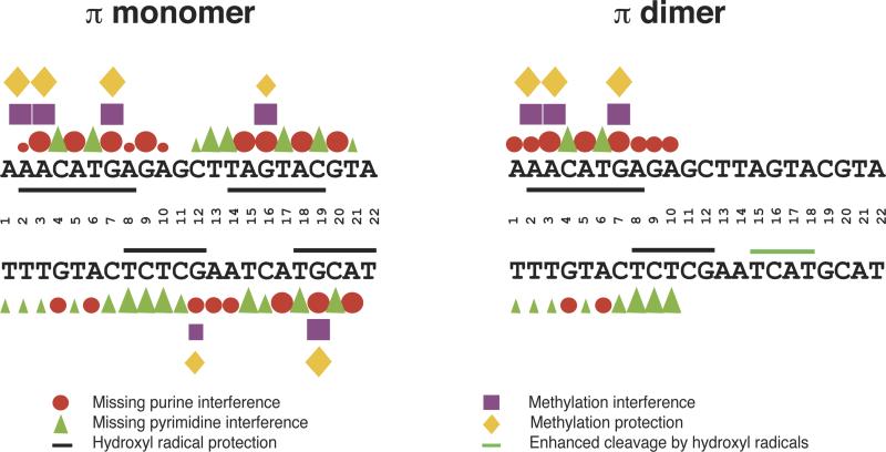
Figure 3.
Iteron DNA sequence (22-bp) and the compilation of base-specific contact probing data for π binding to the iteron. Red circles (purines) and green triangles (pyrimidines) represent bases that affect π binding when removed. Purple squares represent purines whose modification by DMS weakens π binding. Yellow diamonds represent purines that are protected, by bound π protein, from methylation by DMS. In each case, large symbols are indicative of strong effects; medium and small symbols indicate moderate and weak effects, respectively. Black lines represent protection of the DNA backbone, by π, from hydroxyl radical cleavage, and a green line represents enhanced cleavage by hydroxyl radicals. (republished from Kunnimalaiyaan et al., 2004).

