Abstract
Background:
One-stage surgery with immediate loading is possible, with good clinical results. Many types of dental implants are available in the market. Zimmer Dental Implants (ZDIs) have been used since the nineties, but few reports have analyzed the clinical outcome of these fixtures. We planned a retrospective study on a series 566 ZDIs, to evaluate their clinical outcome.
Materials and Methods:
In the period between January 2007 and June 2011, 125 patients were treatetd with ZDIs. The last check-up was performed in June 2012, with a mean follow-up period of 17 ± 9 months (minimum – maximum, 8-4 months). ZDIs were inserted as follows: 295 (53.1%) in the maxilla and 261 (46.9%) in the mandible. There were 480 (86.3%) Screw-vents, 51 (9.2%) Swiss Plusses, and 25 (4.5%) Splines. Sixteen, 355, 34, 90, 55, and six fixtures had a diameter of 3.25, 3.7, 3.75, 4.1, 4.7, and 4.8 mm, respectively. Twenty-eight, 145, 5, 217, 8, 141, and 12 implants hade a length of 8, 10, 11, 11.5, 12, 13, and 14 mm, respectively. The implants were inserted to replace 136 (24.5%) incisors, 80 (14.4%) cuspids, 198 (35.6%) premolars, and 142 (25.5%) molars.
Results:
No implants were lost (i.e., SRV = 100%). Among the studied variables, only those for the jaws were statistically significant, with a better outcome for implants inserted in the maxilla (P = 0.017).
Conclusions:
ZDIs are reliable devices to be used in implantology, althougth a higher marginal bone loss has to be expected when these implants are inserted in mandible.
Keywords: Denture, fixture, implant, prosthesis, restoration
INTRODUCTION
Osseointegrated dental implants have proven to be predictably successful when appropriate guidelines are followed. Traditionally, implant treatment of edentulous patients was based on a two-stage surgical protocol, with a healing period of three to six months, during which the implants were submerged to achieve osseointegration.[1] This approach was considered to be an essential step for successful implant treatment, as it was believed that the micromovement of the implants, due to functional forces at the bone-implant interface during wound healing, could induce the formation of fibrous tissue rather than bone, leading to failure.[1] Several aspects of the implant morphology have been studied for years.[2,3,4,5,6,7,8] Among these, a submerged implant was thought necessary to prevent infection and epithelial downgrowth.[1]
More recently, several reports have demonstrated that one-stage surgery, with immediate loading, is possible, with good clinical results.[9,10,11]
Many types of dental implants are available in the market. Among them are 566 Zimmer Dental Implants (ZDIs, Treviso, Italy). Although ZDIs have been used since the nineties, there are few reports focusing on the clinical outcome of these fixtures. Consequently, we decided to perform a retrospective study on a large series of ZDIs, to identify the variables statistically associated with the clinical outcome.
MATERIALS AND METHODS
Patients
In the period between January 2007 and June 2011, 125 patients were treatetd with ZDIs by two surgeons (CMS and EZ). The last check-up was performed in June 2012, with a mean follow-up period of 17 ± 9 months (minimum−maximum, 8-64 months).
The subjects were screened according to the following inclusion criteria
Controlled oral hygiene, the absence of any lesions in the oral cavity, and sufficient residual bone volume to receive implants at least 3.25 mm in diameter and 8.0 mm in length. In addition, the patients had to agree to participate in a post*`-operative check-up program.
The exclusion criteria were as follows
Insufficient bone volume, a high degree of bruxism, smoking more than 20 cigarettes/day and excessive consumption of alcohol, localized radiation therapy of the oral cavity, antitumor chemotherapy, liver, blood, and kidney diseases, immunosupressed patients, patients taking corticosteroids, pregnant women, patients with inflammatory and autoimmune diseases of the oral cavity, and poor oral hygiene.
Data collection
Before surgery, radiographic examinations were done with the use of an orthopantomograph and computed tomography (CT) scans.
In each patient, the peri-implant crestal bone levels were evaluated by a calibrated examination of the ortopantomograph X-rays. Measurements were recorded before surgery; after surgery, and at the end of the follow-up period. The measurements were carried out mesially and distally to each implant, calculating the distance between the edge of the implant and the most coronal point of contact between the bone and the implant. The bone level recorded just after the surgical insertion of the implant was the reference point for the following measurements. The measurement was rounded off to the nearest 0.1 mm. A peak Scale Loupe with a magnifying factor of seven times and a scale graduated in 0.1 mm was used.
Peri-implant probing was not performed because controversy still existed with regard to the correlation between the probing depth and implant success rates.[12]
The implant success rate (SCR − i.e., good clinical, radiological, and esthetic outcomes) was evaluated according to the following criteria: (1) Absence of persisting pain or dysesthesia; (2) absence of peri-implant infection with suppuration; (3) absence of mobility; and (4) absence of persisting peri-implant bone resorption greater than 1.5 mm during the first year of loading and 0.2 mm/year during the following years.[13]
Implants
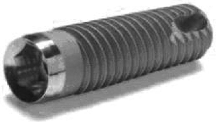
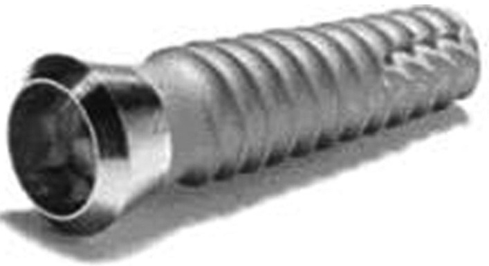
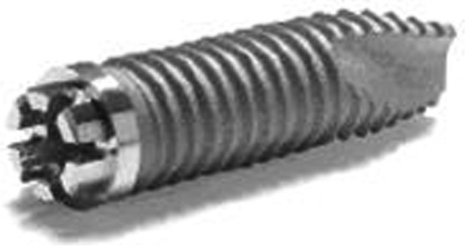
In 125 patients a total of 556 plants were inserted: Two hundred and ninety-five (53.1%) in the maxilla and 261 (46.9%) in the mandible. There were 480 (86.3%) Screw-Vents [Figure 1], 51 (9.2%) Swiss-Plusses [Figure 2], and 25 (4.5%) Splines [Figure 3]. Sixteen, 355, 34, 90, 55, and 6 fixtures had a diameter of 3.25, 3.7, 3.75, 4.1, 4.7, and 4.8 mm, respectively. Twenty-eight, 145, 5, 217, 8, 141, and 12 implants had a length of 8, 10, 11, 11.5, 12, 13, and 14 mm, respectively. The implants were inserted to replace 136 (24.5%) incisors, 80 (14.4%) cuspids, 198 (35.6%) premolars, and 142 (25.5%) molars.
Figure 1.

Screw-vent implant
Figure 2.

Swiss-pluss implant
Figure 3.

Spline implant
Two surgeons inserted the implants: CMS 324 (58.3%) and EZ 232 (41.7%). Three hundred and twenty-seven (58.8.%) were inserted in females and 229 (41.2 %) in males. Seven (1.3%) implants were inserted in diabetic patients and 80 (14.4%) in subjects treated with anti-hypertensive drugs. Two hundred and twenty-nine (41.2) were immediately loaded. Thirty-four (6.1) were inclinated as they were used for an all-on-four technique and 179 (32.2%) were inserted in smokers (less than 20 cigarettes per day). Eighty-three (14.9%) were inserted in grafted maxillary sinuses.
Surgical and prosthetic technique
All patients underwent the same surgical protocol. An antimicrobial prophylaxis was administered with 1 g Amoxycillin twice daily for five days, starting one hour before surgery. Local anesthesia was induced by infiltration of articaine/epinephrine and postsurgical analgesic treatment was performed with 100 mg Nimesulid, twice daily, for three days. Oral hygiene instructions were provided.
After making a crestal incision a mucoperiosteal flap was elevated. The implants were inserted according to the procedures recommended. The implant platform was positioned at the alveolar crest level. The sutures were removed seven days after surgery. In case of a two–stage procedure, following 24 weeks of implant insertion, the provisional prosthesis was provided, and the final restoration was usually delivered within an additional eight weeks. The number of prosthetic units (i.e., implant/crown ratio) was about 0.6. The mean age was 62 ± 10 years (minimum−maximum 23-88 years).
Two hundred and thirty-two (41.7%) implants were insered after the use of piezo, laser, and platelet-rich plasma derivates. Fourteen (2.5%) were covered with resorbable membranes and 244 (43.9%) were inserted in post-extractive sockets. All patients were included in a strict hygiene recall.
Statistical analysis
As no implants were lost (i.e., SRV = 100%) and no statistical differences were detected among the studied variables, no or reduced crestal bone resorption was considered as an indicator of SCR, to evaluate the effect of several host-, implant-, and occlusion-related factors.
The difference between the implant abutment junction and the bone crestal level was defined as the Implant Abutment Junction (IAJ) and calculated at the time of operation and during follow-up. Delta IAJ was the difference between IAJ at the last check-up and IAJ recorded just after the operation. The delta IAJ medians were stratified according to the variables of interest.
The Pearson Chi-sqaure test was used to detect the variables most associated with implant success.
RESULTS
No implant was lost (SVR = 100%). The mean peri-implant bone resorption was 0.7 ± 0.6 mm (minimum-maximum 0-5.7 mm). Twenty-seven fixtures had a peri-implant bone resorption greater than the cut-off values (SCR = 95.1%), and therefore, were used to detect those variables statistically related to an augmented peri-implant bone resorption.
Among the studied variables (i.e., implant type, diameter, length, gender, smoke, diabetes, drugs, platelet-rich plasma, sinus augmentation, membrane, immediate loading, fixture inclination, laser, piezo, post-extraction socket, surgeon, tooth position, and jaws) only jaws was statistically significant, with a better outcome for implants inserted in the maxilla (P = 0.017).
DISCUSSION
ZDIs have been used since the nineties, but few reports analyzed the clinical outcome of these fixtures.
In 2001, Khayat et al.[14] reported a study on a wide diameter Screw-Vent. A total of 131 wide implants were placed. All patients were recalled one year after loading. One hundred and eleven implants were evaluated at the recall examination. Almost all implants (109) supported a fixed partial prosthesis. The mean loading time was 17 months. No implants were lost during the loading period.
In 2002, Arlin analyzed 435 Screw-Vent implants.[15] The focus group was compared to a mixed implant design group, with a variety of abutment connections and surfaces from several other manufacturers. The cumulative survival rates were 94.2% (n = 435) for the focus group and 90.1% (n = 2339) for the reference group.
In 2005, Minichetti et al.[16] reported the results of implants placed into extraction sites grafted with particulate mineralized bone allograft (Puros). A total of 313 extraction sites were grafted with mineralized bone graft during a 36-month period. A total of 252 Screw-Vent implants were placed into the grafted extraction sites after a four- to seven-month healing period. All re-entries revealed a bony hard structure acceptable for osteotomy preparation. A total of 244 implants were restored with fixed prosthesis and six with removable overdentures, for a total of 250 loaded implants. A total of six implants failed, which required their removal (two implants before load and four after loading), resulting in a 97.6% implant success rate.
In 2007, Khayat et al.[17] evaluated 835 Screw-Vent implants. A total of 835 implants, with diameters of 3.7 mm (9%), 4.7 mm (76%), and 6.0 mm (15%) were placed in 328 patients, using a single-stage, delayed-loading protocol. The implants were restored with a variety of prostheses and monitored over two years of functional loading. Five implants failed and were removed before loading. Cumulative implant survival was 99.4% (n = 835); differences between mandibular (99.0%, n = 408) and maxillary (99.8%, n = 427) implants were not statistically significant. Mean marginal bone resorption was 1.66 mm (± 0.13 mm). Six implants failed to meet the success criteria by sustaining mesial and distal bone loss below the first implant thread; however, they remained stable and continued functioning without pain or inflammation. Cumulative implant success was 98.6% (n = 835); the differences between the maxillary (98.6%) and mandibular (98.8%) implants were not statistically significant. The success rates by implant diameter were 98.6% (3.7 mm), 98.4% (4.7 mm), and 100% (6 mm). The authors concluded that after two years of functional loading, the survival and success rates for Screw-Vent implants placed in a non-submerged protocol, equaled or surpassed those of single-thread, straight-walled implant historical controls.
One year later, Minichetti et al.[18] reported the results of implants placed in the maxillary sinuses grafted with particulate mineralized cancellous bone allograft alone or in combination with resorbable hydroxyapatite, over a three-year period. A total of 56 sinuses were grafted, and 136 dental implants were placed into the grafted sites after a four- to eight-month healing period. All re-entries revealed a bony hard structure acceptable for osteotomy preparation. Of these implants, 124 had been restored with fixed prosthesis and 12 with removable overdentures for a total of 136 loaded implants. A total of three implants required removal (failure) resulting in a 97.7% implant success rate (2.3% failure rate). The authors concluded that a mineralized human allograft placed in a lateral window sinus elevation, was a clinically predicable method, acceptable for implant placement and restoration. Luritano et al. evaluated the immediate loading implant and possible effects correlated with peri-implants.[19,20,21]
In 2008, Ormianer et al.,[22] performed a non-randomized, uncontrolled, retrospective study to evaluate the clinical outcomes of treatment with tapered, multithreaded implants, with a special emphasis on peri-implant crestal bone status. Chart reviews were conducted of 60 patients who had been treated with 267 implants for the placement of one or more missing and/or unsalvageable teeth, and who met the general inclusion criteria for dental implant therapy. In all cases, marginal bone changes were calculated from the cementoenamel junction or the implant neck to the crestal bone level, with standardized radiographs taken at the implant placement (baseline) and during annual follow-up. After a mean follow-up of 7.5 years, the implant survival was 98.5% (263/267) for all implants placed, and the implant success was 96.2% (253/263) for all surviving implants. No discernible bone loss was evident in 88% of the surviving implants. Crestal bone loss was observed in 25% (15/60) of the total study subjects and in 12% (32/263) of all surviving implants: Twenty-nine implants exhibited 1 mm of bone loss and three implants lost 2 mm of bone. Low-density maxillary jawbone and more extensive bone remodeling, which were required around the implants immediately placed into extraction sockets, were the probable causes of the observed bone loss in this study. The implants exhibited excellent long-term outcomes, with little or no bone loss.
CONCLUSION
Here we have demonstrated that ZDIs have a high survival and success rate. Most of the studied variables (i.e., implant type, diameter, length, gender, smoke, diabetes, drugs, platelet-rich plasma, sinus augmentetion, membrane, immediate loading, fixture inclination, laser, piezo, post-extraction socket, surgeon, and tooth position) have no impact on the clinical outcome. Implants inserted in the mandible have a slight, but a statistically significant worse outcome, with a higher peri-implant bone resorption, compared to those inserted in the maxilla. In conclusion, ZDIs are reliable devices to use in implantology.
ACKNOWLEDGEMENTS
This work was supported by the University of Ferrara (F.C.), Ferrara, Italy and by PRIN 2008 (20089MANHH_004).
Footnotes
Source of Support: This work was supported by the University of Ferrara (F.C.), Ferrara, Italy and by PRIN 2008 (20089MANHH_004)
Conflict of Interest: None declared
REFERENCES
- 1.Adell R, Lekholm U, Rockler B, Branemark PI. A 15-year study of osseointegrated implants in the treatment of the edentulous jaw. Int J Oral Surg. 1981;10:387–416. doi: 10.1016/s0300-9785(81)80077-4. [DOI] [PubMed] [Google Scholar]
- 2.Fanali S, Carinci F, Zollino I, Brugnati C, Lauritano D. One-piece implants installed in restored mandible: A retrospective study. Eur J Inflamm. 2012;10:37–41. [Google Scholar]
- 3.Fanali S, Carinci F, Zollino I, Brugnati C, Lauritano D. A retrospective study on 83 one-piece implants installed in resorbed maxilla. Eur J Inflamm. 2012;10:55–8. [Google Scholar]
- 4.Fanali S, Carinci F, Zollino I, Brunelli G, Minguzzi R. Effect of distance between one piece implants on crestal bone resorption. Eur J Inflamm. 2011;9:1–6. [Google Scholar]
- 5.Fanali S, Carinci F, Zollino I, Brunelli G, Minguzzi R. Effect of one-piece implant diameter on clinical outcome. Eur J Inflamm. 2011;9:7–12. [Google Scholar]
- 6.Fanali S, Carinci F, Zollino I, Brunelli G, Minguzzi R. Impact of one-piece implant lenght on clinical outcome. Eur J Inflamm. 2011;9:13–8. [Google Scholar]
- 7.Fanali S, Carinci F, Zollino I, Brunelli G, Minguzzi R. Welding improbe the success rate of one-piece implants. Eur J Inflamm. 2011;9:19–24. [Google Scholar]
- 8.Fanali S, Carinci F, Zollino I, Brunelli G, Minguzzi R. Bio-grip and machined titanium stimulate dental pulp stem cells towords osteoblast differentiation. Eur J Inflamm. 2011;9:25–30. [Google Scholar]
- 9.Danza M, Guidi R, Carinci F. Spiral family implants inserted in postextraction bone sites. Implant Dent. 2009;18:270–8. doi: 10.1097/ID.0b013e3181a4584c. [DOI] [PubMed] [Google Scholar]
- 10.Grecchi F, Zollino I, Parafioriti A, Mineo G, Pricolo A, Carinci F. One-step oral rehabilitation by means of implants’ insertion, Le Fort I, grafts, and immediate loading. J Craniofac Surg. 2009;20:2205–10. doi: 10.1097/SCS.0b013e3181bf8487. [DOI] [PubMed] [Google Scholar]
- 11.Danza M, Carinci F. Flapless surgery and immediately loaded implants: A retrospective comparison between implantation with and without computer-assisted planned surgical stent. Stomatologija. 2010;12:35–41. [PubMed] [Google Scholar]
- 12.Quirynen M, Naert I, van Steenberghe D, Teerlinck J, Dekeyser C, Theuniers G. Periodontal aspects of osseointegrated fixtures supporting an overdenture. A 4-year retrospective study. J Clin Periodontol. 1991;18:719–28. doi: 10.1111/j.1600-051x.1991.tb00063.x. [DOI] [PubMed] [Google Scholar]
- 13.Albrektsson T, Zarb GA. Determinants of correct clinical reporting. Int J Prosthodont. 1998;11:517–21. [PubMed] [Google Scholar]
- 14.Khayat PG, Hallage PG, Toledo RA. An investigation of 131 consecutively placed wide screw-vent implants. Int J Oral Maxillofac Implants. 2001;16:827–32. [PubMed] [Google Scholar]
- 15.Arlin ML. Analysis of 435 screw-vent dental implants placed in 161 patients: Software enhancement of clinical evaluation. Implant Dent. 2002;11:58–66. doi: 10.1097/00008505-200201000-00015. [DOI] [PubMed] [Google Scholar]
- 16.Minichetti JC, D’Amore JC, Hong AY. Three-year analysis of Tapered Screw-Vent implants placed into extraction sockets grafted with mineralized bone allograft. J Oral Implantol. 2005;31:283–93. doi: 10.1563/0-757.1. [DOI] [PubMed] [Google Scholar]
- 17.Khayat PG, Milliez SN. Prospective clinical evaluation of 835 multithreaded tapered screw-vent implants: Results after two years of functional loading. J Oral Implantol. 2007;33:225–31. doi: 10.1563/1548-1336(2007)33[225:PCEOMT]2.0.CO;2. [DOI] [PubMed] [Google Scholar]
- 18.Minichetti JC, D’Amore JC, Hong AY. Three-year analysis of tapered screw vent implants placed into maxillary sinuses grafted with mineralized bone allograft. J Oral Implantol. 2008;34:135–41. doi: 10.1563/1548-1336(2008)34[135:TAOTSV]2.0.CO;2. [DOI] [PubMed] [Google Scholar]
- 19.Scarano A, Murmura G, Carinci F, Lauritano D. Immediately loaded small diameter dental implants: Evaluation of retention, stability and comfort for the edentulous patient. Eur J Inflam. 2012;10:19–24. [Google Scholar]
- 20.Brunelli G, Carinci F, Zollino I, Candotto V, Scarano A, Lauritano D. Peri-Implantitis: A case report and literature review. Eur J Inflamm. 2012;10:1–6. [Google Scholar]
- 21.Brunelli CF, Zollino I, Candotto V, Scarano A, Lauritano D. SEM evaluation of 10 infected implants retrived from man. Eur J Inflamm. 2012;10:7–12. [Google Scholar]
- 22.Ormianer Z, Palti A. Retrospective clinical evaluation of tapered screw-vent implants: Results after up to eight years of clinical function. J Oral Implantol. 2008;34:150–60. doi: 10.1563/1548-1336(2008)34[150:RCEOTS]2.0.CO;2. [DOI] [PubMed] [Google Scholar]


