Abstract
Recently, we reported that African swine fever virus (ASFV) multigene family (MGF) 360 and 530 genes are significant swine macrophage host range determinants that function by promoting infected-cell survival. To examine the function of these genes in ASFV's arthropod host, Ornithodoros porcinus porcinus, an MGF360/530 gene deletion mutant (Pr4Δ35) was constructed from an ASFV isolate of tick origin, Pr4. Pr4Δ35 exhibited a significant growth defect in ticks. The deletion of six MGF360 and two MGF530 genes from Pr4 markedly reduced viral replication in infected ticks 100- to 1,000-fold. To define the minimal set of MGF360/530 genes required for tick host range, additional gene deletion mutants lacking individual or multiple MGF genes were constructed. The deletion mutant Pr4Δ3-C2, which lacked three MGF360 genes (3HL, 3Il, and 3LL), exhibited reduced viral growth in ticks. Pr4Δ3-C2 virus titers in ticks were significantly reduced 100- to 1,000-fold compared to control values at various times postinfection. In contrast to the parental virus, with which high levels of virus replication were observed in the tissues of infected adults, Pr4Δ3-C2 replication was not detected in the midgut, hemolymph, salivary gland, coxal gland, or reproductive organs at 15 weeks postinfection. These data indicate that ASFV MGF360 genes are significant tick host range determinants and that they are required for efficient virus replication and generalization of infection. The impaired virus replication of Pr4Δ3-C2 in the tick midgut likely accounts for the absence of the generalized infection that is necessary for the natural transmission of virus from ticks to pigs.
African swine fever (ASF) is a highly lethal, hemorrhagic disease of domestic pigs for which animal slaughter and area quarantine are the only methods of disease control. African swine fever virus (ASFV), the causative agent of ASF, is a large, enveloped, double-stranded DNA virus which is the only member of the newly named Asfarviridae (11). Although the icosahedral morphology of the ASFV virion resembles that of iridoviruses, both the genomic organization, which includes terminal cross-links and inverted terminal repeats, and its cytoplasmic replication strategy suggest some relationship with the Poxviridae (16, 28, 41).
In domestic pigs, ASF occurs in several disease forms, ranging from highly lethal to subclinical infections, depending on contributing viral and host factors (7, 23, 34). ASFV infects cells of the mononuclear-phagocyte system, including fixed-tissue macrophages, and specific lineages of reticular cells in the spleen, lymph node, lung, kidney, and liver (7, 21-23, 25). This ability to replicate and induce marked cytopathology in these cell types in vivo appears to be critical for ASFV virulence. The viral and host factors responsible for the differing outcomes of infection with highly virulent strains and strains of lesser virulence are largely unknown.
ASFV is the only known DNA arbovirus (6, 8). The perpetuation and transmission of this virus in nature involve the cycling of virus between two highly adapted hosts, Ornithodoros ticks and wild pigs (warthogs and bushpigs) in sub-Saharan Africa (30, 31, 45, 49). In areas of ASF enzoocity, adult warthogs are typically nonviremic, although most are seropositive (19, 29, 31, 40, 44), and virus has been isolated from lymph nodes (19, 31). Young warthogs are most likely infected through feeding by infected Ornithodoros porcinus porcinus ticks. Infection in young warthogs is subclinical, with viremic titers ranging from 2 to 3 log10 50% hemadsorption doses (HAD50)/ml (45, 46), a level sufficient to infect a low percentage of naïve ticks (33, 44). The sylvatic ASFV cycle is further maintained by transovarial and venereal transmission in ticks (32, 37). The mechanism of ASFV transmission from the sylvatic cycle to domestic pigs is most likely through infected Ornithodoros ticks feeding on pigs (31, 44), since direct contact with infected warthogs rarely results in transmission to pigs (10, 19, 24, 44).
Previous studies have described experimental infections of O. porcinus porcinus ticks with different ASFV isolates (17, 18, 20, 33, 36). Infection is characterized by the establishment of a long-term, persistent infection with relatively high levels of virus replication occurring in a number of different tissues and organs. These data suggest that ASFV infection of its natural arthropod host represents a well-adapted and possibly coevolved biological system. However, differences in infection rates, infectious doses, and persistent infections have been observed when ticks were exposed to different ASFV isolates (17, 33). The viral and host factors and the mechanisms responsible for the differing outcomes of infections with different ASFV isolates are unknown.
ASFV is genetically complex; its genome of 170 to 190 kbp contains 160 or more open reading frames (ORFs), and approximately 100 proteins have been observed in virus-infected cells (2, 8, 12, 13, 39, 43). The ASFV genome contains a central conserved region and variable terminal regions (5, 16, 41, 42). Swine virulence and host range genes, including the NL, UK, and MGF genes, have been identified in the terminal variable regions of the ASFV genome. The terminal variable regions comprise the left 46-kbp and the right 12.5-kbp ends of the genome and contain at least five multigene families (MGFs): MGF100, MGF110, MGF300, MGF360, and MGF530 (3, 4, 9, 15, 47, 50).
MGF360 and MGF530 genes do not show similarity to other genes or motifs in current databases. Individual MGF genes are conserved among ASFV isolates (with 45 to 100% corresponding amino acid identity), and they are transcribed early in infection (38, 50). The functions of individual MGF genes or the different multigene families in virus-host interactions remain poorly understood.
It was recently shown that MGF360 and MGF530 genes perform an essential macrophage host range function that involves the promotion of infected-cell survival (52), and Neilan et al. reported that MGF360 and MGF530 genes also function in pig virulence (27).
Here, we show that ASFV MGF360 genes are significant tick host range determinants and are required for the replication and generalization of infection in the tick host. Impaired virus replication of the MGF360 gene deletion mutant Pr4Δ3-C2 in the tick midgut likely accounts for the absence of the generalized infection that is necessary for the natural transmission of virus from tick to pig.
MATERIALS AND METHODS
Cell cultures and viruses.
Primary porcine blood macrophage cell cultures were prepared from defibrinated swine blood as previously described (14). Briefly, heparin-treated swine blood was incubated at 37°C for 1 h to allow sedimentation of the erythrocyte fraction. Mononuclear leukocytes were separated by flotation over a Ficoll-Paque (Pharmacia, Piscataway, N.J.) density gradient (1.079 specific gravity). The monocyte/macrophage cell fraction was cultured in plastic Primaria tissue culture flasks (Falcon; Becton Dickinson Labware, Franklin Lakes, N.J.) containing RPMI 1640 medium with 30% L929 supernatant and 20% fetal bovine serum for 48 h (37°C in 5% CO2). Adherent cells were detached from the plastic by using 10 mM EDTA in phosphate-buffered saline and then reseeded into Primaria T25 6- or 96-well dishes at a density of 5 × 10⁶ cells per ml for use in assays 24 h later.
The pathogenic ASFV strain Pretoriuskop/96/4 (Pr4) was isolated from O. porcinus porcinus ticks collected from warthog burrows in Kruger National Park, Republic of South Africa, in 1996 (20). The ASFV gene deletion mutant Pr4Δ35 was constructed as previously described (52).
Construction of recombinant ASFV Pr4 viruses containing deletions in MGF360 and MGF530 genes.
Gene deletion mutants Pr4Δ3-C1, Pr4Δ3-C2, Pr4Δ5-1, and Pr4Δ5-2 were generated by homologous recombination of ASFV Pr4 genomes and recombination transfer vectors following infection and transfection of macrophage cell cultures (53). Flanking DNA fragments to the left and right of the MGF360 and MGF530 ORFs were amplified by using primer sets, each of which introduced a BamHI restriction site adjacent to the MGF360/530 ORFs and a BglII restriction site (left- or right-flank fragment) at the opposite end. The primer set for Pr4Δ3-C1 was as follows: left-flank forward primer, 5′-TTGCTTAAGATCCTTTAGATCCTT-3′; left-flank reverse primer, 5′-TGGATCCACGTATGTTAAAAGATTATCATTC-3′; right-flank forward primer, 5′-GAGGGATCCCAGGGAAGGCAACAT-3′; and right-flank reverse primer, 5′-AAGATCTTCCTTACCTACGATT-3′. The primer set for Pr4Δ3-C2 was as follows: left-flank forward primer, 5′-AAGATCTACGTCTATATCGTTTGG-3′; left-flank reverse primer, 5′-ATATGAGGATCCTCCTTACCTACG-3′; right-flank forward primer, 5′-TGGATCCAACGTTTGTAAAGACAACAT-3′; and right-flank reverse primer, 5′-TCACGCGCTAGATGATGATCATGC-3′. The primer set for Pr4Δ5-1 was as follows: left-flank forward primer, 5′-AAGATCTGCTTTCTTTTAACGTTAA-3′; left-flank reverse primer, 5′-TGGATCCTCAGGAAGTTTACAGTCAGGTAAT-3′; right-flank forward primer, 5′-ACGCTCAGGATCCTACTAATATCA-3′; and right-flank reverse primer, 5′-AAAACAACTACAACCTTATAAAAC-3′. The primer set for Pr4Δ5-2 was as follows: left-flank forward primer, 5′-TAGAAAGATTCATGCCATAATCGA-5′; left-flank reverse primer, 5′-AAACGGATCCCCCTACCTCATTAA-3′; right-flank forward primer, 5′-CCACCGGATCCAGAGACATTTGTA-3′; and right-flank reverse primer, 5′-ATCATAAGATCTGGTCAAATAGTT-3′. Introduced restriction sites are in boldface.
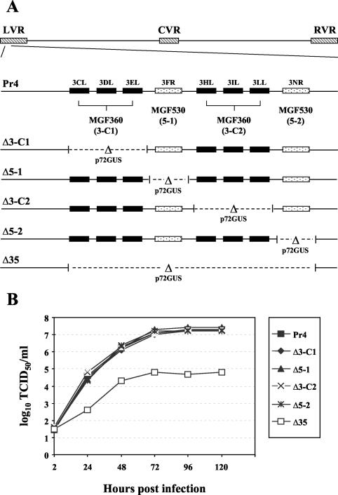
The fragments were digested with the appropriate restriction enzymes and cloned into pCR2.1 to give pPr4.C1, pPr4.C2, pPr4.5-1, and pPr4.5-2. A reporter gene cassette containing the β-glucuronidase (GUS) gene with the ASFV p72 late promoter, p72GUS (53), was inserted into BamHI-digested pPr4 plasmids to yield the transfer vectors p72GUSΔ3-C1, p72GUSΔ3-C2, p72GUSΔ5-1, and p72GUSΔ5-2, creating deletions of the MGF360 genes 3CL, 3DL, and 3EL, the MGF360 genes 3HL, 3IL, and 3LL, the MGF530 gene 3FR, and the MGF530 gene 3NR, respectively (Fig. 1). The primary porcine macrophage cell cultures were infected with Pr4 (multiplicity of infection [MOI], 10) and transfected with p72GUSΔ360/530 transfer vectors. Recombinant viruses representing independent primary plaques were purified to homogeneity by plaque assay and verified as products of a double-crossover recombination event by using PCR and Southern blot analyses, as described previously (51, 53).
FIG. 1.
Characterization of ASFV MGF360 and MGF530 gene deletion mutants Pr4Δ3-C1, Pr4Δ5-1, Pr4Δ3-C2, Pr4Δ5-2, and Pr4Δ35. (A) Diagram of MGF360 and MGF530 gene regions in parental Pr4 and deletion mutants. Transfer vectors and recombinants with gene deletions were constructed as described in Materials and Methods. LVR, left variable region; CVR, central variable region; RVR, right variable region. (B) Growth characteristics of ASFV Pr4 and recombinant viruses Pr4Δ3-C1, Pr4Δ5-1, Pr4Δ3-C2, Pr4Δ5-2, and Pr4Δ35 in swine macrophage cell cultures. Primary macrophage cell cultures were infected (MOI = 1), and at the indicated times postinfection, duplicate samples were titrated for virus yield. These data are the means of results of two independent experiments. TCID50, 50% tissue culture infectious dose.
Infection of ticks.
Groups of O. porcinus porcinus ticks (48) were exposed to ASFV Pr4 wild-type or recombinant viruses by allowing them to feed on an artificial membrane feeder placed in heparinized pig blood containing virus with a known titer. Ticks were allowed to feed to repletion before being removed from the membrane and placed in a holding container. Only fully fed ticks were used for subsequent experiments.
Virus titration.
Individual whole ticks were ground in 0.5 ml of RPMI 1640 medium supplemented with twice the normal level of antibiotics in sterile tubes with plastic pestles (Pellet; Kontes). Samples were stored at −70°C. Immediately prior to titration, the samples were thawed at 37°C, sonicated for 1 min, and centrifuged for 3 min at 10,000 × g. Supernatants were serially diluted and then added to porcine macrophage cell cultures. Titers were calculated by the method of Reed and Muench (35).
Adult ticks were dissected, and tissues were collected. Hemolymph was obtained by clipping the distal tarsus of the second leg of each tick and collecting 1 to 2 μl of hemolymph in a sterile tube. All samples were diluted to 0.5 ml with RPMI 1640 medium and titrated as previously described (20).
To obtain the data presented in Table 2, individual ticks were allowed to feed on a membrane feeding apparatus as previously described (20). Following feeding, coxal fluid and blood from beneath the membrane (containing salivary secretions) were collected, diluted to 1 ml with RPMI 1640 medium, and titrated as described above.
TABLE 2.
Virus titers in tissues from O. porcinus porcinus ticks infected with Pr4 and MGF360/530 gene deletion recombinants
| Source of tissue samples (n = 3) | Mean virus titer in tissue (log10 HAD50/ml) ± SEM of virus strain
|
||||
|---|---|---|---|---|---|
| Pr4 | Pr4Δ3-C1 | Pr4Δ3-C2 | Pr4Δ5-1 | Pr4Δ5-2 | |
| Hemolympha | 4.26 ± 0.12 | 4.24 ± 0.16 | <1.0 | 4.32 ± 0.16 | 4.24 ± 0.08 |
| Midgutb | 6.32 ± 0.40 | 6.32 ± 0.43 | <1.0 | 6.65 ± 0.18 | 6.23 ± 0.08 |
| Reproductive tissueb | 6.26 ± 0.48 | 5.83 ± 0.31 | <1.0 | 5.86 ± 0.17 | 5.90 ± 0.60 |
| Coxal glandb | 5.26 ± 0.35 | 5.57 ± 0.25 | <1.0 | 5.73 ± 0.07 | 4.57 ± 0.72 |
| Salivary glandb | 5.96 ± 0.20 | 4.32 ± 0.23 | <1.0 | 6.43 ± 0.18 | 6.32 ± 0.08 |
| Coxal fluidc | 2.66 ± 0.10 | 3.66 ± 0.15 | <1.0 | 3.66 ± 0.15 | 2.66 ± 0.10 |
Sample taken at 42 days postfeeding.
Tissue dissected at 108 days postfeeding.
Sample taken at 267 days postfeeding.
Reverse transcription-PCR analysis.
A group of stage N1 ticks (n = 2,000) was exposed to blood meals containing 108 HAD50 of Pr4 or Pr4Δ3-C2 virus/ml. At 10 days postfeeding, ticks were snap-frozen in liquid nitrogen, and total RNA was extracted by using a ToTALLY RNA kit (Ambion, Austin, Tex.). Poly(A)+ RNA was purified with a MicroPoly(A) kit (Ambion) and reverse transcribed with a RETROscript kit (Ambion) according to the manufacturer's instructions. The resulting cDNAs were then amplified by PCR for 30 cycles (94°C for 10 s; 50°C for 1 min; 60°C for 1 min) with a final 10-min incubation at 72°C. The primers used were as follows: 3HL forward primer, 5′-GTTGTGGTCTTTGATGCAGG-3′; 3HL reverse primer, 5′-CTCGACCAAAAAGGTGTTGG-3′; 3IL forward primer, 5′-TCGCCATTCATTAAGATCCG-3′; 3IL reverse primer, 5′-CCCTCGTCAAAAAGACGGTA-3′; 3LL forward primer, 5′-CGGCTGAAAAGCTGCTTTACTA-3′; and 3LL reverse primer, 5′-TGTTGTCTTTACAAACGTTGG-3′.
Amplified products were run on a 0.7% agarose gel under standard conditions and cloned into the TA cloning vector pCR 2.1 (Invitrogen). Eight independent clones of each amplification product were completely sequenced with an Applied Biosystems 377 automated DNA sequencer.
Ultrastructural analysis.
Nymphal ticks (stage N3) were orally exposed to high titers (108 HAD50/ml) of Pr4Δ3-C1 and Pr4Δ3-C2. At 7, 21, 42, and 90 days postfeeding, three ticks from each group were processed for transmission electron microscopy as previously described (20). Seventy- to 90-nm sections were collected on single-slot grids coated with Formvar, stabilized with carbon (Electron Microscopy Sciences, Fort Washington, Pa.), and photographed with a Philips 410 electron microscope operated at 80 kV. At least five different sagittal sections from each of the three ticks were examined. The ticks were sampled most extensively from the anterior hemocoel, which contained large portions of the midgut, salivary glands, and coxal glands.
RESULTS
Construction and growth characteristics of recombinant ASFV MGF360 and MGF530 gene deletion mutants in porcine macrophage cell cultures.
ASFV MGF360 and MGF530 gene deletion mutants were constructed from the pathogenic African isolate Pr4 by homologous recombination of the parental genome and engineered recombination transfer vectors in primary swine macrophage cell cultures, as described in Materials and Methods. The deletions introduced into Pr4 removed genomic regions from the left variable region, which contained members of the MGF360 and/or MGF530 genes, and inserted in their place a 2.4-kbp p72GUS reporter gene cassette (Fig. 1A). Deletion mutant Pr4Δ35 was constructed by deleting a 10,163-bp region from Pr4 as previously described (52). The deletion removed six MGF360 ORFs (3CL, 3DL, 3EL, 3HL, 3IL, and 3LL) and two MGF530 ORFs (3FR and 3NR). In Pr4Δ5-1 and Pr4Δ5-2, the deletions removed a single MGF530 ORF, 3FR and 3NR, respectively. Recombinant viruses Pr4Δ3-C1 and Pr4Δ3-C2 contained deletions of three MGF360 genes, 3CL, 3DL, and 3EL and 3HL, 3IL, and 3LL, respectively. Recombinant viruses expressing GUS were obtained at a frequency of approximately 1 in 5,000. Independent primary plaques were purified to homogeneity. Genomic DNAs from parental virus and deletion mutants were analyzed by Southern blotting and PCR to characterize genomic changes. Results obtained verified the predicted genomic structure of the recombinant Pr4 viruses and showed that the mutants were free of contaminating parental virus (data not shown).
The growth kinetics and viral yields of ASFV Pr4 MGF360 and MGF530 gene deletion mutants were compared to those of the Pr4 parental virus by infecting primary porcine macrophage cell cultures (MOI = 1). Unlike recombinant virus Pr4Δ35, which exhibited a significant 100- to 1,000-fold growth defect compared with the growth of the parental strain Pr4 (52), deletion mutants Pr4Δ3-C1, Pr4Δ3-C2, Pr4Δ5-1, and Pr4Δ5-2 exhibited unaltered growth characteristics in macrophage cell cultures (Fig. 1B).
MGF360 genes 3HL, 3IL, and 3LL affect growth of ASFV in O. porcinus porcinus ticks.
To determine the roles of ASFV MGF360 and MGF530 genes in tick host range, Ornithodoros ticks were infected with mutants Pr4Δ35, Pr4Δ3-C1, Pr4Δ3-C2, Pr4Δ5-1, and Pr4Δ5-2. Nymphal O. porcinus porcinus ticks (stages N1 and N3) were fed blood meals containing 106 HAD50 of mutant or parental ASFV/ml. At various times postinfection, whole ticks (n = 8) were titrated for infectious virus. Pr4Δ35 exhibited a reduced infection rate (66 versus 100% for parental virus) and a significant growth defect in Ornithodoros ticks (Table 1, experiment 1). In contrast to parental virus Pr4, where a 100-fold increase over the initial titer was observed at 3 weeks postinfection, viral titers in Pr4Δ35-infected ticks showed no increase at 5 weeks postinfection. Low levels of Pr4Δ35 replication (∼105 HAD50/ml) were detected at 77 days postinfection (DPI); however, these titers were 100-fold lower than those observed for Pr4-infected ticks. These data indicate that ASFV MGF360 and MGF530 genes contained within this region affect viral replication in O. porcinus porcinus ticks.
TABLE 1.
ASFV titers in O. porcinus porcinus ticks following oral inoculation
| Expt and virusa | Mean ASFV titer (log10 HAD50/ml) ± SEM (% positive ticks) at:
|
|||||||
|---|---|---|---|---|---|---|---|---|
| 1 DPI | 7 DPI | 14 DPI | 21 DPI | 28 DPI | 35 DPI | 56 DPI | 77 DPI | |
| Expt 1 | ||||||||
| Pr4 | 3.8 ± 0.3 | 5.8 ± 0.4 | 6.0 ± 0.5 | 7.0 ± 1.0 | ||||
| Pr4Δ35 | 3.9 ± 0.2 | 3.9 ± 0.3 (66) | 4.0 ± 0.2 (66) | 4.8 ± 0.6 (66) | ||||
| Expt 2 | ||||||||
| Pr4 | 2.9 ± 0.3 | 5.5 ± 0.2 | 6.3 ± 0.1 | |||||
| Pr4Δ5-1 | 2.5 ± 0.3 | 4.8 ± 0.2 | 6.5 ± 0.1 | |||||
| Pr4Δ5-2 | 2.5 ± 0.2 | 6.2 ± 0.1 | 6.4 ± 0.1 | |||||
| Pr4Δ3-C1 | 2.3 ± 0.7 | 4.8 ± 0.4 | 6.6 ± 0.0 | |||||
| Pr4Δ3-C2 | 2.8 ± 0.3 | 3.2 ± 0.1 (63) | 4.3 ± 0.1 (75) | |||||
| Expt 3 | ||||||||
| Pr4 | 2.8 ± 0.4 | 4.7 ± 0.1 | 4.2 ± 0.1 | 6.3 ± 0.2 | ||||
| Pr4Δ3-C2 | 3.3 ± 0.2 | 4.0 ± 0.2 | 3.1 ± 0.1 | 4.3 ± 0.1 | ||||
| Expt 4 | ||||||||
| Pr4 | 4.9 ± 0.1 | 5.4 ± 0.1 | 6.8 ± 0.2 | |||||
| Pr4Δ3-C2 | 4.9 ± 0.9 | 4.0 ± 0.2 | 5.0 ± 0.4 | |||||
A virus dose of 106 HAD50/ml was used for each experiment except experiment 4, in which a dose of 108 HAD50/ml was used.
To further define specific MGF360/530 genes within the left variable region of the ASFV genome responsible for this growth defect, viral deletion mutants lacking individual MGF530 genes or multiple MGF360 genes were tested for their ability to infect ticks. Groups of N1 ticks were infected with 2 × 10⁶ HAD50 of either recombinant virus Pr4Δ3-C1, Pr4Δ3-C2, Pr4Δ5-1, or Pr4Δ5-2 or parental virus Pr4 per ml. Titration of individual whole ticks (n = 8) at 1, 28, and 56 DPI demonstrated indistinguishable growth characteristics for wild-type Pr4 and deletion mutants Pr4Δ3-C1, Pr4Δ5-1, and Pr4Δ5-2 (Table 1, experiment 2). In these groups, mean virus titers increased to approximately 5 log10 HAD50/ml at 28 DPI, reaching a peak titer of 6.5 log10 HAD50/ml at 56 DPI. In contrast, MGF360 gene deletion mutant Pr4Δ3-C2 exhibited a significant growth defect. At 28 and 56 DPI, Pr4Δ3-C2 titers were 100- to 500-fold lower than those observed for Pr4 and the other deletion mutant viruses. It is interesting that, as for ASFV Pr4Δ35, Pr4Δ3-C2 titers increased only at a late time postinfection (56 DPI). In two independent experiments, Pr4Δ3-C2 exhibited a significant growth defect as early as 7 DPI (Table 1, experiments 3 and 4). In both experiments, Pr4Δ3-C2 titers showed little to no increase whereas Pr4 titers increased 50- to 100-fold by 7 DPI. Thus, ASFV MGF360 genes 3HL, 3IL, and 3LL perform a tick host range function affecting early aspects of virus replication in the tick.
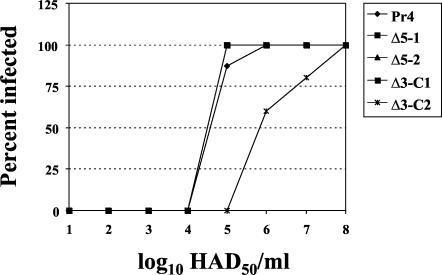
Fifty percent tick infectious doses (TID50) were determined for the wild-type and recombinant Pr4 viruses (Fig. 2). Ticks (stage N1) were exposed to blood meals containing serial 10-fold dilutions of ASFV, and virus isolation and/or titration was performed at 28 DPI. The TID50 for the Pr4, Pr4Δ3-C1, Pr4Δ5-1, and Pr4Δ5-2 viruses was 4.5 log10, while the TID50 for the Pr4Δ3-C2 gene deletion mutant was significantly increased 10- to 25-fold (5.8 log10).
FIG. 2.
TID50 of Pr4 and MGF360/530 gene deletion mutants. N1 ticks (n = 8) were fed blood meals containing serial 10-fold dilutions of ASFVs, and virus isolation and/or titration was performed at 28 DPI. TID50 were expressed as log10 HAD50 of virus per milliliter that resulted in a 50% infection rate.
The expression of MGF360 genes 3HL, 3IL, and 3LL during tick infection was evaluated by reverse transcription-PCR with RNA extracted from Pr4- and Pr4Δ3-C2-infected ticks at 10 DPI. PCR amplification of Pr4 cDNA with specific primers for the MGF360 genes 3HL, 3IL, and 3LL resulted in products of the expected sizes of 720, 600, and 940 bp, respectively. Sequence analysis confirmed that these amplicons corresponded to the expected Pr4 MGF360 genes (data not shown). As expected, PCR amplification products were not detected in ticks infected with Pr4Δ3-C2. These data indicate that the MGF360 genes 3HL, 3IL, and 3LL are transcribed in ASFV-infected ticks within the first 10 DPI, a time that is critical for efficient establishment of infection (20).
MGF360 genes 3HL, 3IL, and 3LL are required for efficient persistence and generalization of ASFV infection in O. porcinus porcinus ticks.
To determine the level of host restriction observed for Pr4Δ3-C2, generalization of infection, virus transmission, and long-term persistence of infection were examined in Ornithodoros ticks. Groups of adult ticks (n = 10) were infected with parental virus Pr4 and MGF360/530 gene deletion mutants (106 HAD50/ml). The mean weight of the tick blood meals was 140 ± 10 mg, indicating a mean exposure dose of 1.1 × 10⁵ HAD50 of ASFV per tick per ml. At various times postinfection, tick tissues (three ticks per group) were dissected and titrated. Virus titers in tissues are shown in Table 2. Tissue involvement and mean virus titers were similar for Pr4 and gene deletion mutants Pr4Δ3-C1, Pr4Δ5-1, and Pr4Δ5-2. In contrast, Pr4Δ3-C2 was not detected in any tissues examined. At 42 DPI, hemolymph samples contained on average 4.2 log10 HAD50 of virus per ml in all tick groups (except those exposed to Pr4Δ3-C2), indicating successful generalization of infection. The failure to isolate virus from the hemolymph of Pr4Δ3-C2-infected ticks is consistent with the low level of virus replication in whole ticks described above. These data indicate a defect in Pr4Δ3-C2 in critical early events in the midgut that are necessary for successful generalization of ASFV infection in Ornithodoros ticks (20). The absence of detectable Pr4Δ3-C2 in the salivary and coxal glands, coxal fluid, and salivary secretion suggests that low levels of primary virus replication in the midgut were not sufficient for successful generalization of infection.
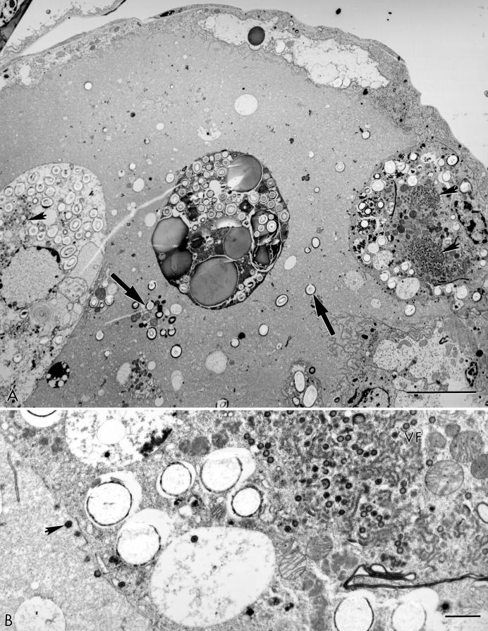
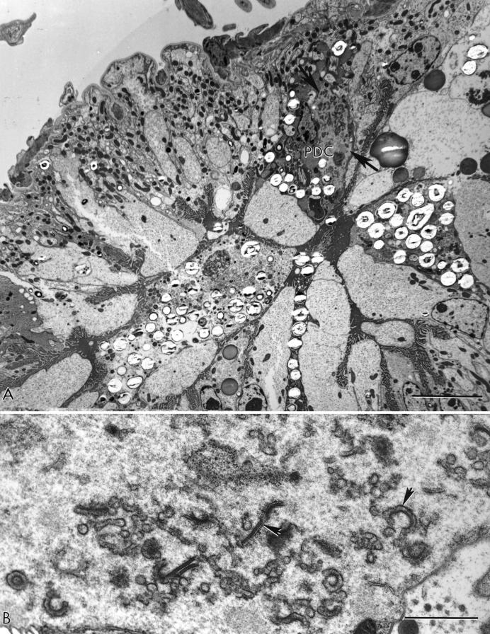
The Pr4Δ3-C2 growth defect in ticks could not be rescued by increasing the infectious dose 1,000-fold (108 HAD50/ml). High-dose infection of ticks with Pr4Δ3-C2 was atypical. Ultrastructural analysis demonstrated significant differences in the cellular distributions of mature virus particles in midgut cells. At 21 DPI, both the phagocytic digestive cells (PDC) and less-differentiated epithelial cells showed the presence of virus factories (31 positive cells of 185 examined) with large numbers of mature virions in Pr4-infected ticks (Table 3 and Fig. 3). In contrast, in Pr4Δ3-C2-infected ticks, only a small number of virus-containing PDC were observed (3 positive cells of 398 examined), and no evidence of Pr4Δ3-C2 virus infection was found in undifferentiated midgut epithelial cells (Table 3 and Fig. 4). Differences were seen in Pr4Δ3-C2-infected salivary and coxal glands at 90 DPI. Significantly, no virus particles were observed in the coxal glands of Pr4Δ3-C2-infected ticks, while in Pr4Δ3-C1-infected tissues, mature virions with an intact plasma membrane appeared in the extracellular space of the filtration membrane and along the internal membranes of connecting tubule cells (data not shown). Interestingly, mature Pr4Δ3-C2 particles were present in the salivary gland; however, virions were localized exclusively in the connective tissue surrounding the salivary gland. In Pr4Δ3-C1-infected ticks, virus particles were present not only in the connective tissue but also in electron-dense secretory granules. These data indicate a role for MGF360 gene function in other aspects of generalized infection and tick tissue tropism.
TABLE 3.
Midgut distribution of mature virions after high-dose infection of ticks with Pr4 and Pr4Δ3-C2a
| Time postfeeding and virus strain | No. of phagocytic digestive cells examined | No. of phagocytic digestive cells with virus (no. of virions) | No. of undifferentiated cells examined | No. of undifferentiated cells with virus (no. of virions) | Virus titer in whole tick (log10 HAD50/ml) (mean ± SEM) |
|---|---|---|---|---|---|
| 7 days | |||||
| Pr4 | 300 | 2 (43) | 230 | 0 | 5.4 ± 0.10 |
| Pr4Δ3-C2 | 818 | 14 (106) | 786 | 0 | 4.0 ± 0.26 |
| 21 days | |||||
| Pr4 | 185 | 31 (999) | 760 | 7 (72) | 6.8 ± 0.20 |
| Pr4Δ3-C2 | 398 | 3 (43) | 770 | 0 | 5.0 ± 0.44 |
Nymphal ticks (stage N3) were exposed to virus doses of 108 HAD50/ml.
FIG.3.
(A) Low-power electron micrograph of an N2 tick midgut after exposure to a blood meal containing 108 TCID50 of Pr4/ml at 3 weeks postfeeding. Two ASFV-infected PDC with virus factories (arrowheads) are present in the midgut lumen. Free granules (large arrows) are remnants of disrupted PDC. (B) A high-magnification view of the infected PDC attached to the midgut wall with an extensive virus factory (VF) and mature virus particles (arrowhead). Bars, 10 (A) and 1 (B) μm.
FIG.4.
(A) Low-power electron micrograph of an N2 tick after exposure to a blood meal containing 108 TCID50 of Pr4Δ3-C2/ml at 3 weeks postfeeding. A single infected PDC is found among growing and expanding undifferentiated cells. The typical electron-lucent agranular area of a virus factory (arrows) is present. (B) A higher magnification of the factory region showing developing virus forms (arrowheads) but no mature virions. Bars, 10 (A) and 1 (B) μm.
DISCUSSION
Here we have shown that MGF360 genes in the left variable region of the ASFV genome encode a novel tick host range determinant required for efficient replication and generalization of infection in the tick host.
The ASFV MGF360 and MGF530 gene deletion mutant Pr4Δ35 failed to replicate efficiently in Ornithodoros ticks. Pr4Δ35, which lacks six MGF360 and two MGF530 genes in the left variable region of the genome, exhibited a 100- to 1,000-fold reduction in viral replication in infected ticks (Table 1), indicating that the MGF360 and MGF530 genes within this region affect tick host range.
By using deletion mutants lacking individual or multiple MGF genes, the minimal set of MGF360/530 genes required for tick host range was mapped. The deletion mutant Pr4Δ3-C2, which lacked three MGF360 genes, exhibited markedly reduced viral replication in ticks. In addition, Pr4Δ3-C2 replication was not observed in the midgut, hemolymph, salivary glands, coxal glands, or reproductive organs of infected adults. These findings indicate that, while the MGF360 genes 3HL, 3IL, and 3LL are dispensable for growth in macrophage cell cultures, they are required for efficient viral replication in the tick host. To our knowledge, these are the first ASFV genes directly associated with Ornithodoros tick host range.
Previous studies demonstrated that the midgut is the initial site of virus replication in the tick host (17, 20) and that high virus titers in the midgut were likely required for subsequent generalization of infection in the tick (20). The results presented here indicate that efficient virus replication in the midgut is critical for the generalization of virus infection and that the MGF360 genes 3HL, 3IL, and 3LL function in this early event. The impaired virus replication of Pr4Δ3-C2 in tick midgut likely accounts for the observed lack of generalized infection.
The Pr4Δ3-C2 growth defect in ticks could not be rescued by increasing the infectious viral dose. Interestingly, some generalization of infection was observed in ticks infected with a high dose of Pr4Δ3-C2; however, infection was atypical with virus titers significantly lower than those for parental Pr4. Thus, ASFV MGF360 genes may perform critical functions in other tick cell types as well.
ASFV MGF360 and MGF530 genes do not show similarity to other known genes or motifs in current databases. Individual MGF genes are conserved among ASFV isolates (with 45 to 100% corresponding amino acid identity) (27). Amino-terminal regions of predicted MGF360 proteins do share similarity with corresponding regions of MGF530 ORFs (50). The alignments of the predicted amino acid sequences for all the MGF360 and MGF530 genes exhibit three conserved regions at the amino termini of these proteins (50). These conserved regions suggest a common ancestral relationship among the MGF genes and roles for these genes in common or related pathways. Given the lack of homology of the products of MGF360 genes to other known proteins, it is difficult to speculate about their function in virus-cell interactions. It was recently shown that MGF360 and MGF530 genes perform an essential macrophage host range function(s) that involves the promotion of infected-cell survival (52). In addition, a region within the left variable region of the ASFV genome containing MGF360 and MGF530 genes has been shown to encode a previously unrecognized virulence determinant for domestic swine (27). Although the virulence phenotype associated with the swine virulence determinant is independent of the macrophage host range phenotype, both determinants share four MGF360 genes. Interestingly, in the studies described here, three of the four MGF360 ORFs (3HL, 3IL, and 3LL) were identified as determinants for tick host range.
ASFV induces apoptosis in primary swine macrophage cell cultures at late times postinfection (26). Previous data indicate that MGF360 and MGF530 genes promote infected-cell survival, allowing efficient virus replication in macrophages, and that they are of significance for viral virulence (27, 52). Initial ASFV replication in the tick occurs in PDC of the midgut epithelium (20). Interestingly, there are significant morphological and functional similarities between macrophages and tick PDC (20). Recent data indicate that ASFV MGF360 and MGF530 genes suppress interferon (IFN) response genes in primary swine macrophage cultures (1). The inability of MGF360/530 mutant Pr4Δ35 to suppress an IFN response in infected cells may account for its growth defect in macrophage cell cultures. Although IFN response had not been associated with viral resistance in arthropods, these genes may be involved directly or indirectly in a yet-to-be-defined IFN-related tick host response to viral infections. The ability of virus to replicate efficiently in both cell types appears to be critical for ASFV virulence in swine and for successful infection of ticks. It is tempting to speculate that ASFV MGF genes perform a similar function, involving the maintenance of infected-cell survival in both swine macrophages and tick PDC.
In summary, the results reported here indicate that the MGF360 genes 3HL, 3IL, and 3LL are significant tick host range determinants. Future studies will examine how these novel genes function in the infection of both hosts.
Acknowledgments
We thank Aniko Zsak, Eric Shedlosky, and the PIADC animal care staff for excellent technical assistance.
REFERENCES
- 1.Afonso, C. L., M. E. Piccone, K. M. Zaffuto, J. G. Neilan, G. F. Kutish, Z. Lu, C. A. Balinsky, T. R. Gibb, T. J. Bean, L. Zsak, and D. L. Rock. 2004. African swine fever virus multigene family 360 and 530 genes affect host interferon response. J. Virol. 78:1858-1864. [DOI] [PMC free article] [PubMed]
- 2.Alcaraz, C., B. Pasamontes, G. F. Ruiz, and J. M. Escribano. 1989. African swine fever virus-induced proteins on the plasma membranes of infected cells. Virology 168:406-408. [DOI] [PubMed] [Google Scholar]
- 3.Almazán, F., J. M. Rodríguez, G. Andrés, R. Pérez, E. Viñuela, and J. F. Rodriguez. 1992. Transcriptional analysis of multigene family 110 of African swine fever virus. J. Virol. 66:6655-6667. [DOI] [PMC free article] [PubMed] [Google Scholar]
- 4.Almendral, J. M., F. Almazán, R. Blasco, and E. Viñuela. 1990. Multigene families in African swine fever virus: family 110. J. Virol. 64:2064-2072. [DOI] [PMC free article] [PubMed] [Google Scholar]
- 5.Blasco, R., I. de la Vega, F. Almazán, A. Agüero, and E. Viñuela. 1989. Genetic variation of African swine fever virus: variable regions near the ends of the viral DNA. Virology 173:251-257. [DOI] [PubMed] [Google Scholar]
- 6.Brown, F. 1986. The classification and nomenclature of viruses: summary of results of meetings of the International Committee on Taxonomy of Viruses in Sendai, September 1984. Intervirology 25:141-143. [DOI] [PubMed] [Google Scholar]
- 7.Colgrove, G. S., E. O. Haelterman, and L. Coggins. 1969. Pathogenesis of African swine fever in young pigs. Am. J. Vet. Res. 30:1343-1359. [PubMed] [Google Scholar]
- 8.Costa, J. V. 1990. African swine fever virus, p. 247-270. In G. Darai (ed.), Molecular biology of iridoviruses. Kluwer Academic Publishers, Norwell, Mass.
- 9.de la Vega, I., E. Viñuela, and R. Blasco. 1990. Genetic variation and multigene families in African swine fever virus. Virology 179:234-246. [DOI] [PubMed] [Google Scholar]
- 10.DeTray, D. E. 1963. African swine fever. Adv. Vet. Sci. Comp. Med. 8:299-333. [PubMed] [Google Scholar]
- 11.Dixon, L. K., J. V. Costa, J. M. Escribano, D. L. Rock, E. Vinuela, and P. J. Wilkinson. 1999. Family Asfarviridae, p. 159-165. In M. H. V. van Regenmortel, C. M. Fauquet, D. H. L. Bishop, E. B. Carstens, M. K. Estes, S. M. Lemon, et al. (ed.), Virus taxonomy. Seventh report of the International Committee on Taxonomy of Viruses. Academic Press, San Diego, Calif.
- 12.Escribano, J. M., and E. Tabarés. 1987. Proteins specified by African swine fever virus. V. Identification of immediate early, early and late proteins. Arch. Virol. 92:221-238. [DOI] [PubMed] [Google Scholar]
- 13.Estevez, A., M. I. Marquez, and J. V. Costa. 1986. Two-dimensional analysis of African swine fever virus proteins and proteins induced in infected cells. Virology 152:192-206. [DOI] [PubMed] [Google Scholar]
- 14.Genovesi, E. V., F. Villinger, D. J. Gerstner, T. C. Whyard, and R. C. Knudsen. 1990. Effect of macrophage-specific colony-stimulating factor (CSF-1) on swine monocyte/macrophage susceptibility to in vitro infection by African swine fever virus. Vet. Microbiol. 25:153-176. [DOI] [PubMed] [Google Scholar]
- 15.González, A., V. Calvo, F. Almazán, J. M. Almendral, J. C. Ramírez, I. de la Vega, R. Blasco, and E. Viñuela. 1990. Multigene families in African swine fever virus: family 360. J. Virol. 64:2073-2081. [DOI] [PMC free article] [PubMed] [Google Scholar]
- 16.González, A., A. Talavera, J. M. Almendral, and E. Viñuela. 1986. Hairpin loop structure of African swine fever virus DNA. Nucleic Acids Res. 14:6835-6844. [DOI] [PMC free article] [PubMed] [Google Scholar]
- 17.Greig, A. 1972. The localization of African swine fever virus in the tick Ornithodoros moubata porcinus. Arch. Gesamte Virusforsch. 39:240-247. [DOI] [PubMed] [Google Scholar]
- 18.Hess, W. R., R. G. Endris, A. Lousa, and J. M. Caiado. 1989. Clearance of African swine fever virus from infected tick (Acari) colonies. J. Med. Entomol. 26:314-317. [DOI] [PubMed] [Google Scholar]
- 19.Heuschele, W. P., and L. Coggins. 1969. Epizootiology of African swine fever in warthogs. Bull. Epizoot. Dis. Afr. 17:179-183. [PubMed] [Google Scholar]
- 20.Kleiboeker, S. B., T. G. Burrage, G. A. Scoles, D. Fish, and D. L. Rock. 1998. African swine fever virus infection in the argasid host, Ornithodoros porcinus porcinus. J. Virol. 72:1711-1724. [DOI] [PMC free article] [PubMed] [Google Scholar]
- 21.Konno, S., W. D. Taylor, and A. H. Dardiri. 1971. Acute African swine fever. Proliferative phase in lymphoreticular tissue and the reticuloendothelial system. Cornell Vet. 61:71-84. [PubMed] [Google Scholar]
- 22.Konno, S., W. D. Taylor, W. R. Hess, and W. P. Heuschele. 1971. Liver pathology in African swine fever. Cornell Vet. 61:125-150. [PubMed] [Google Scholar]
- 23.Mebus, C. A. 1988. African swine fever. Adv. Virus Res. 35:251-269. [DOI] [PubMed] [Google Scholar]
- 24.Montgomery, R. E. 1921. On a form of swine fever occurring in British East Africa (Kenya colony). J. Comp. Pathol. 34:159-191. [Google Scholar]
- 25.Moulton, J., and L. Coggins. 1968. Comparison of lesions in acute and chronic African swine fever. Cornell Vet. 58:364-388. [PubMed] [Google Scholar]
- 26.Neilan, J. G., Z. Lu, G. F. Kutish, L. Zsak, T. G. Burrage, M. V. Borca, C. Carrillo, and D. L. Rock. 1997. A BIR motif containing gene of African swine fever virus, 4CL, is nonessential for growth in vitro and viral virulence. Virology 230:252-264. [DOI] [PubMed] [Google Scholar]
- 27.Neilan, J. G., L. Zsak, Z. Lu, G. F. Kutish, C. L. Afonso, and D. L. Rock. 2002. Novel swine virulence determinant in the left variable region of the African swine fever virus genome. J. Virol. 76:3095-3104. [DOI] [PMC free article] [PubMed] [Google Scholar]
- 28.Ortín, J., L. Enjuanes, and E. Viñuela. 1979. Cross-links in African swine fever virus DNA. J. Virol. 31:579-583. [DOI] [PMC free article] [PubMed] [Google Scholar]
- 29.Plowright, W. 1981. African swine fever, p. 178-190. In J. W. Davis, L. H. Karstad, and D. O. Trainer (ed.), Infectious diseases of wild mammals, 2nd ed. Iowa State University Press, Ames.
- 30.Plowright, W., J. Parker, and M. A. Peirce. 1969. African swine fever virus in ticks (Ornithodoros moubata, murray) collected from animal burrows in Tanzania. Nature (London) 221:1071-1073. [DOI] [PubMed] [Google Scholar]
- 31.Plowright, W., J. Parker, and M. A. Pierce. 1969. The epizootiology of African swine fever in Africa. Vet. Rec. 85:668-674. [PubMed] [Google Scholar]
- 32.Plowright, W., C. T. Perry, and M. A. Peirce. 1970. Transovarial infection with African swine fever virus in the argasid tick, Ornithodoros moubata porcinus, Walton. Res. Vet. Sci. 11:582-584. [PubMed] [Google Scholar]
- 33.Plowright, W., C. T. Perry, M. A. Peirce, and J. Parker. 1970. Experimental infection of the argasid tick, Ornithodoros moubata porcinus, with African swine fever virus. Arch. Gesamte Virusforsch. 31:33-50. [DOI] [PubMed] [Google Scholar]
- 34.Plowright, W., G. R. Thomson, and J. A. Neser. 1994. African swine fever, p. 568-599. In J. A. W. Coetzer, G. R. Thomson, and R. C. Tustin (ed.), Infectious diseases in livestock with special reference to South Africa, vol. 1. Oxford University Press, Oxford, United Kingdom.
- 35.Reed, L. J., and H. Muench. 1938. A simple method of estimating fifty per cent endpoints. Am. J. Hyg. 27:493-497. [Google Scholar]
- 36.Rennie, L., P. J. Wilkinson, and P. S. Mellor. 2000. Effects of infection of the tick Ornithodoros moubata with African swine fever virus. Med. Vet. Entomol. 14:355-360. [DOI] [PubMed] [Google Scholar]
- 37.Rennie, L., P. J. Wilkinson, and P. S. Mellor. 2001. Transovarial transmission of African swine fever virus in the argasid tick Ornithodoros moubata. Med. Vet. Entomol. 15:140-146. [DOI] [PubMed] [Google Scholar]
- 38.Rodriguez, J. M., R. J. Yañez, R. Pan, J. F. Rodriguez, M. L. Salas, and E. Viñuela. 1994. Multigene families in African swine fever virus: family 505. J. Virol. 68:2746-2751. [DOI] [PMC free article] [PubMed] [Google Scholar]
- 39.Santarén, J. F., and E. Viñuela. 1986. African swine fever virus-induced polypeptides in Vero cells. Virus Res. 5:391-405. [DOI] [PubMed] [Google Scholar]
- 40.Simpson, V. R., and N. Drager. 1979. African swine fever antibody detection in warthogs. Vet. Rec. 105:61. [DOI] [PubMed] [Google Scholar]
- 41.Sogo, J. M., J. M. Almendral, A. Talavera, and E. Viñuela. 1984. Terminal and internal inverted repetitions in African swine fever virus DNA. Virology 133:271-275. [DOI] [PubMed] [Google Scholar]
- 42.Sumption, K. J., G. H. Hutchings, P. J. Wilkinson, and L. K. Dixon. 1990. Variable regions on the genome of Malawi isolates of African swine fever virus. J. Gen. Virol. 71:2331-2340. [DOI] [PubMed] [Google Scholar]
- 43.Tabarés, E. 1987. Characterization of African swine fever virus proteins, p. 51-61. In Y. Becker (ed.), African swine fever. Martinus Nijhoff, Boston, Mass.
- 44.Thomson, G. R. 1985. The epidemiology of African swine fever: the role of free-living hosts in Africa. Onderstepoort J. Vet. Res. 52:201-209. [PubMed] [Google Scholar]
- 45.Thomson, G. R., M. Gainaru, A. Lewis, H. Biggs, E. Nevill, M. Van Der Pypekamp, L. Gerbes, J. Esterhuysen, R. Bengis, D. Bezuidenhout, and J. Condy. 1983. The relationship between ASFV, the warthog and Ornithodoros species in southern Africa, p. 85-100. In P. J. Wilkinson (ed.), ASF, EUR 8466 EN, Proceedings of CEC/FAO Research Seminar, Sardinia, Italy, September 1981. Commission of the European Communities, Rome, Italy.
- 46.Thomson, G. R., M. D. Gainaru, and A. F. V. Dellen. 1980. Experimental infection of warthog (Phacochoerus aethiopicus) with African swine fever virus. Onderstepoort J. Vet. Res. 47:19-22. [PubMed] [Google Scholar]
- 47.Vydelingum, S., S. A. Baylis, C. Bristow, G. L. Smith, and L. K. Dixon. 1993. Duplicated genes within the variable right end of the genome of a pathogenic isolate of African swine fever virus. J. Gen. Virol. 74:2125-2130. [DOI] [PubMed] [Google Scholar]
- 48.Walton, G. A. 1979. A taxonomic review of the Ornithodoros moubata (murray) 1877 (sensu Walton. 1962) species group in Africa. Recent Adv. Acarol. 11:491-500. [Google Scholar]
- 49.Wilkinson, P. J. 1989. African swine fever virus, p. 17-35. In M. B. Pensaert (ed.), Virus infections of porcines. Elsevier Science Publishers, Amsterdam, The Netherlands.
- 50.Yozawa, T., G. F. Kutish, C. L. Afonso, Z. Lu, and D. L. Rock. 1994. Two novel multigene families, 530 and 300, in the terminal variable regions of African swine fever virus genome. Virology 202:997-1002. [DOI] [PubMed] [Google Scholar]
- 51.Zsak, L., E. Caler, Z. Lu, G. F. Kutish, J. G. Neilan, and D. L. Rock. 1998. A nonessential African swine fever virus gene UK is a significant virulence determinant in domestic swine. J. Virol. 72:1028-1035. [DOI] [PMC free article] [PubMed] [Google Scholar]
- 52.Zsak, L., Z. Lu, T. G. Burrage, J. G. Neilan, G. F. Kutish, D. M. Moore, and D. L. Rock. 2001. African swine fever virus multigene family 360 and 530 genes are novel macrophage host range determinants. J. Virol. 75:3066-3076. [DOI] [PMC free article] [PubMed] [Google Scholar]
- 53.Zsak, L., Z. Lu, G. F. Kutish, J. G. Neilan, and D. L. Rock. 1996. An African swine fever virus virulence-associated gene NL-S with similarity to the herpes simplex virus ICP34.5 gene. J. Virol. 70:8865-8871. [DOI] [PMC free article] [PubMed] [Google Scholar]


