Abstract
Listeria monocytogenes, a foodborne bacterial pathogen, is comprised of four phylogenetic lineages that vary with regard to their serotypes and distribution among sources. In order to characterize lineage-specific genomic diversity within L. monocytogenes, we sequenced the genomes of eight strains from several lineages and serotypes, and characterized the accessory genome, which was hypothesized to contribute to phenotypic differences across lineages. The eight L. monocytogenes genomes sequenced range in size from 2.85–3.14 Mb, encode 2,822–3,187 genes, and include the first publicly available sequenced representatives of serotypes 1/2c, 3a and 4c. Mapping of the distribution of accessory genes revealed two distinct regions of the L. monocytogenes chromosome: an accessory-rich region in the first 65° adjacent to the origin of replication and a more stable region in the remaining 295°. This pattern of genome organization is distinct from that of related bacteria Staphylococcus aureus and Bacillus cereus. The accessory genome of all lineages is enriched for cell surface-related genes and phosphotransferase systems, and transcriptional regulators, highlighting the selective pressures faced by contemporary strains from their hosts, other microbes, and their environment. Phylogenetic analysis of O-antigen genes and gene clusters predicts that serotype 4 was ancestral in L. monocytogenes and serotype 1/2 associated gene clusters were putatively introduced through horizontal gene transfer in the ancestral population of L. monocytogenes lineage I and II.
Introduction
In this study we focus on the evolution and dynamics of the accessory genome of the foodborne pathogen Listeria monocytogenes. L. monocytogenes is a saprotrophic Firmicute, which can be commonly found in the environment. In case of an (usually foodborne) infection of a susceptible host, it switches from a saprotrophic to an intracellular pathogenic lifestyle and can cause a severe systemic infection termed listeriosis [1]. Current population genetic and phylogenetic data show that L. monocytogenes can be subdivided into four phylogenetic lineages, designated Lineage I, II, III and IV, which seem to differ in ecology, recombination rates and genomic content [2]. Lineage I strains seem to be overrepresented among human clinical cases in many countries, while lineage II strains are common in foods and seem to be widespread in natural and farm environments [2]. Lineage III and IV strains are rare among human clinical cases and in foods compared to strains of the other lineages and have been associated with animal clinical cases [3].
Traditional subtyping of L. monocytogenes has relied on serotyping [4]. L. monocytogenes serotypes are predominantly determined by somatic (O-) antigens, with 12 recognized O-antigens, which are highly variable between serotypes. Flagellar (H-) antigens are less abundant (only four antigens in L. monocytogenes) and are conserved in the majority of the L. monocytogenes serotypes [5]. Serotypes 4b and 1/2b are the dominant serotypes in lineage I, while serotypes 1/2a and 3a are the most common serotypes in lineage II [2], [6]. Lineages III and IV contain the serotypes 4a, 4b and 4c [3]. O-antigenic variation is correlated to the biochemistry of the wall teichoic acids, components of the cell wall, which are exposed to the external milieu [7]–[9]. In particular the decoration of the wall teichoic acids seems to be correlated to serotype, ranging from no decoration in serotype 7, rhamnose based decorations in serotypes 1/2, and glucose and galactose based decorations in variants of serotypes 4, 5 and 6 [7], [10]. To our knowledge, only rhamnose has so far been proven experimentally to be a major antigenic determinant, for serotype 1/2a [9]. The major antigenic determinants for the other serotypes still have to be experimentally confirmed. The prevailing hypothesis based on population genetic research of L. monocytogenes has been that the most recent common ancestor of L. monocytogenes had a 1/2b serotype, and that the 4b serotype arose only recently from a 1/2b ancestor [6], [11]. The number of serotypes recognized within L. monocytogenes is very small as compared to pathogens such as Salmonella enterica, which has more than 2,600 distinct serotypes [12], [13]. This suggests that cell surface-related proteins responsible for antigen variation in L. monocytogenes may be under less diversifying selection as compared to other pathogens.
Comparative genomic research on L. monocytogenes has previously focused on pan/core genome size estimates and the role of recombination and positive selection in the evolution of the core genome [14]–[16]. The pan-genome (the collection of all genes) of a given bacterial taxonomical unit (TU; usually a species or genus) can be subdivided into the accessory genome (the collection of genes found in a subset of strains but not all strains of the TU) and the core genome (the collection of genes found among all strains of the TU). The core genome can be used to identify the specific genomic characteristics of a given TU, while the size, content and dynamics of the accessory genome can be an indicator of the plasticity or adaptability of a given TU [17]. The accessory genome of different populations within a bacterial species can differ significantly due to selective pressures experienced in different environments [18]. Therefore knowledge of the content and dynamics of the accessory genome of individual populations within a species may elucidate the kind of selective pressures experienced by these populations and increase our understanding of the ecology of a species. Here, we sequenced the genomes of 8 strains of L. monocytogenes, including representatives of lineages I, II, and III, and previously unsequenced serotypes 3a and 1/2c. We used these data to characterize the evolutionary dynamics of the accessory genome of L. monocytogenes to gain a better understanding of the genome organization of this pathogen and further focus on the evolution of O-antigen associated genes.
Materials and Methods
Bacterial Strains and Genome Sequencing
Bacterial strains used in this analysis and basic assembly information of each strain can be found in Table 1. In addition to the newly sequenced genomes presented in this paper we added representative published genomes [19]–[25] to the analysis. Sanger sequences were generated from three whole genome shotgun sequencing libraries for each strain (two plasmid libraries (4 kb and 10 kb inserts) and a Fosmid library (40 kb inserts)), using ABI 3730 machines as described previously [26]. The remaining sequences were generated using 454 [27] and Illumina [28] technology. Genome sequences of strains J0161, 10403S, FSL R2-561, and Finland 1998 were assembled with HybridAssemble from the September 2008 version of the Arachne assembly package [29] using both Sanger and 454 sequences. Assemblies for the other strains were created with Newbler 1.1.03.19 (http://454.com/products/analysis-software/index.asp) using 454 data and were then improved using SolexaPoly (from the September 2008 Arachne assembly package), which uses Illumina sequence data to correct 454 errors.
Table 1. Genomes and strains used for analyses.
| Strain | Sero-type | Line-age | Source | Sequencing center | Coverage & Status | Accession |
| L. monocytogenes | ||||||
| 10403S | 1/2a | II | Streptomycin resistant lab strain, USA | Broad Institute v6 | 48×finished | AARZ00000000.3 |
| Finland1998 | 3a | II | Butter associated outbreak, Finland | Broad Institute v3 | 48×finished | AART00000000.2 |
| FSL R2-561 | 1/2c | II | Human sporadic case, England | Broad Institute v2 | 48×finished | AARS00000000.2 |
| J0161 | 1/2a | II | Human clinical case, USA | Broad Institute v82 | 29×finished | AARW00000000.3 |
| F6900 | 1/2a | II | Human clinical case, USA | Broad Institute v32 | 26×draft | AARU00000000.2 |
| FSL J1-194 | 1/2b | I | Human sporadic case, USA | Broad Institute v32 | 22×draft | AARJ00000000.2 |
| FSL J2-071 | 4c | III | Bovine clinical case, USA | Broad Institute v5 | 21×draft | AARN00000000.4 |
| FSL N1-017 | -1 | I | Food processing environment, USA | Broad Institute v5 | 22×draft | AARP00000000.4 |
| FSL R2-503 | 1/2b | I | Human clinical case, USA | Broad Institute v3 | 35×draft | AARR00000000.2 |
| FSL N3-165 | 1/2a | II | Farm environment, USA | Broad Institute v3 | 22×draft | AARQ00000000.2 |
| Aureli1997(HPB2262) | 4b | I | Human clinical case, Italy | Broad Institute v3 | 20×draft | AARL00000000.2 |
| J2818 | 1/2a | II | Food, outbreak related, USA | Broad Institute v32 | 24×draft | AARX00000000.2 |
| EGD-e | 1/2a | II | Lab strain, origin unknown | Institut Pasteur | finished3 | NC_003210 |
| H7858 | 4b | I | Food, outbreak related, USA | TIGR | 8×draft | AADR00000000 |
| F6854 | 1/2a | II | Food (Turkey Franks), USA | TIGR | 8×draft | AADQ00000000 |
| F2365 | 4b | I | Food, USA | TIGR | 8×finished | AE017262 |
| CLIP80459 | 4b | I | Human clinical case, France | Institut Pasteur | finished3 | NC_012488 |
| 08_5578 | 1/2a | II | Human clinical case, Canada | Public Health Agency of Canada | 40×finished | CP001602 |
| 08_5923 | 1/2a | II | Human clinical case, Canada | Public Health Agency of Canada | 36×finished | CP001604 |
| HCC23 | 4a | III | Healthy catfish, USA | Mississippi State University | 15×finished | NC_011660 |
| FSL J1-208 | 4a | IV | Caprine clinical case, outbreak related | Cornell University/Broad Institute | 200×draft | AEIS01000000 |
| L. marthii | ||||||
| FSL S4-1204 | 6a | Forest soil, NY, USA, 2001 | Cornell University/Life Technologies | 200×draft | CM001047.1 | |
| L. innocua | ||||||
| CLIP11262 | 6a | Wild type lab strain | Institut Pasteur | finished3 | NC_003212 | |
| FSL S4-3784 | 4ab | Puddle of water, NY, USA, 2002 | Cornell University/Life Technologies | 200×draft | NZ_CM001048.1 | |
| FSL J1-0234 | 4b | Obtained from Qualicon | Cornell University/Life Technologies | 200×draft | NZ_CM001049.1 | |
| L. welshimeri | ||||||
| SLCC5334 | 6b | Decaying vegetation, USA | Justus-Liebig-University | 6.4×finished | AM263198 | |
| L. seeligeri | ||||||
| SLCC3954 | 1/2b | Soil, Germany | Justus-Liebig-University | 7×finished | NC_013891 | |
| FSL N1-0674 | 7 | Food processing plant | Cornell University/Life Technologies | 200×draft | NZ_CM001051.1 | |
| FSL S4-1714 | 4c | Urban environment, NY, USA, 2001 | Cornell University/Life Technologies | 200×draft | NZ_CM001052.1 | |
| L. ivanovii | ||||||
| PAM 554 | 5 | Sheep, Spain | Institut Pasteur | 8×finished | NC_016011.1 | |
| ATCC 499544 | 5 | Food, France | Cornell University/Life Technologies | 200×draft | NZ_CM001050.1 | |
While FSL N1-017 has been reported to be serotype 4b (http://www.broadinstitute.org/annotation/genome/listeria_group/GenomeDescriptions.html), gene content analysis suggests that it should be serotype 1/2b.
These genomes were previously sequenced [56], but here the assemblies were improved and the genomes were re-annotated.
Coverage information for these genome sequences could not be determined.
Genome sequences only used in O-antigen related analyses. Serotypes for these strains, with the exception of L. ivanovii, were newly determined in this study.
Serotyping
Classical serotyping was performed for a select number of isolates of Listeria innocua (FSL S4-378, FSL J1-023), Listeria seeligeri (FSL N1-067, FSL S4-171) and L. marthii (FSL S4-120), using antisera from Denka Seiken (Denka Seiken Co Ltd, Tokyo, Japan).
Annotation
Protein-coding genes were predicted using a combination of ab initio, synteny-based, and homology-based gene prediction methods. For ab initio gene predictions, ORFs were predicted using Glimmer3 with default parameters [30], MetaGene with default parameters [31], and GeneMark trained with the 500 longest ORFs predicted by Glimmer3 [32]. Synteny-based gene prediction was conducted as previously described [33], using default parameters for both Nucmer [34] and LAGAN [35] alignments and strains EGD-e and F2365 as reference genomes. In regions without ab initio or synteny-based gene models, homology-based gene models were constructed from BLAST hits to the non-redundant protein database with an e-value cutoff of 1×10⁻¹⁰. Gene product names were assigned based on BLAST hits to the UniRef90 database and hmmer hits to TIGRfam and PFAM, and every gene was assigned a unique locus number of the form xxxG_#####. Ribosomal RNAs were identified with RNAmmer [36], tRNA features were identified using tRNAScan [37], and other non-coding features were identified with RFAM [38].
Gene Ontology and Enrichment Analysis
Overrepresentation (enrichment) of certain Gene Ontology (GO) categories in the core versus the accessory genome and in the region around the chromosomal origin of replication was tested using a Bonferroni corrected Fisher’s exact test. Gene Ontology terms were assigned to each gene using Blast2GO [39] with an e-value cutoff of 1×10⁻¹⁰.
Gene Clustering and Evolutionary Analyses
Orthology assignment was performed with OrthoMCL 1 [40] with a Markov inflation index of 1.5 and a maximum e-value of 1e-5, using the default parameter settings. We defined core genes as those present in all 10 finished genomes and accessory genes as those missing from at least 1 finished genome. Sequences of these clusters were aligned using MUSCLE [41], poorly aligned regions were trimmed using trimAl under default settings [42], and individual gene phylogenies were estimated using FastTree [43]. We then calculated dN/dS for each cluster using the CODEML program of the PAML package (version 4.4) using the model of a single omega for all branches [44]. To generate an organismal phylogeny we concatenated alignments of the 2,086 genes that were present as single copies in all genomes, and estimated a phylogeny using the GTRMIX model in RAxML [45]. The tree was made ultrametric using PathD8 [46] for ease of visualization.
Insertion/Deletion Hot Spot Analyses
Insertion/deletion hotspot maps were created as described in Touchon et al. [47]. In short, genes present in all strains (single copy core genes) were plotted on the X-axis, while genes that were present in the insertion/deletion regions (the accessory genome) were plotted on the y-axis at the position relative to the adjacent core genome genes. To test if two groups (L. monocytogenes vs Staphylococcus aureus) have different accessory gene distributions across the chromosome, we plotted the cumulative distribution of accessory genes over the chromosome. Positions of the accessory genes on the genome were transformed to degrees (following the formula given in [48]) to allow comparisons of the distribution of the accessory genome among genomes, even in the presence of frequent genome rearrangements. Prophage related genes were excluded from this analysis. We then identified the degree position that divided the genome into two regions and maximized the χ2 value of the difference in the distributions of core and accessory genes.
Evolution of Genes Associated with O-antigen Variation
Genes with phylogenetic histories discordant with the major lineage divisions were identified using a previously described method [49]. Briefly, each gene was assigned a value based on its position within the phylogenetic tree of its orthologs within L. monocytogenes. These values were mapped to a gradient ranging from dark red (groups solely with lineage I, III, and IV) to dark blue (groups solely with lineage II). Then each gene of each genome was plotted by color against the reference genome of strain F2365, using Circos [50]. Nucleotide sequences of genes from discordant regions in L. monocytogenes, along with genes from additional Listeria species (L. innocua CLIP11262 serotype 6a, FSL S4-378 serotype 4ab, FSL J1-023 serotype 4b; L. seeligeri SLCC3954 serotype 1/2b, FSL S4-171 serotype 4c, FSL N1-067 serotype 7; L. marthii FSL S4-120 6a; L. ivanovii subsp. ivanovii PAM and L. ivanovii subsp. londoniensis; both serotype 5) were aligned using MUSCLE version 3.8.31 [41]. Phylogenetic trees were inferred from these alignments using the maximum likelihood criterion in PHYML version 3 [51], with 100 bootstrap replicates. Maximum likelihood trees were inspected and categorized into two groups; (i) trees primarily clustering according to the organismal tree (that is the phylogenetic relationships are congruent to the inter- and intraspecific phylogenies of Listeria as inferred in [52]) and (ii) trees that cluster according to serotype. To reconcile individual gene trees with the organismal tree, AnGST [53] (http://almlab.mit.edu/angst/) and Mowgli [54] (http://www.atgc-montpellier.fr/Mowgli/) were used. AnGST was run using the event penalties recommended by the authors of the software (horizontal gene transfer: 3, gene duplication: 2, gene loss: 1, and speciation: 0), Mowgli was ran using the default parameters, with the exception that nearest neighbor editing was allowed for branches with a bootstrap support <60.
Recombination Analysis Tool (RAT: [55]) was used to detect putative recombination breakpoints in gene clusters.
Results
L. monocytogenes Genomes are Highly Conserved
We sequenced the genomes of eight strains of L. monocytogenes (Table 1), yielding three finished (single scaffold) and five high-quality draft (coverage ≥20X, multiple scaffolds) genomes. Furthermore, we generated improved assemblies for four previously published genomes [56], resulting in one additional finished and three high-quality draft genomes. All genomes were annotated (see methods) and resulting statistics are shown in Table 2. In Table 1, we compare these genomes to an additional six finished and three annotated draft genomes already available in Genbank. Genome size in L. monocytogenes genomes is tightly conserved, ranging from the 2.74 Mb genome of FSL J1-208 to the 3.14 Mb genome of FSL N1-017, and is not correlated with lineage membership. As expected, the largest and smallest genomes also had the fewest and most genes, 2,765 and 3,187, respectively.
Table 2. Genome statistics of L. monocytogenes genome sequences used in this study.
| Strain | No. of Scaffolds | Genome Size (Mb) | No. of Genes | Contig N50 (kb)1 | Q40 bases (%)2 | No. of Accessory Genes | No. of alignable Non-Phage Acc. Genes | No. of Unique Genes |
| 10403S | 1 | 2.90 | 2828 | N/A | 99.9 | 389 | 336 | 12 |
| F6900 | 23 | 2.97 | 3005 | 524 | 99.9 | 567 | 439 | 5 |
| Finland1998 | 1 | 2.87 | 2762 | N/A | 98.8 | 323 | 323 | 11 |
| FSL J1-194 | 30 | 2.99 | 3013 | 354 | 99.8 | 577 | 430 | 32 |
| FSL J2-071 | 53 | 2.85 | 2822 | 145 | 99.3 | 398 | 334 | 72 |
| FSL N1-017 | 79 | 3.14 | 3187 | 92 | 99.5 | 753 | 448 | 134 |
| FSL R2-503 | 55 | 2.99 | 3027 | 173 | 99.5 | 592 | 426 | 47 |
| FSL N3-165 | 39 | 2.88 | 2890 | 147 | 99.7 | 451 | 441 | 40 |
| FSL R2-561 | 1 | 2.97 | 2910 | N/A | 99.7 | 471 | 347 | 29 |
| Aureli1997 (HPB2262) | 79 | 2.99 | 3053 | 143 | 99.6 | 616 | 434 | 71 |
| J0161 | 1 | 3.00 | 2973 | N/A | 99.9 | 534 | 403 | 3 |
| J2818 | 24 | 2.97 | 3084 | 489 | 99.8 | 646 | 503 | 38 |
| EGDe | 1 | 2.94 | 2864 | N/A | – | 425 | 344 | 8 |
| H7858 | 181 | 2.97 | 3111 | N/A | – | 700 | 530 | 227 |
| F6854 | 133 | 2.95 | 2967 | N/A | – | 568 | 455 | 67 |
| F2365 | 1 | 2.91 | 2821 | N/A | – | 382 | 382 | 31 |
| CLIP80459 | 1 | 2.91 | 2766 | N/A | – | 327 | 327 | 12 |
| 08_5578 | 1 | 3.03 | 3010 | N/A | – | 571 | 404 | 33 |
| 08_5923 | 1 | 3.00 | 2966 | N/A | – | 527 | 406 | 5 |
| HCC23 | 1 | 2.98 | 2974 | N/A | – | 535 | 338 | 127 |
| FSL J1-208 | 8 | 2.74 | 2765 | 527 | – | 401 | 401 | 96 |
Contig N50 values are given for draft (unfinished) genomes assembled here, while the percent Q40 bases is given for all genomes assembled here.
N/A = not available, because the genome sequence is closed.
Percentage Q40 bases is only given for genome sequences newly presented in this publication.
OrthoMCL [40] was used to identify clusters of orthologous genes across all Listeria genomes. We identified 2,439 L. monocytogenes core genes present in all 10 completely sequenced genomes, similar to previously estimated size (between 2,330 and 2,465 genes) of the core genome of L. monocytogenes [26], [62]. The accessory genome represents a small fraction of L. monocytogenes gene content relative to the core genomes (12–23%). Therefore, while there is substantial variation in the size of the accessory genome (which ranges from 323 to 753 genes per strain), genome size and the total number of genes are highly conserved across L. monocytogenes strains. Variation in accessory genome size can be due to many factors, including biological factors such as the presence/absence of various prophages in the L. monocytogenes genomes (as previously shown in Den Bakker et al. [24]) or artifacts such as completeness of the genome assemblies.
The First 65 Degrees Adjacent to the Origin of Replication of L. monocytogenes are Significantly Enriched for Accessory Genes
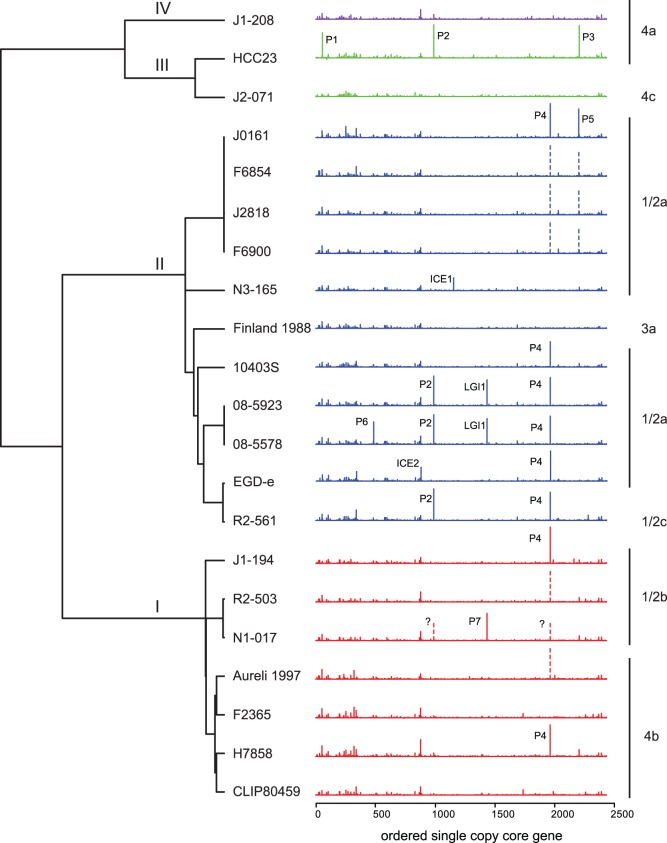
Utilizing the 2,086 genes identified as orthologous across all Listeria species, we constructed a phylogeny of L. monocytogenes genomes rooted with outgroup genomes of the closely related species L. innocua, L. welshimeri, and L. seeligeri (Fig. 1, outgroups not shown). This phylogeny agrees with previous phylogenetic analyses [52], [57] and divides L. monocytogenes into its four major lineages. To examine the positioning of the accessory genes along L. monocytogenes genomes in a phylogenetic context, the number of accessory genes between each core gene was plotted for each genome (Fig. 1). Positioning of accessory gene clusters is conserved across L. monocytogenes genomes, as was observed by Touchon et al. in E. coli [47]. The distribution of accessory gene clusters over the chromosome in L. monocytogenes seems to differ from that of E. coli in that in L. monocytogenes there is a high concentration of these accessory gene clusters close to the origin of replication. This is particularly true in the region spanning the first approximately 500 Kb of the chromosome. While this paper was under review, Kuenne et al. [58] published an analysis of accessory gene distribution using a largely non-overlapping set of L. monocytogenes strains, and also found genes clustered into insertion-deletion hotspots. Independent confirmation of these insertion-deletion hotspots in different sets of genomes by Kuenne et al. [58] and this study show that these hotspots are highly conserved among L. monocytogenes strains. Concentration of hotspots to the right of the origin of replication is also supported by Kuenne et al. [58], who found eight out of nine insertion deletion hotspots identified in their study to be positioned in the right replichore. In our work, however, we noted that genomic change is not restricted to these hotspots, but that the whole region of the chromosome adjacent to the origin of DNA replication is prone to insertion and deletion events (see below) and can be considered a ‘hot region’.
Figure 1. L. monocytogenes phylogenetic tree and accessory genome distribution plots.
Plots show the number of accessory genes in between each core gene as ordered in the reference strain EGDe. Insertion sites of prophages (P), integrated conjugative elements (ICE), and Listeria genomic islands (LGI) as detailed in Table 4 are indicated above each accessory genome distribution plot. Vertical dotted lines with a question mark indicate prophages, which are not assembled in a single contiguous piece, but are hypothesized to be present in the location based on presence of the appropriate phage genes in unalignable fraction of the assembly. Plots are colored by lineage: I, red, II, blue, III, green, IV, purple. Serotypes are shown to the right of each plot. The phylogenetic tree is based on a maximum likelihood analysis of the concatenated alignments of 2,086 core genes.
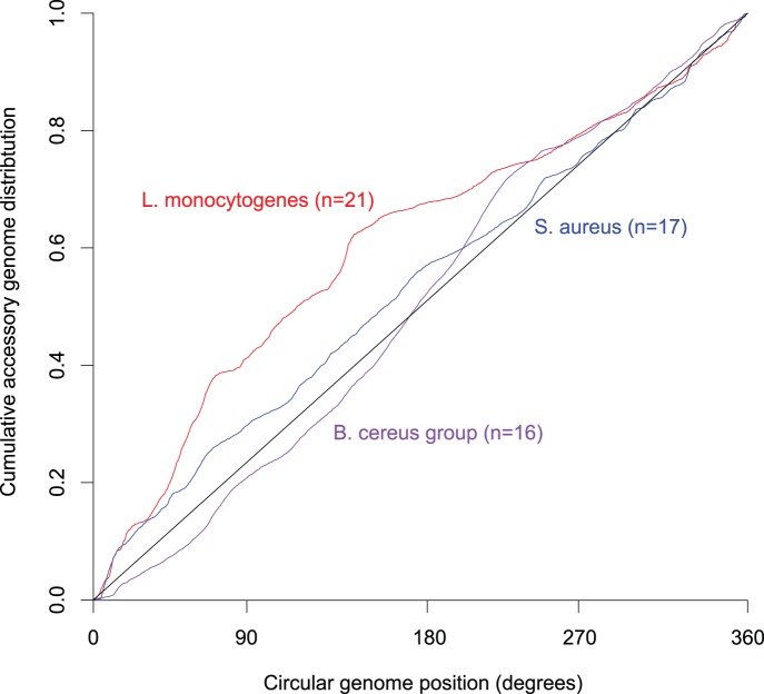
To test if this distribution is uniquely found in L. monocytogenes we plotted the cumulative distribution of accessory genes along the chromosome for L. monocytogenes, and the phylogenetically closely related species Staphylococcus aureus and the Bacillus cereus group, with the exclusion of prophage regions. L. monocytogenes shows a highly unequal distribution with 38% of the accessory genes found within the first 65° (approximately a 0.5 Mb region) from the origin of replication (χ2 = 2411, p<0.0001), while the accessory genomes of S. aureus and the B. cereus group are more evenly distributed over the chromosome (Fig. 2). The distributions of accessory genes in S. aureus and B. cereus were significantly different from that of L. monocytogenes (P<0.0001, Kruskal-Wallis test), confirming the uniqueness of the pattern found in L. monocytogenes.
Figure 2. Cumulative distribution of the accessory genome throughout the chromosome in L. monocytogenes (n = 21), Staphylococcus aureus (n = 17) and strains of the Bacillus cereus group (n = 16).
The circular genome position starts at the origin of replication, which is at 0 degrees.
To evaluate whether the strength of selection differs between the different regions of the genome and between core and accessory genes, we calculated dN/dS for all genes shared by at least two L. monocytogenes strains. As expected, we found that genes in the accessory genome are less selectively constrained than those in the core genome (median dN/dS = 0.131 and 0.036, respectively, p<0.001, Wilcoxon test). However we also found that core genes in the first 65° of the genome experience significantly less purifying selection than core genes in the last 295° of the genome (median dN/dS = 0.045 and 0.035, respectively, p<0.001, Wilcoxon test). The same pattern was also found for accessory genes (median dN/dS = 0.133 and 0.128, respectively, p = 0.003, Wilcoxon test). This suggests that irrespective of designation as a core or accessory, genes in the first 65° of the genome are more rapidly evolving than those in the last 295°.
We also found differences in the length of intergenic regions. Intergenic regions in the first 65° of the genome are significantly longer than intergenic regions in the last 295° of the genome (p<0.0001, Wilcoxon test). This difference in intergenic length distributions is the result of comparably long regions between neighboring accessory and core genes (median length 85 bp); intergenic regions between accessory and core genes are significantly more common in the first 65° relative to the last 295° (p<0.0001, chi-square test) of the chromosome. Interestingly, the core-core intergenic regions (median length = 45 bp) were found to be significantly longer than accessory-accessory intergenic regions (median length = 24 bp; p<0.0001, Wilcoxon test).
The Accessory Genome of L. monocytogenes is Enriched for Phosphotransferase Systems, Cell Surface Genes, and Prophages
Eight functional categories were found significantly overrepresented in the accessory genome of L. monocytogenes and were represented by more than 100 genes in each category (Table 3). These categories relate to four broad classes of genes: (i) phosphotransferase system (PTS) components (involved in sugar transport), (ii) cell wall components, (iii) transcriptional regulators (represented by the sequence-specific DNA binding term), and (iv) mobile elements (represented by the DNA integration term). The enrichment for mobile elements is likely reflective of the numerous large prophages that are unequally distributed across the different L. monocytogenes strains (Fig. 1; Table 4). The over-representation of genes corresponding to the remaining three categories likely represents a response to the diverse environmental pressures faced by L. monocytogenes.
Table 3. Top 25 most abundant Gene Ontology (GO) terms which are significantly enriched in the accessory genome versus the core genome of Listeria monocytogenes.
| Original GO term | No. of genes in | P value | GO description | |
| Core genome | Accessory genome | |||
| GO:0009401 | 1690 | 411 | 2.04E-45 | phosphoenolpyruvate-dependent sugar phosphotransferase system |
| GO:0008982 | 1235 | 335 | 1.10E-44 | protein-N(PI)-phosphohistidine-sugar phosphotransferase activity |
| GO:0005351 | 1020 | 333 | 1.35E-59 | sugar:hydrogen symporter activity |
| GO:0009986 | 302 | 246 | 2.81E-103 | cell surface |
| GO:0005618 | 363 | 233 | 1.25E-82 | cell wall |
| GO:0043565 | 533 | 150 | 6.87E-20 | sequence-specific DNA binding |
| GO:0009273 | 178 | 124 | 1.52E-45 | peptidoglycan-based cell wall biogenesis |
| GO:0015074 | 42 | 101 | 4.78E-67 | DNA integration |
| GO:0005576 | 169 | 70 | 4.30E-15 | extracellular region |
| GO:0004802 | 0 | 68 | 1.48E-67 | transketolase activity |
| GO:0005518 | 42 | 64 | 2.42E-35 | collagen binding |
| GO:0005529 | 250 | 60 | 1.53E-04 | sugar binding |
| GO:0004803 | 20 | 53 | 3.15E-35 | transposase activity |
| GO:0006313 | 62 | 53 | 2.95E-21 | transposition, DNA-mediated |
| GO:0004351 | 0 | 51 | 5.75E-50 | glutamate decarboxylase activity |
| GO:0006536 | 42 | 51 | 5.63E-25 | glutamate metabolic process |
| GO:0015755 | 104 | 48 | 3.86E-11 | fructose transport |
| GO:0015573 | 125 | 45 | 2.12E-07 | beta-glucoside transmembrane transporter activity |
| GO:0008861 | 0 | 42 | 1.15E-40 | formate C-acetyltransferase activity |
| GO:0000150 | 0 | 41 | 1.24E-39 | recombinase activity |
| GO:0008706 | 42 | 40 | 1.04E-16 | 6-phospho-beta-glucosidase activity |
| GO:0043624 | 126 | 40 | 4.22E-05 | cellular protein complex disassembly |
| GO:0047632 | 0 | 38 | 1.56E-36 | agmatine deiminase activity |
| GO:0006306 | 21 | 36 | 5.30E-20 | DNA methylation |
| GO:0006323 | 42 | 35 | 3.70E-13 | DNA packaging |
P values were calculated using Fisher’s exact test and Bonferroni corrected.
Table 4. Overview of prophage and Inserted Conjugative Elements (ICE) insertion sites in L. monocytogenes.
| Mobile element integration site in Fig. 1 | Type of element | gene closest to integration site (attC) | Insertion site occupied in1: |
| ICE1 | ICE | FlaR (lmo1412) | FSL N3-165 |
| ICE2 | Integrated conjugative element (ICE) | guaA (lmo1096) | EGD-e (tn916-like: ICELm1) |
| LGI1 | Genomic Island | RNA methyltransferase, TrmA family-Fosfomycin resistance protein FosX | 08-5578, 08-5923 |
| P1 | Prophage | tRNA-Lys | L99, HCC23 (PSA-like) |
| P2 | Prophage | tRNA-Arg | 08-5578, 08-5923, FSL R2-561, FSL N1-017 (B025-like) |
| P3 | Prophage | ribosomal protein S9 | L99, HCC23 (A500-like) |
| P4 | Prophage | ComK | J0161, F6854, J2818, F6900, 10403S, 08-5578, 08-5923, EGD-e, FSL R2-561, FSL J1-194, FSL R2-503, FSL N1-017.Aureli 1997, H7858 (A118-like) |
| P5 | Prophage | tRNA-Thr-4 | J0161, F6854, J2818, F6900 (A118-like) |
| P6 | Prophage | tRNA-Ser | 08-5578, 08-5923 (A118-like) |
| P7 | Prophage | hypothetical protein LMHG_11046 | FSL N1-017 (B054-like) |
Selection of strains in which this insertion site is occupied by a mobile element or prophage. Between parenthesis resemblance to sequenced phages is indicated.
To further examine evolutionary changes in the accessory genome, we identified accessory loci that distinguish the two major lineages of L. monocytogenes, I and II (Table 5). Lineage II has significantly more distinguishing genes than lineage I (38 vs. 21; p = 0.03, chi-square test). Most functional categories from the enrichment analysis are represented within the lineage specific operons – both lineages have specific PTS operons (including transcriptional regulators) and cell-wall anchored proteins (including internalins). Furthermore, each lineage had a specific antimicrobial resistance-related operon/gene (Table 5; lineage I, anti-microbial peptide ABC-type transport system; lineage II, bacteriocin immunity protein). Despite inclusion of only two representatives of lineage III in our analysis (HCC23 and J2-071), this lineage showed a large degree of variation with respect to presence/absence of loci it from distinguishing lineages I and II, consistent with a previous array-based study [14].
Table 5. Accessory genome loci that distinguish lineages I and II.
| Presence in Lineage1 | |||||
| I | II | III | IV | Loci | Putative Function2 |
| + | – | + | + | LMOf2365_0374 | Internalin |
| + | – | +/− | + | LMOf2365_0413-0417 | ABC-type antimicrobial peptide transport system, cell-wall-anchored protein |
| + | – | – | + | LMOf2365_0693-0694 | Cell-wall-anchored proteins |
| + | – | + | + | LMOf2365_1131 | Unknown |
| + | – | + | + | LMOf2365_1142-1143 | Unknown |
| + | – | +/− | +/− | LMOf2365_1252-1254 | Internalin, cell-wall-anchored |
| + | – | +/− | – | LMOf2365_1681-1683 | N-acetylmuramic acid specific PTS |
| + | – | – | – | LMOf2365_2059 | Regulatory protein |
| + | – | – | + | LMOf2365_2361 | cAMP-binding protein |
| + | – | + | + | LMOf2365_2416 | Internalin |
| + | – | – | – | LMOf2365_2638 | Cell-wall-anchored protein |
| – | + | +/− | – | lmo0147 | Unknown |
| – | + | – | + | lmo0171 | Internalin, cell-wall-anchored |
| – | + | – | – | lmo0332 | Unknown |
| – | + | +/− | – | lmo0341 | Bacteriocin Immunity protein |
| – | + | +/− | – | lmo0421-0423 | Cell division, lineage-specific thermal regulator protein, RNA polymerase factor sigma C |
| lmo0525 | Unknown | ||||
| – | + | – | – | lmo0734-0739 | PTS, putative pentose phosphate specific |
| – | + | +/− | – | lmo0749-750 | putative regulatory protein |
| – | + | + | – | lmo0780 | Unknown |
| – | + | – | – | lmo1060-1063 | heavy metal associated two component response system and ABC transporter |
| – | + | + | + | lmo1125 | Unknown |
| – | + | + | + | lmo1289 | Internalin-like protein, Cell-wall-anchored |
| – | + | + | + | lmo1307 | Unknown |
| – | + | – | – | lmo1968-1974 | Creatinine amidohydrolase, KDPG and KHGaldolase, L-ascorbate specific PTS |
| – | + | + | – | lmo2169 | Unknown |
| – | + | +/− | – | lmo2576 | Putative collagen adhesion protein, cell-wall-anchored |
| – | + | + | + | lmo2644 | Regulation of 1,3-beta-glucan synthase |
| – | + | – | – | lmo2686 | Unknown |
| – | + | +/− | – | lmo2786-2788 | glucose–glucoside (Glc) family PTS |
+ = present, – = absent, +/− = present in some strains.
The putative function is inferred from the initial gene annotation.
Presence/absence of orthologs in each of the four lineages is listed, as well as putative function and the locus identifier(s) in the reference genome, either F2365 (lineage I) or EGDe (lineage II).
O-antigen Associated Genes seem to Follow a Serotype Specific Phylogenetic Pattern and show Several Instances of Horizontal Gene Transfer
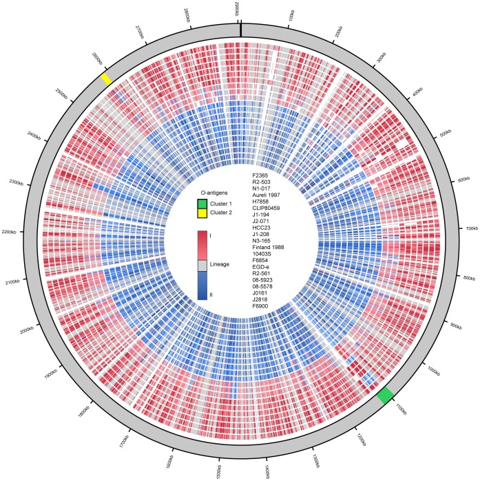
A phylogenetic approach to identify genes with evolutionary histories that deviate from the organismal phylogeny identified two gene clusters: (i) a cluster corresponding to lmo1074–1091 in L. monocytogenes EGD-e (cluster 1), and (ii) a cluster (cluster 2) corresponding to lmo2549-2558 in L. monocytogenes EGD-e (Fig. 3). These clusters are found in distinct regions of the genome; however, they both contain genes implicated in the biosynthesis of wall teichoic and lipoteichoic acids. Wall teichoic acids are associated with O-antigen variation [7], [59], [60] and because of this putative involvement, we will refer to these clusters as O-antigen clusters 1 and 2. For these clusters, the lineage I serotype 1/2b strains appear to have genes that are much more closely related to their orthologs in lineage II, which includes all the 1/2a and 3c strains, than to their orthologs in other lineage I strains (Fig.3). The phylogenetic distribution of serotype 1/2 related genes is incongruent with the organismal phylogeny (Fig. 1), and therefore horizontal transfer of these clusters from lineage II into lineage I could explain the occurrence of 1/2 serotypes in both lineages.
Figure 3. Clade membership plot of individual genes plotted against the genome of L. monocytogenes F2365.
The order of genome rings is listed in the circle center, with F2365 being the outermost ring. The 7 outermost rings represent lineage I (serotype 4b and 1/2b), the next three rings represent lineage III and lineage IV strains (serotype 4a and 4c), and the last 11 rings represent lineage II strains (serotype 1/2a, 1/2c, and 3a). Clade membership of the individual genes is indicated by color; blue indicates lineage II, red indicates lineage I, and gray is unresolved membership. The two O-antigen gene clusters are highlighted in green and yellow. Genes in these clusters found in serotype 1/2b lineage I cluster phylogenetically with orthologs found in lineage II clade.
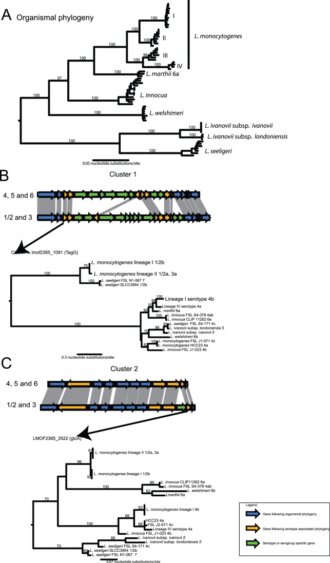
Within a serotype (1/2 or 4, irrespective of alphabetical designation), all L. monocytogenes strains have largely the same gene content and order across both clusters (Fig. 4A, Figs. S1 and S2). Exceptions are a hypothetical protein (LMOf2365_1098 in strain F2365) in cluster 1 of lineage I serotype 4b strains and the lineage IV serotype 4 strain FSL J1-208. Between serotypes, O-antigen clusters 1 and 2 substantially differ in gene content (Fig. 4A, Fig. S1 and 2). The genomes of newly sequenced serotype 3a and 1/2c strains have identical gene content in the two serotype clusters as 1/2a strains, consistent with the phylogeny based on the concatenated alignments of the 2,086 core genes, which places the serotype 3a and 1/2c genomes among lineage II 1/2a strains (Fig. 1).
Figure 4. Synteny and gene-specific phylogenetic history of the two O-antigen specific gene clusters.
The organismal phylogeny of the genus Listeria is shown in the upper panel (A), while the syntenic relationships of the two O-antigen gene clusters between the two major serotype divisions and the phylogenetic tree based on a representative serotype specific gene are shown in the two lower panels (B and C). Genes are colored by their phylogenetic histories: Serotype-specific genes (i.e., genes found only in specific serotypes) are colored green, while genes displaying an organismal phylogeny across the Listera genus are colored blue. Genes which follow a serotype-related phylogeny across Listeria are shown in orange. Values on the branches represent bootstrap values based on 100 bootstrap replicates. The organismal tree is based on a 10 locus multi-locus sequence analysis as described in Den Bakker et al. [52]. The topology of this tree is congruent with a tree based on the MLST scheme used in Ragon et al. [6].
To determine if the phylogeny of O-antigen cluster genes is discordant with the organismal phylogeny across the entire Listeria genus we analyzed the gene content and synteny for both clusters in non-L. monocytogenes Listeria species for which genome sequences are available. In addition, we investigated other genes outside the two clusters which displayed a serotype related phylogenetic pattern, genes that were uniquely found within one serotype or the other, and genes that had been implicated in L. monocytogenes O-antigen variation in previous publications [20], [61], [62] (see supplemental Table S1 for key results). To aid in the analysis we also serotyped five additional Listeria strains (see Table 1). Gene content and gene order in cluster 1 was found to be highly similar between serotypes 1/2 (found in L. monocytogenes and L. seeligeri), 3 and 7 (found in L. seeligeri FSL N1-067 and in L. monocytogenes [58]), irrespective of the species in which the cluster was found (Fig. S1). While gene content and gene order in cluster 1 in serotypes 1/2, 3 and 7 are extremely similar among L. monocytogenes strains and even between species (L. seeligeri versus L. monocytogenes), we found this cluster to display subtle differences when serotypes 4, 5 and 6 were compared. Cluster 1 in L. innocua CLIP 11262 (serotype 6a) was found to be identical in gene content and gene order to L. monocytogenes serotype 4b and L. monocytogenes FSL J1-208 (serotype 4a). Gene content and gene order in cluster 1 of L. welshimeri SLCC5334 serotype 6b was found to be identical to L. monocytogenes serotype 4a (strain HCC23) and serotype 4c (strain FSL J2-071). We further found homologs of gltA and gltB in cluster 1 in L. innocua FSL J1-023 serotype 4b and in L. ivanovii serotype 5 (see Fig. S1). The gltA-gltB gene cassette was previously reported to be serotype 4b specific and involved in wall teichoic acid glycosylation [61]. This gene cassette is found in a region approximately 1.6 Mb removed from cluster 1 in L. monocytogenes serotype 4b isolates such as F2365 (LMOf2365_2740 and LMOf2365_2741).
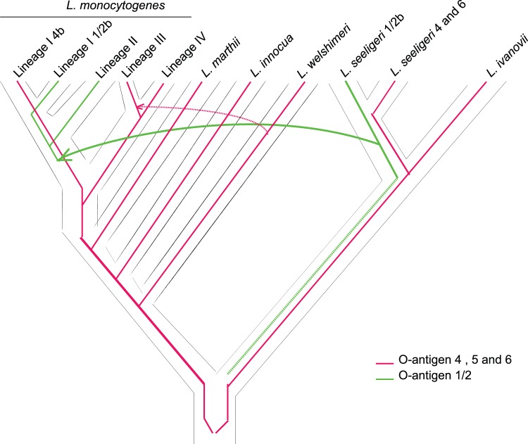
To further probe the evolution of the two O-antigen clusters, we constructed gene phylogenies for genes, within these clusters, that had orthologs in both serotypes 1/2 and 4. Two phylogenetic patterns could be found among the shared genes in O-antigen cluster 1 (Fig. 4B): (i) a serotype-specific pattern, showing a clade consisting of serotypes 1/2, 3 and 7 and a clade consisting of serotypes 4, 5, and 6, (Fig. 4B, orange pattern; seven genes), and (ii) a pattern mirroring the organismal phylogeny of Listeria (Fig. 4B, blue pattern). The shared genes in cluster 2 also showed two distinct phylogenetic patterns (Fig. 4C): (i) a phylogenetic pattern reminiscent of the organismal phylogeny of Listeria and similar to that seen in cluster 1 (Fig. 4C, blue pattern), and (ii) a serotype-associated pattern for L. monocytogenes, L. innocua, L. welshimeri and L. marthii, but a non-serotype specific pattern for L. seeligeri and L. ivanovii (Fig. 4C, orange pattern; three genes). Cluster 1 genes with a serotype specific phylogenetic pattern were tagG (LMOf2365_1091) and tagH (LMOf2365_1092), an UTP-glucose-1-phosphate uridylyltransferase (homologous to rfbA: LMOf2365_1099), a glycosyl transferase (LMOf2365_1100), ribitol-5-phosphate cytidylyltransferase (LMOf2365_1101), tagB (CDP-glycerol:N-acetyl-beta-D-mannosaminyl-1,4-N-acetyl-D-glucosaminyldiphosphoundecaprenylglycerophosphotransferase: LMOf2365_1104) and a putative sorbitol dehydrogenase (LMOf2365_1105). Shared genes with a serotype specific phylogenetic pattern in cluster 2 were an autolysin (LMOf2365_2530), a gene annotated as UDP-N-acetylglucosamine 1-carboxyvinyltransferase (LMOf2365_2524), a transcription termination factor (LMOf2365_2523), and the cell wall teichoic acid glycosylation protein GtcA (LMOf2365_2522). Most of these shared genes with a serotype-associated phylogenetic pattern are homologous to genes implicated in basic functions in wall teichoic acid synthesis in other Firmicutes [63], [64], and in L. monocytogenes [60], [65], [66]. All wall teichoic acid associated genes that display a serotype-associated phylogenetic pattern show a high nucleotide divergence (e.g., 8.2–40%) between homologous genes of lineage I L. monocytogenes serotype 4b and L. monocytogenes 1/2b strains, while the nucleotide divergence between L. monocytogenes 1/2a (lineage II) and L. monocytogenes 1/2b (lineage I) strains is between 1.0 and 2.7%. The high nucleotide divergence suggests that 1/2- and 4- like serotypes predate the most common ancestor of L. monocytogenes. The fact that L. monocytogenes lineage III and IV, and closely related species such as L. marthii and L. innocua display 4 and 6 like serotypes, suggests that the most recent common ancestor of L. monocytogenes putatively was of serotype 4, and the 1/2-like serotypes were introduced, through horizontal gene transfer, in the ancestral population of L. monocytogenes lineage I and II. Alternatively both 1/2-like and 4-like serotypes could have been present in the ancestral L. monocytogenes population, and 4-like serotypes were subsequently lost in lineage II.
To reconstruct the putative evolutionary history of serotypes in L. monocytogenes we reconciled the gene trees with serotype-specific patterns (Fig. 4B, red and orange patterns) with the organismal tree of the genus Listeria (similar to Fig. 4B, blue pattern) using the AnGST [53] and Mowgli [54] algorithms. Both algorithms simultaneously account for gene loss, gene duplications and horizontal gene transfer. The majority of the reconciliations for both cluster 1 genes (6/7 genes) and cluster 2 genes (3/3 genes) support a scenario in which horizontal gene transfer was responsible for the introduction of the 1/2 serotypes in the ancestral population of L. monocytogenes lineage I and II (Fig. 5). In the case of cluster 1, the putative donor of the genes encoding expression of the L. monocytogenes 1/2 serotypes was the ancestral population of L. seeligeri. Reconciliations of the cluster 2 genes suggest that the 1/2 serotypes arose once, either in the ancestral populations of L. welshimeri or L. seeligeri. The gene cluster was then transferred from these populations into the ancestral population of L. monocytogenes lineage I and II, and were subsequently lost in the donor populations.
Figure 5. Phylogenetic reconstruction of serotype evolution in Listeria. Serotype 4 is shown in red while serotype 1/2 is shown in green.
This construction suggests that serotype 1/2 genes were horizontally transferred from L. seeligeri to an ancestor of L. monocytogenes lineages I and II. The origin of the serotype 1/2 cluster is unclear, we hypothesize that this cluster putatively originated in the most recent common ancestor of the L. seeligeri and L. ivanovii clade (as indicated by the dashed line). Serotype 4 genes appear to be largely inherited by vertical descent, except for a lateral transfer of genes from L. welshimeri into some strains of L. monocytogenes lineage III (dotted red line).
In contrast to genes of the serotype 1/2 gene clusters, the serotype 4 O-antigen clusters followed a largely vertical descent through Listeria species (Fig. 5). The one exception to this mode of inheritance appears to be a replacement, in lineage III serotype 4a and 4c strains, of part of the ancestral O-antigen cluster 1 with a L. welshimeri type O-antigen cluster 1 through horizontal transfer. Horizontal transfer of the O-antigen cluster 1 into lineage III serotype 4a and 4c strains is further supported by the similarity in synteny of this cluster in both donor (L. welshimeri SLCC5334) and recipient (L. monocytogenes lineage III serotype 4a and 4c; see Fig. S1). All gene tree reconciliations support a most recent common ancestor of L. monocytogenes, which had serotype 4.
The phylogenetic patterns detailed above suggest the occurrence of homologous recombination within cluster 2 between L. monocytogenes donors and recipients. To test for homologous recombination and sequence tracts involved in these recombination events we used RAT [55] to detect putative breakpoints. We subjected sequences representing the entire cluster 2 (minus large indel regions) of L. monocytogenes serotypes 1/2b, lineage I 4b, and 1/2a to this analysis. The results of this analysis suggest that two sequence tracts within cluster 2 were putatively introduced into the lineage I serotype 1/2b strains from a lineage II serotype 1/2a donor. These tracts include (i) a tract encoding part of a homoserine dehydrogenase (lmo2547), the entire 50S ribosomal protein L31, gtcA, transcription termination factor Rho, UDP-N-acetylglucosamine 1-carboxyvinyltransferase, a hypothetical protein (lmo2555 homolog) and a glycosyl transferase, and (ii) a tract encoding an autolysin.
Discussion
L. monocytogenes genomes are highly conserved and free of major genomic rearrangements even when compared to closely related Listeria species [67]. However, our work here suggests that this picture does not fully represent what appears to be an unappreciated property of this species; the Listerial genomes show evidence for uneven vulnerability to the gain of, or tolerance for, horizontal transfer based on position in the genome. The first 65° of the chromosome is enriched for accessory genes, while the last 295° is enriched for core genes; this genome compartmentalization is absent from the closely related bacteria such as S. aureus and B. cereus. There could be an adaptive value in such a behavior although the molecular mechanism responsible for this is unresolved. We also find a series of genes, which cluster phylogenetically according to serotype, but not according to the organismal phylogeny. The majority of these genes is organized in two gene clusters, and reconstruction of the putative evolutionary history of these clusters shows these genes have a complex evolutionary history, involving multiple instances of horizontal gene transfer.
The enrichment of the first 65° degrees of the genome for accessory genes can only be partly attributed to the eight hotspots recently described by Kuenne et al. [58] for this chromosomal region, as less than 25% of the accessory genome could be attributed to these hotspots. Overall, we found 38% of the accessory genome (prophage related genes not included) in the first 65° degrees of the genome. Kuenne et al. [58] used a strict definition of an insertion deletion hotspot (‘hotspots were defined by the localization of at least three non-homologous insertions between mutually conserved core genes’). We find that a large part of the accessory genome found in the first 65° degrees is found outside of the eight hotspots identified previously [58] and in the work reported here. We thus propose that this portion of the chromosome may be more accurately described as a "hot region" for the gain of horizontally acquired information.
The genome partitions we find in L. monocytogenes appear to stem from differences in selective pressures and different rates of gene insertion. The former is supported by the finding in L. monocytogenes genomes that core genes in the first 65° of the genome are under less purifying selection than genes in the last 295°, indicating that to some extent, the position of a gene within the genome may affect its rate of evolution regardless of whether the gene is part of the core or accessory genome. The size of intergenic regions is thought to be driven by, and reflective of, the balance between insertions and deletions [68]. The longer intergenic distances in accessory-rich region of the genome may reflect the dynamic nature of this region where the balance is tipped toward insertions of new accessory operons.
What molecular mechanism could account for one region becoming more prone to the accretion of foreign DNA? One possible explanation could involve systems that physically sequester regions of the genome. For example in E. coli the terminus region is physically and functionally gathered together through the action of the MatP protein that recognizes a series of sites (matS) in this region of the chromosome [69]. This region containing matS sites is constrained by another protein that seems to allow the terminus region to interact with the division machinery [70]). If a similar system worked in the first 65 degrees of the L. monocytogenes chromosome it could conceivable render this region differentially accessible for new DNA sequences that enter the cell. Interestingly the terminus region of the E. coli chromosome appears to evolve differently from the rest of the genome displaying lower rates of recombination without higher mutation rates [47].
Alternatively, as suggested previously for E. coli [47], one could also imagine a series of "domino" effects that follow the acquisition of a very large segment of DNA. If beneficial gene products were encoded in this DNA segment it could encourage maintenance of the new large DNA segment. However, genes on this same stretch of DNA that were under negative selection or were neutral would allow (if not encourage) the acquisition of more insertions. This entire new region would then be active for gain and loss of genes for a protracted period of time as deletions also occurred across the regions under negative or neutral selection. Eventually the original genes that allowed the new DNA to become fixed in the population would be unrecognizable from other core genes from the species, but the process of gaining more genetic information in the region and winnowing of the sequences under negative and neutral selection could occur over a much longer period of time. The net result would be a mosaic of core and accessory genes without any necessary association to mobile elements. Interestingly only one complete prophage can be found in the first 65° of the chromosome. Core genes found in this region may have only relatively recently become fixed in the population (or part of the core genome), which may explain why this region is more rapidly evolving compared to the rest of the chromosome.
Regardless of the mechanism that accounts for the regional effect suggested by our analyses, the compartmentalization of the Listeria chromosome into accessory gene rich and poor regions could provide an evolutionary risk management strategy analogous to one recently described in E. coli, where the chromosome is divided into mutational hot and cold spots [71]. In E. coli, mutational cold spots (regions with a lower mutation rate) coincide with highly expressed genes and genes under strong purifying selection, thereby reducing the risk of deleterious mutations in these regions [71].
Functional enrichment of transcriptional regulators, cell surface genes, and phosphotransferase systems in the accessory genome highlights the selective pressures faced by contemporary strains of L. monocytogenes. The complex regulation potentially required for networks of auxiliary or core genes to respond to these pressures may explain the abundance of transcription factors among the auxiliary genome. Enrichment of cell surface-related genes in the accessory genome of suggests that there is sustained selective pressure on L. monocytogenes to continually remodel the cell surface, playing a putative role in host specificity, host interactions, and the evasion of predators such as bacteriophages and protists in the non-host environment. Enrichment of cell surface-related genes in L. monocytogenes was also found in previous array based studies [14], [72]. These cell wall-enriched accessory genes include internalins, a class of genes that also encodes well characterized virulence factors such as internalin A, internalin B and internalin C [73]. The finding that phosphotransferase systems are enriched in the auxiliary genome suggests a selective pressure for L. monocytogenes to maintain a diverse repertoire of sugar transporters to cope with the diverse carbon sources in both hosts and the environment [74]. Another explanation for the diversification of phosphotransferase systems could involve interaction with other microbes, as it has been shown that certain phosphotransferase systems in L. monocytogenes are putative targets for bacteriocins [75]. A high diversity of phosphotransferase systems, combined with functional redundancy, may be a way to reduce bacteriocin sensitivity within host microbial communities.
While most genes in the L. monocytogenes genome follow a pattern of vertical descent, O-antigen associated genes and gene clusters seem to have distinct phylogenetic histories suggesting lateral transfers. A gene-by-gene gene-tree reconciliation approach suggests lateral transfer of O-antigen cluster 1 from a serotype 1/2 or 7 L. seeligeri ancestor into the serotype 4 L. monocytogenes ancestor. A putative change of function of O-antigen associated genes in cluster 2 in the L. seeligeri donor could explain the discrepancy between the phylogenetic patterns of cluster 1 and cluster 2 genes, where cluster 1 genes show a serotype specific pattern across Listeria species and cluster 2 genes only show a serotype-specific pattern within L monocytogenes. The fact that O-antigen cluster 2 genes in L. seeligeri 1/2b or 7 do not phylogenetically cluster according to serotype, suggests that genes in O-antigen cluster 1 are probably the most important determinants of O-antigen serotype. A break point analysis of L. monocytogenes cluster 2 suggests that Lineage I 1/2b serotype strains only recently acquired the serotype 1/2 gene fragments from Lineage II 1/2a donors. Further experimental work will be needed to clarify the role of cluster 1 and cluster 2 genes in serotype expression in different L. monocytogenes and Listeria species serotypes.
While serotype 1/2 was previously hypothesized to be the ancestral serotype in L. monocytogenes [6], our data support the alternative hypothesis, proposed here for the first time that 4-like serotypes were present in the ancestral population of L. monocytogenes lineages. This hypothesis seems to be supported by the observation that both lineage III and IV display 4-like serotypes, while the species most closely related to L. monocytogenes (i.e., L. innocua and L. marthii) also have 4-like serotypes. Based on the current data it is hard to refute the possibility that genes encoding serotype 1/2 expression (i.e., the clusters associated with this O-antigens) were introduced in the ancestor of both lineages I and II, and subsequently replaced by serotype 4 genes in a subset of lineage I. Additionally, while our gene tree reconciliations suggest that L. seeligeri was a donor of clusters 1/2, the reverse transfer cannot be excluded at this stage. More research on the function and evolution of these O-antigen related genes is necessary to unravel their complex evolutionary history and involvement in host-pathogen and bacteriophage interactions.
Supporting Information
Comparison of O-antigen cluster 1 in L. monocytogenes and closely related Listeria species.
(EPS)
Comparison of O-antigen cluster 2 in L. monocytogenes and closely related Listeria species.
(PDF)
Summary of phylogenetic patterns found for wall teichoic and lipoteichoic acid associated genes in Listeria .
(PDF)
Acknowledgments
We acknowledge the contribution of the Broad Institute Genome Sequencing Platform. We would like to thank Dr. Zuzana Kecerova and Dr. Cheryl L. Tarr from the Centers for Disease Control and Prevention, Atlanta GA for serotyping L. innocua, L. marthii and L. seeligeri isolates. We thank Renato H. Orsi and Jennifer Wortman for their helpful comments on the manuscript.
Funding Statement
This project has been funded in whole or in part with Federal funds from the National Institute of Allergy and Infectious Diseases, National Institutes of Health, Department of Health and Human Services, under Contract No.: HHSN272200900018C. The funders had no role in study design, data collection and analysis, decision to publish, or preparation of the manuscript.
References
- 1. Gray MJ, Freitag NE, Boor KJ (2006) How the bacterial pathogen Listeria monocytogenes mediates the switch from environmental Dr. Jekyll to pathogenic Mr. Hyde. Infect Immun 74: 2505–2512 doi:10.1128/IAI.74.5.2505-2512.2006 [DOI] [PMC free article] [PubMed] [Google Scholar]
- 2. Orsi RH, Bakker den HC, Wiedmann M (2011) Listeria monocytogenes lineages: Genomics, evolution, ecology, and phenotypic characteristics. Int J Med Microbiol 301: 79–96 doi:10.1016/j.ijmm.2010.05.002 [DOI] [PubMed] [Google Scholar]
- 3. Roberts AJ, Nightingale KK, Jeffers G, Fortes ED, Kongo JM, et al. (2006) Genetic and phenotypic characterization of Listeria monocytogenes lineage III. Microbiology (Reading, Engl) 152: 685–693 doi:10.1099/mic.0.28503-0 [DOI] [PubMed] [Google Scholar]
- 4. Bille J, Rocourt J (1996) WHO International Multicenter Listeria monocytogenes Subtyping Study–rationale and set-up of the study. Int J Food Microbiol 32: 251–262. [DOI] [PubMed] [Google Scholar]
- 5.Seeliger HPR, Höhne K (1979) Chapter II Serotyping of Listeria monocytogenes and Related Species. In: Norris TBAJR, editor. Methods in Microbiology. Methods in Microbiology. Academic Press, Vol. Volume 13. 31–49T2–. doi: 10.1016/S0580-9517(08)70372-6.
- 6. Ragon M, Wirth T, Hollandt F, Lavenir R, Lecuit M, et al. (2008) A New Perspective on Listeria monocytogenes Evolution. PLoS Pathog 4: e1000146 doi:10.1371/journal.ppat.1000146 [DOI] [PMC free article] [PubMed] [Google Scholar]
- 7. Fiedler F (1988) Biochemistry of the cell surface of Listeria strains: a locating general view. Infection 16 Suppl 2S92–S97. [DOI] [PubMed] [Google Scholar]
- 8. Uchikawa K, Sekikawa I, Azuma I (1986) Structural studies on lipoteichoic acids from four Listeria strains. J Bacteriol 168: 115–122. [DOI] [PMC free article] [PubMed] [Google Scholar]
- 9. Kamisango K, Fujii H, Okumura H, Saiki I, Araki Y, et al. (1983) Structural and immunochemical studies of teichoic acid of Listeria monocytogenes . J Biochem 93: 1401–1409. [DOI] [PubMed] [Google Scholar]
- 10. Uchikawa K, Sekikawa I, Azuma I (1986) Structural studies on teichoic acids in cell walls of several serotypes of Listeria monocytogenes . J Biochem 99: 315–327. [DOI] [PubMed] [Google Scholar]
- 11. Zhang C, Zhang M, Ju J, Nietfeldt J, Wise J, et al. (2003) Genome diversification in phylogenetic lineages I and II of Listeria monocytogenes: identification of segments unique to lineage II populations. J Bacteriol 185: 5573–5584. [DOI] [PMC free article] [PubMed] [Google Scholar]
- 12.Grimont PAD, Weill F (2007) Antigenic formulae of the Salmonella serovars. WHO Collaborating Centre for Reference and Research on Salmonella, Institut Pasteur, Paris, France.
- 13. Guibourdenche M, Roggentin P, Mikoleit M, Fields PI, BockemUhl J, et al. (2010) Supplement 2003–2007 (No. 47) to the White-Kauffmann-Le Minor scheme. Research in Microbiology 161: 26–29 doi:10.1016/j.resmic.2009.10.002 [DOI] [PubMed] [Google Scholar]
- 14. Deng X, Phillippy AM, Li Z, Salzberg SL, Zhang W (2010) Probing the pan-genome of Listeria monocytogenes: new insights into intraspecific niche expansion and genomic diversification. BMC Genomics 11: 500 doi:10.1186/1471-2164-11-500 [DOI] [PMC free article] [PubMed] [Google Scholar]
- 15. Orsi RH, Sun Q, Wiedmann M (2008) Genome-wide analyses reveal lineage specific contributions of positive selection and recombination to the evolution of Listeria monocytogenes . BMC Evol Biol 8: 233 doi:10.1186/1471-2148-8-233 [DOI] [PMC free article] [PubMed] [Google Scholar]
- 16. Dunn KA, Bielawski JP, Ward TJ, Urquhart C, Gu H (2009) Reconciling ecological and genomic divergence among lineages of Listeria under an “extended mosaic genome concept.”. Mol Biol Evol 26: 2605–2615 doi:10.1093/molbev/msp176 [DOI] [PubMed] [Google Scholar]
- 17. Tettelin H, Riley D, Cattuto C, Medini D (2008) Comparative genomics: the bacterial pan-genome. Curr Opin Microbiol 11: 472–477. [DOI] [PubMed] [Google Scholar]
- 18. Coleman ML, Chisholm SW (2010) Ecosystem-specific selection pressures revealed through comparative population genomics. Proc Natl Acad Sci USA 107: 18634–18639 doi:10.1073/pnas.1009480107 [DOI] [PMC free article] [PubMed] [Google Scholar]
- 19. Steele CL, Donaldson JR, Paul D, Banes MM, Arick T, et al. (2011) Genome sequence of lineage III Listeria monocytogenes strain HCC23. J Bacteriol 193: 3679–3680 doi:10.1128/JB.05236-11 [DOI] [PMC free article] [PubMed] [Google Scholar]
- 20. Nelson KE, Fouts DE, Mongodin EF, Ravel J, DeBoy RT, et al. (2004) Whole genome comparisons of serotype 4b and 1/2a strains of the food-borne pathogen Listeria monocytogenes reveal new insights into the core genome components of this species. Nucleic Acids Res 32: 2386–2395 doi:10.1093/nar/gkh562 [DOI] [PMC free article] [PubMed] [Google Scholar]
- 21. Glaser P, Frangeul L, Buchrieser C, Rusniok C, Amend A, et al. (2001) Comparative genomics of Listeria species. Science 294: 849–852 doi:10.1126/science.1063447 [DOI] [PubMed] [Google Scholar]
- 22. Hain T, Steinweg C, Kuenne CT, Billion A, Ghai R, et al. (2006) Whole-genome sequence of Listeria welshimeri reveals common steps in genome reduction with Listeria innocua as compared to Listeria monocytogenes . J Bacteriol 188: 7405–7415 doi:10.1128/JB.00758-06 [DOI] [PMC free article] [PubMed] [Google Scholar]
- 23. Steinweg C, Kuenne CT, Billion A, Mraheil MA, Domann E, et al. (2010) Complete genome sequence of Listeria seeligeri, a nonpathogenic member of the genus Listeria. J Bacteriol 192: 1473–1474 doi:10.1128/JB.01415-09 [DOI] [PMC free article] [PubMed] [Google Scholar]
- 24.Bakker den HC, Bowen BM, Rodriguez-Rivera LD, Wiedmann M (2012) FSL J1–208: a virulent uncommon phylogenetic lineage IV Listeria monocytogenes strain with a small chromosome size and a putative virulence plasmid carrying internalin-like genes. Appl Environ Microbiol. doi:10.1128/AEM.06969-11. [DOI] [PMC free article] [PubMed]
- 25. Gilmour MW, Graham M, van Domselaar G, Tyler S, Kent H, et al. (2010) High-throughput genome sequencing of two Listeria monocytogenes clinical isolates during a large foodborne outbreak. BMC Genomics 11: 120 doi:10.1186/1471-2164-11-120 [DOI] [PMC free article] [PubMed] [Google Scholar]
- 26. Kämper J, Kahmann R, Bölker M, Ma L-J, Brefort T, et al. (2006) Insights from the genome of the biotrophic fungal plant pathogen Ustilago maydis . Nature 444: 97–101 doi:10.1038/nature05248 [DOI] [PubMed] [Google Scholar]
- 27. Margulies M, Egholm M, Altman WE, Attiya S, Bader JS, et al. (2005) Genome sequencing in microfabricated high-density picolitre reactors. Nature 437: 376–380 doi:10.1038/nature03959 [DOI] [PMC free article] [PubMed] [Google Scholar]
- 28. Bentley DR, Balasubramanian S, Swerdlow HP, Smith GP, Milton J, et al. (2008) Accurate whole human genome sequencing using reversible terminator chemistry. Nature 456: 53–59 doi:10.1038/nature07517 [DOI] [PMC free article] [PubMed] [Google Scholar]
- 29. Jaffe DB, Butler J, Gnerre S, Mauceli E, Lindblad-Toh K, et al. (2003) Whole-genome sequence assembly for mammalian genomes: Arachne 2. Genome Res 13: 91–96 doi:10.1101/gr.828403 [DOI] [PMC free article] [PubMed] [Google Scholar]
- 30. Delcher AL, Harmon D, Kasif S, White O, Salzberg SL (1999) Improved microbial gene identification with GLIMMER. Nucleic Acids Res 27: 4636–4641. [DOI] [PMC free article] [PubMed] [Google Scholar]
- 31. Noguchi H, Park J, Takagi T (2006) MetaGene: prokaryotic gene finding from environmental genome shotgun sequences. Nucleic Acids Res 34: 5623–5630 doi:10.1093/nar/gkl723 [DOI] [PMC free article] [PubMed] [Google Scholar]
- 32. Borodovsky M, McIninch J (1993) Recognition of genes in DNA sequence with ambiguities. BioSystems 30: 161–171. [DOI] [PubMed] [Google Scholar]
- 33. Holder JW, Ulrich JC, DeBono AC, Godfrey PA, Desjardins CA, et al. (2011) Comparative and functional genomics of Rhodococcus opacus PD630 for biofuels development. PLoS Genet 7: e1002219 doi:10.1371/journal.pgen.1002219 [DOI] [PMC free article] [PubMed] [Google Scholar]
- 34. Delcher AL, Kasif S, Fleischmann RD, Peterson J, White O, et al. (1999) Alignment of whole genomes. Nucleic Acids Res 27: 2369–2376. [DOI] [PMC free article] [PubMed] [Google Scholar]
- 35. Brudno M, Do CB, Cooper GM, Kim MF, Davydov E, et al. (2003) LAGAN and Multi-LAGAN: efficient tools for large-scale multiple alignment of genomic DNA. Genome Res 13: 721–731 doi:10.1101/gr.926603 [DOI] [PMC free article] [PubMed] [Google Scholar]
- 36. Lagesen K, Hallin P, Rødland EA, Staerfeldt H-H, Rognes T, et al. (2007) RNAmmer: consistent and rapid annotation of ribosomal RNA genes. Nucleic Acids Res 35: 3100–3108 doi:10.1093/nar/gkm160 [DOI] [PMC free article] [PubMed] [Google Scholar]
- 37. Lowe TM, Eddy SR (1997) tRNAscan-SE: a program for improved detection of transfer RNA genes in genomic sequence. Nucleic Acids Res 25: 955–964. [DOI] [PMC free article] [PubMed] [Google Scholar]
- 38. Griffiths-Jones S, Moxon S, Marshall M, Khanna A, Eddy SR, et al. (2005) Rfam: annotating non-coding RNAs in complete genomes. Nucleic Acids Res 33: D121–D124 doi:10.1093/nar/gki081 [DOI] [PMC free article] [PubMed] [Google Scholar]
- 39. Götz S, García-Gómez JM, Terol J, Williams TD, Nagaraj SH, et al. (2008) High-throughput functional annotation and data mining with the Blast2GO suite. Nucleic Acids Res 36: 3420–3435 doi:10.1093/nar/gkn176 [DOI] [PMC free article] [PubMed] [Google Scholar]
- 40. Li L, Stoeckert CJ, Roos DS (2003) OrthoMCL: identification of ortholog groups for eukaryotic genomes. Genome Res 13: 2178–2189 doi:10.1101/gr.1224503 [DOI] [PMC free article] [PubMed] [Google Scholar]
- 41. Edgar RC (2004) MUSCLE: multiple sequence alignment with high accuracy and high throughput. Nucleic Acids Res 32: 1792–1797 doi:10.1093/nar/gkh340 [DOI] [PMC free article] [PubMed] [Google Scholar]
- 42. Capella-Gutiérrez S, Silla-Martínez JM, Gabaldón T (2009) trimAl: a tool for automated alignment trimming in large-scale phylogenetic analyses. Bioinformatics 25: 1972–1973 doi:10.1093/bioinformatics/btp348 [DOI] [PMC free article] [PubMed] [Google Scholar]
- 43. Price MN, Dehal PS, Arkin AP (2009) FastTree: computing large minimum evolution trees with profiles instead of a distance matrix. Mol Biol Evol 26: 1641–1650 doi:10.1093/molbev/msp077 [DOI] [PMC free article] [PubMed] [Google Scholar]
- 44. Yang Z (2007) PAML 4: phylogenetic analysis by maximum likelihood. Mol Biol Evol 24: 1586–1591 doi:10.1093/molbev/msm088 [DOI] [PubMed] [Google Scholar]
- 45. Stamatakis A (2006) RAxML-VI-HPC: maximum likelihood-based phylogenetic analyses with thousands of taxa and mixed models. Bioinformatics 22: 2688–2690 doi:10.1093/bioinformatics/btl446 [DOI] [PubMed] [Google Scholar]
- 46. Britton T, Anderson CL, Jacquet D, Lundqvist S, Bremer K (2007) Estimating divergence times in large phylogenetic trees. Systematic Biology 56: 741–752 doi:10.1080/10635150701613783 [DOI] [PubMed] [Google Scholar]
- 47. Touchon M, Hoede C, Tenaillon O, Barbe V, Baeriswyl S, et al. (2009) Organised genome dynamics in the Escherichia coli species results in highly diverse adaptive paths. PLoS Genet 5: e1000344 doi:10.1371/journal.pgen.1000344 [DOI] [PMC free article] [PubMed] [Google Scholar]
- 48. Janssen PJ, Audit B, Ouzounis CA (2001) Strain-specific genes of Helicobacter pylori: distribution, function and dynamics. Nucleic Acids Res 29: 4395–4404. [DOI] [PMC free article] [PubMed] [Google Scholar]
- 49.Palmer KL, Godfrey P, Griggs A, Kos VN, Zucker J, et al. (2012) Comparative Genomics of Enterococci: Variation in Enterococcus faecalis, Clade Structure in E. faecium, and Defining Characteristics of E. gallinarum and E. casseliflavus. mBio 3. doi:10.1128/mBio.00318-11. [DOI] [PMC free article] [PubMed]
- 50. Krzywinski M, Schein J, Birol I, Connors J, Gascoyne R, et al. (2009) Circos: an information aesthetic for comparative genomics. Genome Res 19: 1639–1645 doi:10.1101/gr.092759.109 [DOI] [PMC free article] [PubMed] [Google Scholar]
- 51. Guindon S, Gascuel O (2003) A simple, fast, and accurate algorithm to estimate large phylogenies by maximum likelihood. Systematic Biology 52: 696–704. [DOI] [PubMed] [Google Scholar]
- 52. Bakker den HC, Bundrant BN, Fortes ED, Orsi RH, Wiedmann M (2010) A population genetics-based and phylogenetic approach to understanding the evolution of virulence in the genus Listeria . Appl Environ Microbiol 76: 6085–6100 doi:10.1128/AEM.00447-10 [DOI] [PMC free article] [PubMed] [Google Scholar]
- 53. David LA, Alm EJ (2011) Rapid evolutionary innovation during an Archaean genetic expansion. Nature 469: 93–96 doi:10.1038/nature09649 [DOI] [PubMed] [Google Scholar]
- 54. Doyon J-P, Ranwez V, Daubin V, Berry V (2011) Models, algorithms and programs for phylogeny reconciliation. Brief Bioinformatics 12: 392–400 doi:10.1093/bib/bbr045 [DOI] [PubMed] [Google Scholar]
- 55. Etherington GJ, Dicks J, Roberts IN (2005) Recombination Analysis Tool (RAT): a program for the high-throughput detection of recombination. Bioinformatics 21: 278–281 doi:10.1093/bioinformatics/bth500 [DOI] [PubMed] [Google Scholar]
- 56. Orsi RH, Borowsky ML, Lauer P, Young SK, Nusbaum C, et al. (2008) Short-term genome evolution of Listeria monocytogenes in a non-controlled environment. BMC Genomics 9: 539 doi:10.1186/1471-2164-9-539 [DOI] [PMC free article] [PubMed] [Google Scholar]
- 57. Ward TJ, Ducey TF, Usgaard T, Dunn KA, Bielawski JP (2008) Multilocus genotyping assays for single nucleotide polymorphism-based subtyping of Listeria monocytogenes isolates. Appl Environ Microbiol 74: 7629–7642 doi:10.1128/AEM.01127-08 [DOI] [PMC free article] [PubMed] [Google Scholar]
- 58. Kuenne C, Billion A, Mraheil MA, Strittmatter A, Daniel R, et al. (2013) Reassessment of the Listeria monocytogenes pan-genome reveals dynamic integration hotspots and mobile genetic elements as major components of the accessory genome. BMC Genomics 14: 47 doi:10.1186/1471-2164-14-47 [DOI] [PMC free article] [PubMed] [Google Scholar]
- 59. Webb AJ, Karatsa-Dodgson M, Gründling A (2009) Two-enzyme systems for glycolipid and polyglycerolphosphate lipoteichoic acid synthesis in Listeria monocytogenes . Mol Microbiol 74: 299–314 doi:10.1111/j.1365-2958.2009.06829.x [DOI] [PMC free article] [PubMed] [Google Scholar]
- 60. Promadej N, Fiedler F, Cossart P, Dramsi S, Kathariou S (1999) Cell wall teichoic acid glycosylation in Listeria monocytogenes serotype 4b requires gtcA, a novel, serogroup-specific gene. J Bacteriol 181: 418–425. [DOI] [PMC free article] [PubMed] [Google Scholar]
- 61. Lei XH, Fiedler F, Lan Z, Kathariou S (2001) A novel serotype-specific gene cassette (gltA-gltB) is required for expression of teichoic acid-associated surface antigens in Listeria monocytogenes of serotype 4b. J Bacteriol 183: 1133–1139 doi:10.1128/JB.183.4.1133-1139.2001 [DOI] [PMC free article] [PubMed] [Google Scholar]
- 62. Cheng Y, Promadej N, Kim J-W, Kathariou S (2008) Teichoic acid glycosylation mediated by gtcA is required for phage adsorption and susceptibility of Listeria monocytogenes serotype 4b. Appl Environ Microbiol 74: 1653–1655 doi:10.1128/AEM.01773-07 [DOI] [PMC free article] [PubMed] [Google Scholar]
- 63. Denapaite D, Brückner R, Hakenbeck R, Vollmer W (2012) Biosynthesis of teichoic acids in Streptococcus pneumoniae and closely related species: lessons from genomes. Microb Drug Resist 18: 344–358 doi:10.1089/mdr.2012.0026 [DOI] [PubMed] [Google Scholar]
- 64. Lazarevic V, Abellan F-X, Möller SB, Karamata D, Mauël C (2002) Comparison of ribitol and glycerol teichoic acid genes in Bacillus subtilis W23 and 168: identical function, similar divergent organization, but different regulation. Microbiology (Reading, Engl) 148: 815–824. [DOI] [PubMed] [Google Scholar]
- 65. Faith N, Kathariou S, Cheng Y, Promadej N, Neudeck BL, et al. (2009) The role of L. monocytogenes serotype 4b gtcA in gastrointestinal listeriosis in A/J mice. Foodborne Pathog Dis 6: 39–48 doi:10.1089/fpd.2008.0154 [DOI] [PubMed] [Google Scholar]
- 66. Lan Z, Fiedler F, Kathariou S (2000) A sheep in wolf’s clothing: Listeria innocua strains with teichoic acid-associated surface antigens and genes characteristic of Listeria monocytogenes serogroup 4. J Bacteriol 182: 6161–6168. [DOI] [PMC free article] [PubMed] [Google Scholar]
- 67. Hain T, Steinweg C, Chakraborty T (2006) Comparative and functional genomics of Listeria spp. J Biotechnol 126: 37–51 doi:10.1016/j.jbiotec.2006.03.047 [DOI] [PubMed] [Google Scholar]
- 68. Mira A, Ochman H, Moran NA (2001) Deletional bias and the evolution of bacterial genomes. Trends Genet 17: 589–596. [DOI] [PubMed] [Google Scholar]
- 69. Mercier R, Petit MA, Schbath S, Robin S, Karoui El M, et al. (2008) The MatP/matS site-specific system organizes the terminus region of the E. coli chromosome into a macrodomain. Cell 135: 475–485 doi:10.1016/j.cell.2008.08.031 [DOI] [PubMed] [Google Scholar]
- 70. Thiel A, Valens M, Vallet-Gely I, Espeli O, Boccard F (2012) Long-range chromosome organization in E. coli: a site-specific system isolates the Ter macrodomain. PLoS Genet 8: e1002672 doi:10.1371/journal.pgen.1002672 [DOI] [PMC free article] [PubMed] [Google Scholar]
- 71.Martincorena I, Seshasayee ASN, Luscombe NM (2012) Evidence of non-random mutation rates suggests an evolutionary risk management strategy. Nature. doi:10.1038/nature10995. [DOI] [PubMed]
- 72. Doumith M, Cazalet C, Simoes N, Frangeul L, Jacquet C, et al. (2004) New aspects regarding evolution and virulence of Listeria monocytogenes revealed by comparative genomics and DNA arrays. Infect Immun 72: 1072–1083. [DOI] [PMC free article] [PubMed] [Google Scholar]
- 73. Bierne H, Sabet C, Personnic N, Cossart P (2007) Internalins: a complex family of leucine-rich repeat-containing proteins in Listeria monocytogenes . Microbes Infect 9: 1156–1166 doi:10.1016/j.micinf.2007.05.003 [DOI] [PubMed] [Google Scholar]
- 74. Stoll R, Goebel W (2010) The major PEP-phosphotransferase systems (PTSs) for glucose, mannose and cellobiose of Listeria monocytogenes, and their significance for extra- and intracellular growth. Microbiology (Reading, Engl) 156: 1069–1083 doi:10.1099/mic.0.034934-0 [DOI] [PubMed] [Google Scholar]
- 75. Kjos M, Salehian Z, Nes IF, Diep DB (2010) An Extracellular Loop of the Mannose Phosphotransferase System Component IIC Is Responsible for Specific Targeting by Class IIa Bacteriocins. J Bacteriol 192: 5906–5913 doi:10.1128/JB.00777-10 [DOI] [PMC free article] [PubMed] [Google Scholar]
Associated Data
This section collects any data citations, data availability statements, or supplementary materials included in this article.
Supplementary Materials
Comparison of O-antigen cluster 1 in L. monocytogenes and closely related Listeria species.
(EPS)
Comparison of O-antigen cluster 2 in L. monocytogenes and closely related Listeria species.
(PDF)
Summary of phylogenetic patterns found for wall teichoic and lipoteichoic acid associated genes in Listeria .
(PDF)


