Abstract
Dorsalis pedis artery on the dorsum of the foot was studied to establish the standard description and any variation from normal in the branching pattern in lower limbs of 30 adult human cadavers. The dorsalis pedis artery was present in all 60 (100%) cases. The branching pattern of the artery had textbook description in 54 (90%) cases. In 6 (10%), variation in branching pattern was observed and typing of branching pattern was done as Type A, B and C. The arcuate artery was present in 55(91.7%) cases. High origin of arcuate artery was seen in 1(1.66%) case. Dorsal metatarsal arteries originated from arcuate artery.
Keywords: Dorsalis pedis artery, Typing, Arcuate artery, High origin, Dorsal metatarsal artery
Introduction
The arcuate artery arises near the medial cuneiform, passing laterally over the metatarsal bases, deep into the tendons of the digital extensors, and then anastomoses with the lateral tarsal and plantar arteries. It supplies the second to fourth dorsal metatarsal arteries, running distally superficial to the corresponding dorsal interosseous muscles. In the interdigital clefts each divides into two dorsal digital branches for the adjoining toes. Proximally in the interosseous spaces, they receive proximal perforating branches from the plantar metatarsal arteries. The fourth dorsal metatarsal sends a branch to the lateral side of the fifth toe [1].
The arcuate artery is described as arising laterally from the dorsalis pedis artery at the level of the first tarso-metatarsal joint and continuing laterally across the bases of metatarsals 2 through 4 to supply the second through fourth dorsal metatarsal arteries [2].
Materials and Methods
Thirty adult human cadavers from the Department of Anatomy, Govt. Medical College, Patiala comprised the study material. Dorsalis pedis artery was identified at the ankle joint lying midway between the two malleoli deep in the superior extensor retinaculum . The artery was traced on the dorsum of the foot along with its branches and its length measured.
Results
Of 60 lower limbs dissected, 28 (46.6%) specimens belonged to the left side and 32 (53.4%) to the right.
There was variation in branching pattern of the dorsalis pedis artery in 6 (10%). For the purpose of observations and description, these variations were grouped under subheadings as Type A, B and C. The remaining 54 limbs (90%) followed the usual textbook description.
Normal Branching Pattern of the Dorsalis Pedis Artery
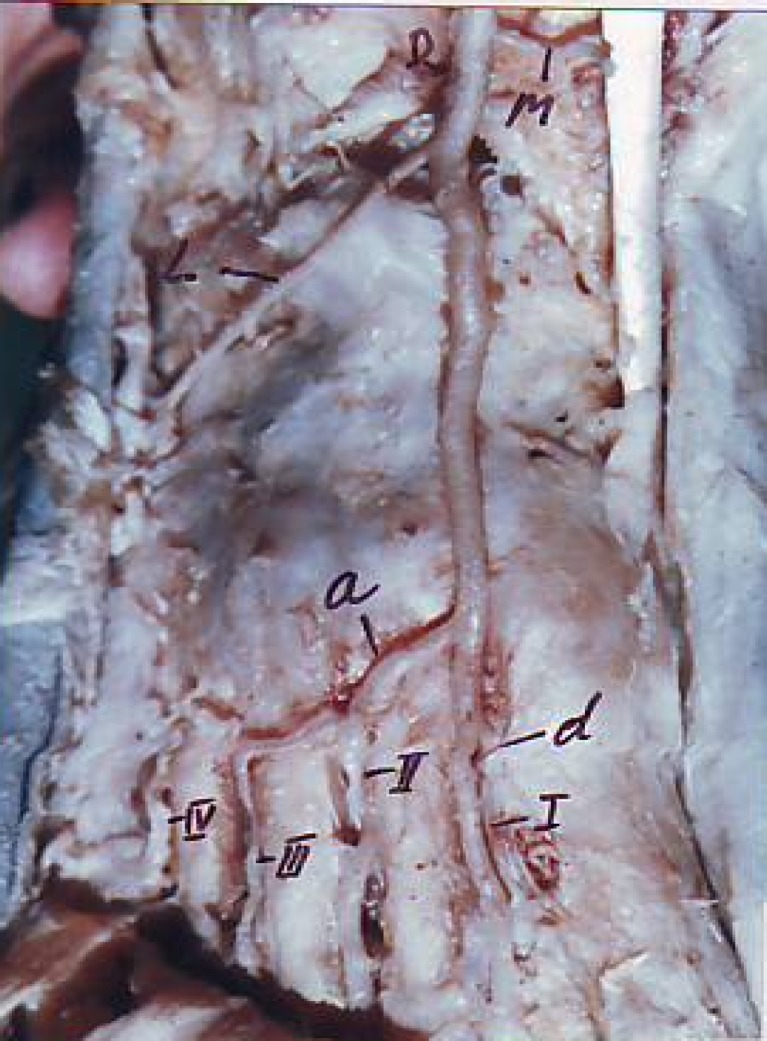
The dorsalis pedis artery was a continuation (photo 1) of the anterior tibial artery distal to the ankle joint in all the 60 limbs. It continued straight downwards on the medial side of the dorsum of foot, undercover of inferior extensor retinaculum between the tendons of long extensors, to reach the posterior end of the first interosseous space. It then passed between the heads of first dorsal interosseous to reach the sole of the foot and anastomoses with the lateral plantar artery to form the plantar arch.
Photo 1.
Normal branching pattern of dorsalis pedis artery
The dorsalis pedis artery branches into the medial and lateral tarsal arteries, and then runs a straight course on the dorsum of the foot. It gives rise to the arcuate artery which further gives the 2nd, 3rd and 4th dorsal metatarsal arteries. The 1st dorsal metatarsal artery is the direct continuation of the dorsalis pedis artery.
The case studied, considered as high arcuate artery, belonged to type C variation. The dorsalis pedis artery is the continuation of anterior tibial artery which after traveling a distance of 3.6 cm (photo 2) divides into medial and lateral branches. The medial branch continues as 1st dorsal metatarsal artery supplying the 1st intermetatarsal space. The lateral branch behaves like an arcuate artery which gives 2nd, 3rd and 4th dorsal metatarsal arteries.
Photo 2.
High origin of arcuate artery
Discussion
The arcuate artery was present in 55 (91.7%) cases. Of these, the high origin of arcuate artery was seen in 1 (1.66%) case.
Normal origin of arcuate artery at tarso-metatarsal joint was seen in 54 (90%) cases and high origin of arcuate artery at cuneo-navicular joint was seen in 1 (1.66%) case.
The arcuate artery was absent in 33%.
It arose directly from the dorsalis pedis artery in 90% and branched off the lateral tarsal artery in 10% [3].
Ontogenic Basis of Variations
Most of the variations are because of the enlargement of certain branches of the fundamental network on the dorsum of the foot and diminution or even disappearance of certain other connecting branches [4].
Knowledge of the variations in the anatomy of foot arteries is important for vascular surgeons as it is useful in deciding whether the absence of pulse in dorsalis pedis artery is due to thrombosis of the vessel or its abnormal course or absence.
References
- 1.William PL, Bannister LH, Berry MM (2000), Arteries of trunk. In: Gray’s Anatomy in arteries of trunk, 38th edn. Churchill Livingstone, New York, 1512–1513
- 2.Dilandro AC, Lilja EC, Lepore FL. The prevalence of arcuate artery–A cadaveric study of 72 feet. J Am Podiatr Med Assoc. 2001;91(6):300–305. doi: 10.7547/87507315-91-6-300. [DOI] [PubMed] [Google Scholar]
- 3.Yamada T, Glovinczki P, Bower TC. Variations of the arterial anatomy of foot. Am J Surg. 1993;166(2):30–136. doi: 10.1016/S0002-9610(05)81043-8. [DOI] [PubMed] [Google Scholar]
- 4.Huber JF. The arterial network supplying the dorsum of the foot. Anat Rec. 1941;81:373–379. doi: 10.1002/ar.1090800307. [DOI] [Google Scholar]


