Abstract
Bezoars are rare entities in medical science. Basically, they are of four types: trichobezoars; phytobezoars, pharmacobezoars, and lactobezoars. Some rare types of bezoars are also known. In this article a unique case of plastic bezoars is presented. It was diagnosed preoperatively by esophagogastroduodenoscopy and was removed surgically. The literature on this case is briefly revised.
Keywords: Esophagogastroduodenoscopy, Plastic bezoars, Huge abdominal lump
Introduction
Bezoars are mainly concretions in the stomach. Mainly trichobezoars and phytobezoars are described.Trichobezoars are almost exclusively found in teenager females with psychiatric comorbidities who eat their own hairs. Phytobezoars are more common and found in patients with diabetes, autonomic neuropathy, gastroparesis, gastric outlet obstruction, and those who have undergone gastric surgery [1]. Pharmacobezoars are undigested medicines and lactobezoars are undigested milk in the form of curd; they are found in young children. The symptoms associated with bezoars are mainly early satiety, pain, vomiting, sometimes a lump, small-bowel obstruction, and gastric outlet obstruction. Bezoars can cause gastric ulceration and perforation. Plastic bezoars are a unique introduction in the bezoar family. Like other bezoars they can be diagnosed preoperatively by esophagogastroduodenoscopy and barium studies, and can be removed surgically [2].
Patients and Methods
A 12-year-old male presented with a large swelling in the upper abdomen and off and on vomiting for the past 1 year.
It began as a small swelling and grew to attain a large size measuring 10 × 26 cm on admission (Fig. 1).
Fig. 1.
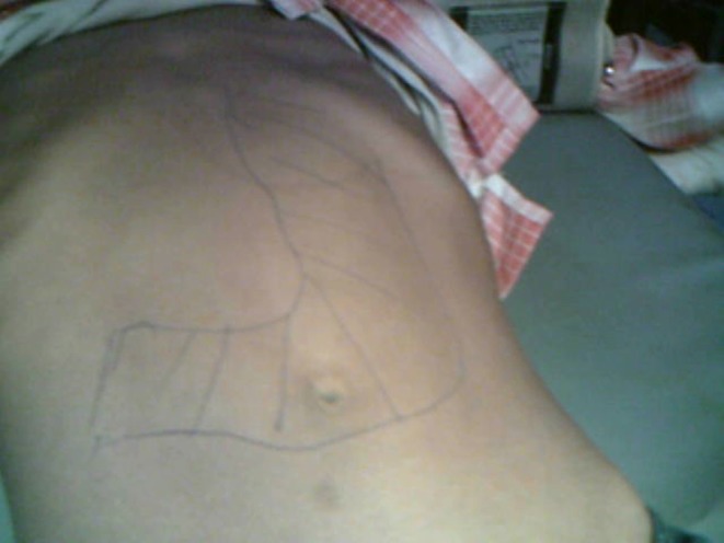
Clinical photograph of case
Abdominal examination revealed a large mass moving a little with respiration, occupying left hypochondrium, epigastrium, umbilical, and left and right flanks. It had a sooth surface and rounded border. The mass was dull on percussion. In history and on clinical examination diagnosis remained uncertain.
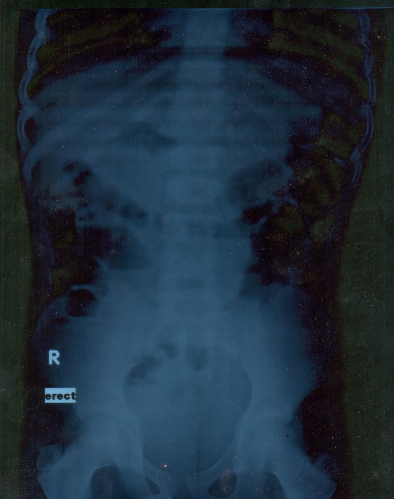
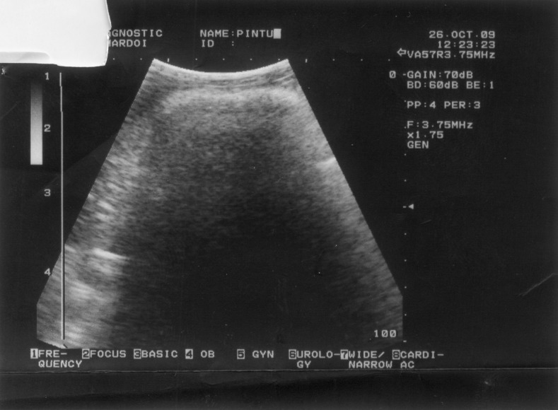
Blood, urine – routine and microscopic – and liver function test results were within normal limits. The X-ray of the abdomen showed the features of intestinal obstruction (Fig. 0). Abdominal sonography revealed a huge homogeneous lump (Fig. 2).
Fig. 2.
X-ray abdomen showing intestinal obstruction
Esophagogastroduodenoscopy was performed in the hospital; endoscope could not be negotiated beyond gastroesophageal junction.
In the endoscopic view, few different colored wires were recognized.
Some of them were removed through the endoscope using biopsy forceps.
The diagnosis of the plastic wires, removed endoscopically, forced for surgical removal.
An exploratory laparotomy was undertaken and two separate lumps of wires were found in the stomach and the jejunum (Fig. 3). An incision was made on the stomach wall, and a lump of messed up plastic wires, casting the shape of the stomach, was removed (Fig. 4).
Fig. 3.
USG abdomen homogeneus lump in upper abdomen
Fig. 4.
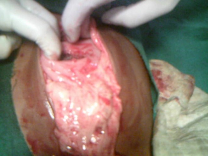
Two separate lump of stomach and jejunum
A few wires were going to the jejunum through the duodenum.
Second incision was made on the jejunal wall and almost a 3-ft-long lump of messed up plastic wire, casting the shape of C loop of the duodenum, jejunum, and ileum, as a whole was removed (Figs. 5 and 6). Retrocolic gastrojejunostomy was also performed. Postoperative period remained uneventful and the patient is presently asymptomatic.
Fig. 5.
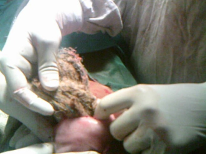
Messed plastic wires being removed from stomach
Fig. 6.
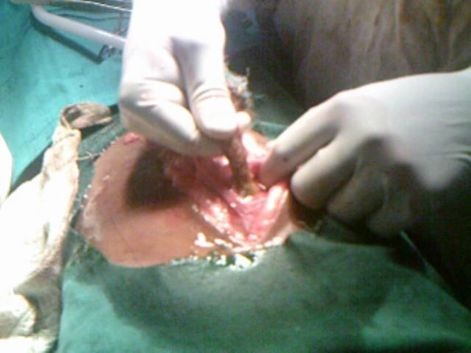
Plastic wire going in to deudenum
Discussion
Bezoars are rare disease. Trichobezoars and phytobezoars are the two most common bezoars described in literature. Other unusual bezoars are pharmacobezoars, lactobezoars, metal bezoars, plastic bezoars, and sand bezoars. Trichobezoars are found in teenagers, especially in female patients with psychiatric problems. Phytobezoars are found more frequently and are incarcerated foodstuffs not digested in the intestine.
The case of a mentally sound teenager male having eaten plastic wires in such a large quantity is unique and astonishing.
Patients usually present with recurrent vomiting, abdominal pain, and sometimes lump in the abdomen. Gastroscopy is diagnostic; foreign material can be directly seen and can be removed with biopsy forceps endoscopically. Barium study is another diagnostic technique used for the detection of bezoars. Bezoars can be treated endoscopically if foreign materials are few in quantity, but usually they require open surgical removal (Figs. 7 and 8).
Fig. 7.
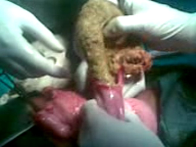
Messed plastic wires being removed from jejumum
Fig. 8.
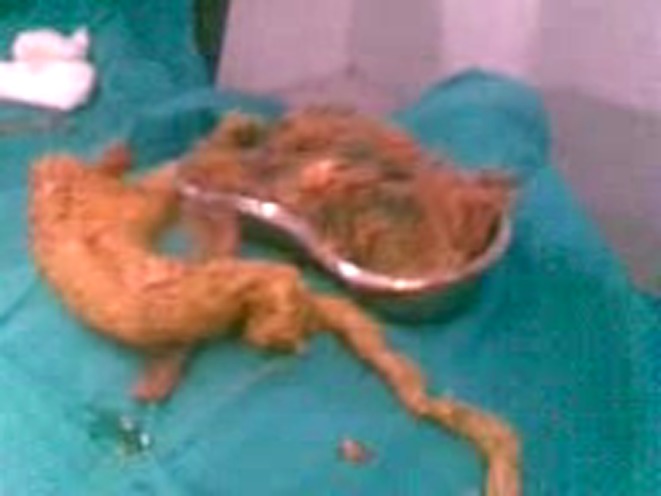
Specimen of removed plastic wires
Footnotes
The literature on this case is briefly revised.
References
- 1.De Bakey M, Oschsner A. Bezoars and concretion: a comprehensive review of literature with an analysis of 303 collected cases and a presentation of 8 additional cases – Part 1. Surgery. 1938;4:934. [Google Scholar]
- 2.De Bakey M, Oschsner A. Bezoars and concretion: a comprehensive review of literature with an analysis of 303 collected cases and a presentation of 8 additional cases – Part II. Surgery. 1939;5:132. [Google Scholar]


