Abstract
In this report, a case of blue toe syndrome related to persistent sciatic artery pseudoaneurysm in a 63-year-old woman, which was diagnosed by selective angiography, is presented. The pseudoaneurysm was successfully treated with coil embolization with good clinical results. A persistent sciatic artery is a rare embryological anomaly that occurs when the sciatic artery fails to regress during fetal development. Therefore, thromboembolisms from persistent sciatic artery aneurysm are rare.
Keywords: Blue toe syndrome, Persistent sciatic artery, Pseudoaneurysm, Coil embolization
Introduction
The development of blue toe syndrome (BTS) in a 63-year-old woman due to embolization from an undiagnosed persistent sciatic artery (PSA) aneurysm is reported.
BTS is a bluish discoloration of the toes as a result of tissue ischemia, which is caused by blockage of small vessels. The most common cause of occlusion of vessels is microembolization from a cardiac or, more commonly, peripheral artery (atherosclerotic arteries or aneurysms) [1].
PSA is a rare congenital vascular anomaly occurring in approximately 0.05 % of the population. PSA is susceptible to atherosclerotic degeneration, resulting in aneurysm formation, occlusive thrombosis, or distal embolization [2].
Case Presentation
A 63-year-old woman was admitted to rheumatology ward because of pain and bluish discoloration of her left second and third toes. She has been complaining of left buttock pain radiating to the posterior part of thigh and leg for 2 years, after a fall. In the first evaluation, the patient’s vital signs were normal. The physical examination was normal, except a tender pulsatile mass with a diameter of approximately 6 cm detected in her left gluteal region. The left foot was cold. Severe pain and bluish discoloration of the second and third left toes were apparent (Fig. 1).
Fig. 1.

Bluish discoloration of toes
All laboratory investigations were normal. Abdominopelvic ultrasound, color Doppler ultrasonography of the lower limbs, ECG, Holter monitoring and transesophageal echocardiography were normal.
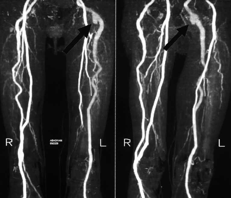
CT-Angiography revealed a PSA with a saccular aneurysm in its proximal part containing thrombosis within it (Fig. 2).
Fig. 2.
CT angiography revealed a persistent sciatic artery instead of the inferior gluteal artery. A clot containing aneurysm in proximal part is present
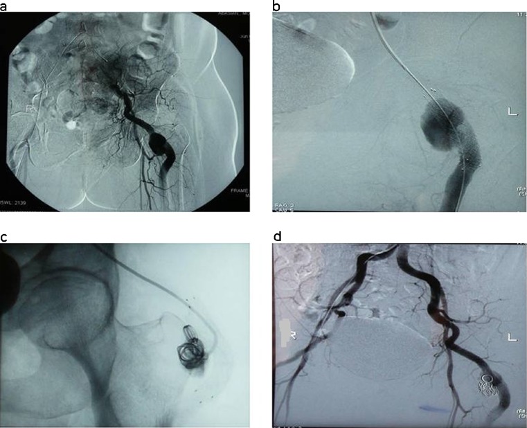
The patient underwent digital subtraction angiography (DSA) with coil embolization. After sterilization under local anesthesia, right common femoral artery puncture was performed with an 18 G needle, and a 6 Fr catheter was introduced over the guide wire through sheath into right common femoral artery. Contrast run revealed a broad-based saccular pseudoaneurysm adjacent to the left femoral neck. Also, it revealed a variation in origination of left superficial femoral artery (LSFA) which was feeding from the left internal iliac artery instead of the left external iliac artery. Because of this anatomic variation, it was not possible to sacrifice the artery proximal to the pseudoaneurysm. So because of tortuosity of the vessel, a bare metallic covered stent was first placed in the artery and then the aneurysm was coiled with 10 different sizes spiral coils. Control angiography after embolization showed the aneurysm had been excluded from the vascular tree and the perfusion of the left lower extremity was completely normal (Fig. 3). The patient’s pain was relieved dramatically.
Fig. 3.
Digital subtraction angiography. A broad-based saccular pseudoaneurysm of persistent sciatic artery (a), placement of a metallic stent in the artery (b), obstruction of pseudoaneurysm with spiral coils (c), control angiography that shows the aneurysm is out of vascular tract and perfusion of the lower extremity is completely normal (d)
Discussion
We described here the development of BTS in a 63-year-old woman due to embolization from an undiagnosed PSA aneurysm, which was successfully treated with coil embolization.
BTS is the cyanotic mottling of toes as a result of small distal arteries occlusion, caused by various conditions (Table 1). However, the most common causes of BTS are atheroembolic diseases. Emboli usually come from cardiac, or more commonly, peripheral arteries (atherosclerosis or aneurysm) [1]. In our case, the clot filling pseudoaneurysm of PSA was the origin of microemboli as the cause of BTS, and the history of falling two years ago was the probable cause of pseudoaneurysm.
Table 1.
Causes of blue toe syndrome
| Mechanical obstruction(secondary to emboli or atherosclerosis) |
| Vasospasm (Due to a primary disease of the blood vessel or secondary to cold, medication or forefoot surgery) |
| Vasculitis |
| Hyperviscosity |
| Hypercoagulability (such as anti-phospholipid syndrome) |
| Calciphylaxis |
| Medication (Warfarin and rarely Steroids) |
In the developing embryo, the sciatic artery develops along with the limb bud as the axial artery. The sciatic artery is then normally superseded and annexed by the femoral artery as it extends off the internal iliac artery. Failure of this process in the first 3 months of embryonic life results in atresia of the superficial femoral artery system and a PSA. Clinically, patients with PSA may present with a buttock pain due to mass effect of the aneurismal sac, or sometimes pulsatile swelling in the gluteal region, or symptoms of sciatic nerve compression. Aneurysm formation occurs in approximately 46 % of cases of PSA. The etiology of PSA aneurysms includes penetrating or blunt trauma to the buttock, atherosclerosis, hypertension, congenital lack of arterial elastic tissue, and infection [2, 3].
Transcatheter embolization has become a reasonable alternative to surgery in the treatment of PSA and it is generally as effective as surgical exclusion and avoids surgical complications [4].
Conclusion
In this case, BTS presented as a manifestation of PSA aneurysm, which is a rare condition. In such a condition, minimally invasive techniques such as selective angiography and coil embolization may be the best diagnostic and therapeutic modalities, respectively.
Also, attention to careful evaluation of posttraumatic swelling of the gluteal region is quite important.
References
- 1.Weinberg I (2010) Blue toe. Evidence based vascular medicine website. http://www.angiologist.com/blue-toe. Accessed April 22, 2011
- 2.Fung HS, Lau S, Chan MK, et al. Persistent sciatic artery complicated by aneurysm formation and thrombosis. Hong Kong Med J. 2008;14(6):492–494. [PubMed] [Google Scholar]
- 3.Mandell VS, Jaques PF, Delaney DJ, Oberheu V. Persistent sciatic artery: clinical, embryologic, and angiographic features. Am J Roentgenol. 1985;144(2):245–249. doi: 10.2214/ajr.144.2.245. [DOI] [PubMed] [Google Scholar]
- 4.Ooka T, Murakami T, Makino Y (2009) Coil embolization of symptomatic persistent sciatic artery aneurysm: a case report. Ann Vasc Surg 23(3):411:e1–e4 [DOI] [PubMed]


