

Abstract
Management of massive liver trauma (grades IV–VI) is associated with high morbidity and mortality. It can pose serious challenges to treating physician and requires multimodality interventions. For a successful outcome, timing of intervention is crucial. We report a case of grade V hepatic injury treated successfully with angioembolization, laparoscopic lavage, and endoscopic stenting during a time period from admission to discharge on the 24th day. Angioembolization was performed at admission after resuscitation as CT scan showed active extravasation in the arterial phase. Laparoscopic lavage and drainage was performed on the fifth day as abdominal inflammatory response failed to respond to medical management. There was biliary component of abdominal fluid noticed during laparoscopy, which manifested by postoperative localized biliary fistula; hence endoscopic stenting of the bile duct was performed on the seventh day. The patient recovered well with timely minimal invasive approach and was fine at 1 year follow-up.
Keywords: Hepatic trauma, Angiographic embolization, Laparoscopic lavage, Biliary fistula
Introduction
Nonoperative management of isolated liver trauma is the standard of care in today’s practice [1]. While grade I–III injuries do not pose problems, management of grade IV–V injuries can be tricky; hemorrhage, peritoneal inflammatory response, and bile leak can complicate the situation. Vigilance, timely interventions by multidisciplinary team approach with availability of specialized facilities to tackle complications is required to decrease morbidity and mortality. We report a case of grade V liver injury with all these complications managed by multimodality minimally invasive approach.
Case report
A 24-year-old woman was brought to the emergency department approximately 60 min after a motorcycle accident. She was in a state of hemorrhagic shock with pulse rate of 160/min, respiratory rate of 40/min, and systolic blood pressure of 70 mmHg. There were multiple bruises on the chest and tenderness in the right hypochondrium. She was resuscitated; four units of packed red blood cells and six units of fresh frozen plasma were transfused. Emergency contrast-enhanced computerized tomography (CECT scan) revealed multiple right posterior rib fractures with hemothorax and large liver parenchyma disruption spanning segments 5, 6, and 7 with active blush of intrahepatic contrast indicating grade V liver injury (Fig. 1).
Fig. 1.

CT scan showing blush of contrast
The patient was intubated in critical care unit, and a right intercostal drainage tube was inserted. In view of active intrahepatic bleeding seen on CT scan, an emergency celiac angiography was performed. Right anterior hepatic artery bleeding and the pseudoaneurysm of right posterior branch were observed. It was successfully embolized with coils and polyvinyl alcohol particles (Fig. 2). On day 5, the patient deteriorated with increase in abdominal girth, tachycardia, and leukocytosis. CECT scan demonstrated localized segment of liver trauma, no extravasation of contrast but significant increase in free fluid in the abdomen (Fig. 3). This was diagnosed as peritoneal inflammatory syndrome, and surgical intervention was considered. Laparoscopy revealed presence of 4.8 L of blood mixed with bile, contused liver segments, and floating pieces of necrotic liver tissue. Saline lavage was given, floating necrotic liver pieces was removed, absence of active bleeding was confirmed, and drains were placed in right, left sub diaphragmatic, and subhepatic spaces (Fig. 4). The patient recovered well, was extubated on day 4 of surgery, and on diet by day 5. Her right subdiaphragmatic drain, however, drained 200–275 mL of bile daily.
Fig. 2.

Celiac angiography: coils in the right hepatic artery branches
Fig. 3.

Day 5 CT scan: gross intra-abdominal fluid
Fig. 4.

Intraoperative liver contusion
On postoperative day 7, she developed fever, tachycardia, and leukocytosis suggestive of either biliary or necrosed liver related sepsis. Contrast-enhanced ultrasonography (CES) documented viable liver tissue with no collection (Fig. 5); hence, sepsis related to biliary fistula was deemed to be the cause. She was subjected to ERCP. It showed tear in right anterior duct, stenting of which decreased the biliary leak (Fig. 6). She stabilized and was discharged on day 24. ERCP stent removal was done after 6 months and the CT scan repeated after a year showed normal regenerated liver.
Fig. 5.
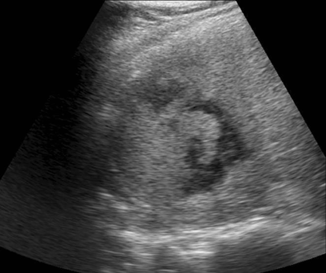
CES: regenerating liver
Fig. 6.

ERCP: bile leak from right posterior duct
Discussion
Liver is the most commonly injured organ in blunt abdominal trauma [2]. Management of liver trauma has witnessed a sea of change since the advent of CT scans and advances in critical-care monitoring. Hepatic injury scale grades liver injuries from grades I to VI and has universally streamlined the management of liver trauma [3]. Grade I–III injuries constitute 80–90 % of all liver injuries and are successfully managed by nonoperative method [2, 4]. Grade VI injuries are avulsion of vascular pedicle and are associated with mortality. Dilemma occurs in management of grades IV–V injuries wherein delay in accurate management leads to poor outcome due to complex issues of bleeding, peritoneal inflammatory syndrome, bile leak, and sepsis [1, 5].
Traditionally, active bleeding into the peritoneal cavity along with homodynamic instability requiring massive transfusion was managed by emergency surgery. Recently, early angiographic embolization has become an important management tool in grade IV and V liver injuries to control active arterial bleeding [6]. This not only prevents emergency exploration and its associated morbidity but also reduces the chances of transfusion-associated lung injury. In the current case, CT scan showed blush of contrast in injured liver, with no coexisting injury; hence, selective angioembolization was done, which stopped the arterial leak instantaneously. Selective embolization decreases the risk of ischemic injury to surrounding healthy liver. However, if significant liver tissue gets completely devitalized and patient has features of septicemia, delayed resection debridement may be needed [3]. Close hemodynamic monitoring and serial blood investigations form an essential tool in management of inflammatory response to liver trauma. Hemoglobin should be maintained at 10 gm%, platelet count >1,00,000/cmm, and coagulation derangements should be rectified [7].
The term “inflammatory syndrome” was coined by Carrillo signifying the importance of strict surveillance. It is secondary to the action of blood and bile in the peritoneum [8]. Surgical intervention in such situations can be accomplished by laparoscopy and should be viewed as part of the minimally invasive approach. Our patient developed fever, abdominal distention with drop in hemoglobin, and leukocytosis on day 5, suggestive of abdominal inflammatory syndrome. Thus, clinical deterioration with CT scan showing massive free fluid in the abdomen warranted surgical intervention. Timely laparoscopic lavage proved very effective, leading to drastic improvement in clinical parameters.
Morbidity in massive liver trauma does not end with surgical intervention. Five percent of liver trauma patients additionally suffer major biliary tract injury leading to bile leak and sepsis [9]. In cases of persistent clinical deterioration and biliary sepsis, delayed resectional debridement of necrosed liver is considered as an option [3, 4]. Our patient developed fever and leukocytosis on the seventh postoperative day. Although CT scan documented no intraabdominal collection, there was a dilemma between ischemic liver vs bile leak as the source of sepsis. CES ascertained that liver was viable, and we attributed sepsis to bile leak. Utility of CES is also echoed in the literature as an important tool both in initial and delayed assessment of liver trauma patients [10]. We subjected the patient to ERCP and stenting, which relieved the patient of any further complications.
Conclusion
Hemorrhage, abdominal inflammatory response, and biliary sepsis are complications occurring in grades III–V liver injuries requiring close monitoring and timely intervention. Availability of technological support and specialized facilities under one roof decreases the morbidity and mortality in complex liver trauma. This report illustrates that it is possible to manage complex liver injuries with minimally invasive techniques. Timely interventions both early and in the delayed setting with good interdepartmental coordination precludes the need for open exploration, thus resulting in decreased hospital stay, blood transfusion requirements, and abdominal infection rates.
Acknowledgments
We thank Dr Ajay Thakker (CE0- Jupiter Hospital) for permitting us to publish hospital data.
Sources of support
None.
Conflicts of interest
None.
Footnotes
Key messages: Complex liver traumas are known for high morbidity and high mortality. Timely intervention by various specialties under one roof is essential for optimum care of these patients.
References
- 1.Croce MA, Fabian TC, Menke PG, Waddle-Smith L, Minard G, Kudsk KA, et al. Nonoperative management of blunt hepatic trauma is the treatment of choice for hemodynamically stable patients. Results of a prospective trial. Ann Surg. 1995;221:744–755. doi: 10.1097/00000658-199506000-00013. [DOI] [PMC free article] [PubMed] [Google Scholar]
- 2.Cothren CC, Moore EE. Hepatic trauma. Eur J Trauma Emg Surg. 2008;34:339–354. doi: 10.1007/s00068-008-8029-5. [DOI] [PubMed] [Google Scholar]
- 3.Cresswell AB, Bowles MJ, Wendon JA (2007) The management of liver trauma. In: Vincent JL (ed) Yearbook of intensive care and emergency medicine, 1st edn. Springer, Berlin, pp 666–77.
- 4.Trunkey DD. Hepatic trauma: contemporary management. Surg Clin N Am. 2004;84:437–450. doi: 10.1016/S0039-6109(03)00228-7. [DOI] [PubMed] [Google Scholar]
- 5.Letoublon C, Chen Y, Arvieux C, Voirin D, Morra I, Broux C, et al. Delayed celiotomy or laparoscopy as part of the nonoperative management of blunt hepatic trauma. World J Surg. 2008;32:1189–1193. doi: 10.1007/s00268-007-9439-y. [DOI] [PubMed] [Google Scholar]
- 6.Monnin V, Sengel C, Thony F, Bricault I, Voirin D, Letoublon C, et al. Place of arterial embolization in severe blunt hepatic trauma: a multidisciplinary approach. Cardiovasc Intervent Radiol. 2008;31:875–882. doi: 10.1007/s00270-007-9277-1. [DOI] [PubMed] [Google Scholar]
- 7.Moore FA, McKinley BA, Moore EE, Nathens AB, West M, Shapiro MB, et al. Inflammation and the host response to injury, a large-scale collaborative project: patient-oriented research core-standard operating procedures for clinical care. III. Guidelines for shock resuscitation. J Trauma. 2006;61:82–89. doi: 10.1097/01.ta.0000225933.08478.65. [DOI] [PubMed] [Google Scholar]
- 8.Carrillo EH, Reed DN, Jr, Gordon L, Spain DA, Richardson JD. Delayed laparoscopy facilitates the management of biliary peritonitis in patients with complex liver injuries. Surg Endosc. 2001;15:319–322. doi: 10.1007/s004640000300. [DOI] [PubMed] [Google Scholar]
- 9.Delgado Millian MA, Deballon PO. Computed tomography, angiography and endoscopic retrograde cholangiopancreatography in the nonoperative management of hepatic and splenic trauma. World J Surg. 2001;25:1397–1402. doi: 10.1007/s00268-001-0139-8. [DOI] [PubMed] [Google Scholar]
- 10.Catalano O, Lobianco R, Raso MM, Siani A. Blunt hepatic trauma: evaluation with contrast-enhanced sonography: sonographic findings and clinical application. J Ultrasound Med. 2005;24:299–310. doi: 10.7863/jum.2005.24.3.299. [DOI] [PubMed] [Google Scholar]