

Abstract
We report a case of a 24-year-old man with complex shoulder defect on the right side following a road traffic accident. The patient was found to have a subclavian artery thrombosis in its second part with good collaterals ensuring limb viability. Free-flap reconstruction was not possible because of lack of recipient vascular pedicle nearby. Pedicled flaps routinely used for this area such as pectoralis major and latissimus dorsi myocutaneous flaps could not be used as their vascular pedicles arise from the second and third parts of the axillary artery distal to the subclavian artery thrombosis. The best option we could think was the trapezius myocutaneous flap as its vascular pedicle arises from the first part of the subclavian artery proximal to the thrombosis. This article further establishes the importance of knowing the vascular supply of flaps before their clinical application. In this case the trapezius flap acts as a lifeboat for coverage of the complex shoulder defect as its vascular supply is proximal to the site of subclavian artery thrombosis.
Keywords: Shoulder defect, Subclavian artery thrombosis, Trapezius flap
Introduction
Complex tissue defects of the shoulder with the exposed shoulder joint following trauma are uncommon. They are more common following excision malignant tumors in this area. Traditionally, these areas have been reconstructed using pedicled flaps such as pectoralis major [1] or latissimus dorsi flaps [2, 3]. Free flaps such as anterolateral thigh free flap or radial artery free flap are an alternative when the recipient pedicle is available nearby. When these alternatives are precluded, the trapezius myocutaneous flap is a good alternative.
Case Report
A 24-year-old man came to us with a complex shoulder defect of the right side with the shoulder bones and joint exposed 2 months following a road traffic accident. He had been managed primarily in the orthopedic and general surgical departments as he also had a hemothorax for which he had an intercostal drainage tube inserted. As his distal pulses on his right upper limb were feeble, he was investigated with an angiography which revealed right-sided subclavian artery thrombosis in its second part with good distal reformation.
No intervention for the subclavian artery thrombosis was advised by the cardiovascular surgeon as the limb was surviving with distal reformation of the vessels through collaterals. When we were called in to provide wound cover for this patient, there were certain important considerations which we would like to highlight.
First, reconstruction of this complex defect with a free flap would be extremely difficult as there were no reliable recipient pedicles. Second, the vascular pedicle of pectoralis major and latissimus dorsi myocutaneous flaps arise from the second and third parts of the axillary artery both of which are distal to site of thrombosis.
The best option in this situation was the trapezius myocutaneous flap as the vascular basis of this flap is based on the transverse cervical artery which arises from the first part of subclavian artery proximal to the thrombosis. The preoperative and postoperative photographs are shown in Figs. 1 and 2. Postoperatively once the flap settled, the patient was asked to mobilize the shoulder joint.
Fig. 1.
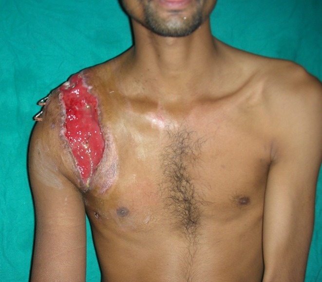
Preoperative exposed shoulder joint
Fig. 2.

Postoperative well-settled trapezius flap
Discussion
Understanding the vascular anatomy of flaps is very important in the clinical application of these flaps. Blockage of the vascular supply of a flap precludes the use of the flap in reconstruction of other areas. This article further establishes the importance of knowing the vascular supply of flaps before their clinical application. In this case the trapezius flap acts as a lifeboat for coverage of the complex shoulder defect as its vascular supply is proximal to the site of subclavian artery thrombosis.
References
- 1.Paterson P, Prinsloo DJ. The pectoralis major muscle flap—a first choice for shoulder disruption injuries. Injury. 2004;35(5):537–540. doi: 10.1016/S0020-1383(02)00353-4. [DOI] [PubMed] [Google Scholar]
- 2.Behnam AB, Chen CM, Pusic AL, Mehrara BJ, Disa JJ, et al. The pedicled latissimus dorsi flap for shoulder reconstruction after sarcoma resection. Ann Surg Oncol. 2007;14(5):1591–1595. doi: 10.1245/s10434-006-9292-5. [DOI] [PubMed] [Google Scholar]
- 3.Schoeller T, Gurunluoglu R, Wechselberger G, Hussl H, Huemer GM. Transfer of pedicled musculocutaneous latissimus dorsi flap for restoration of shoulder contour after neurogenic atrophy. Ann Plast Surg. 2007;58(6):694–697. doi: 10.1097/01.sap.0000250843.29618.7d. [DOI] [PubMed] [Google Scholar]