Abstract
Right-sided Bochdalek hernia in adults is a very rare clinical entity. A case of a 50-year-old female patient is reported, who presented with long history of intermittent breathlessness and right-sided thoracoabdominal pain. The hernia was managed laparoscopically. Contents were colon, omentum, and right kidney. It was successfully repaired using a polypropylene mesh.
Keywords: Right-sided Bochdalek hernia, Laparoscopic repair, Colon, Right kidney
Introduction
Bochdalek hernia, first described in 1848, occurs through a congenital defect in the posterolateral aspect of the diaphragm. It is primarily a disorder of neonates and children and most commonly occurs on the left side of diaphragm. It is very rare in adults and fewer than 20 cases of right-sided Bochdalek hernia have been reported [1]. Among these cases only a few have been treated laparoscopically.
Case Report
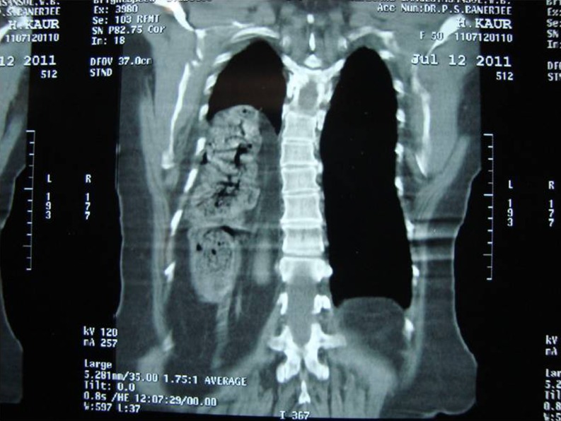
A 50-year-old female patient was admitted with complaints of breathlessness on and off for 25 years, right-sided chest pain and upper abdominal discomfort for 2 weeks. She was a known case of type 2 diabetes mellitus and hypertension. She had undergone conventional appendicectomy 14 years ago. There was no history of trauma or previous thoracoabdominal surgery. Chest X-ray revealed elevated right hemidiaphragm. The patient was diagnosed to have right-sided Bochdalek hernia on CT scan (Fig. 1). Her hematological investigations were normal. She was advised breathing exercises and spirometry preoperatively.
Fig. 1.
AP view of CT Scan showing Rt sided Bochdalek hernia with colon and kidney in the chest
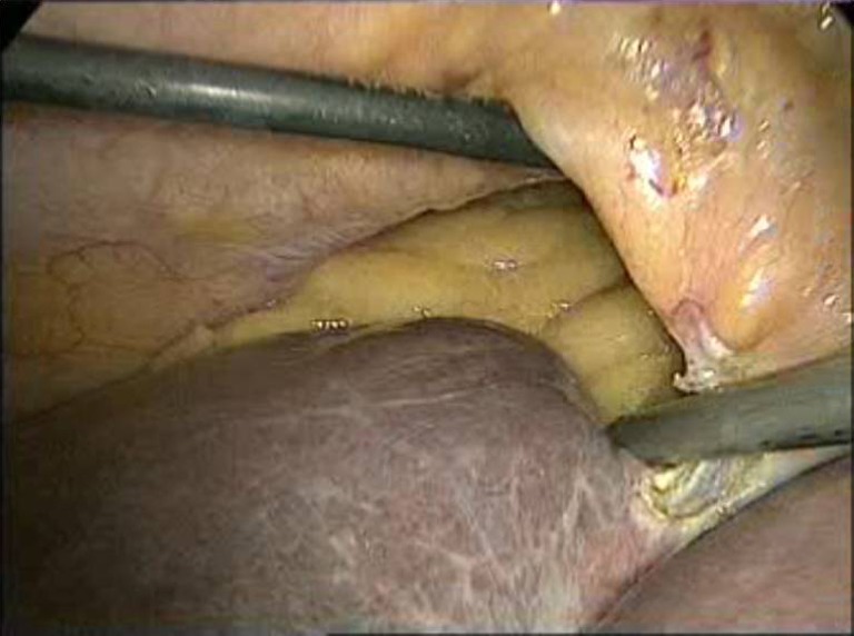
The patient was subjected to laparoscopy under general anesthesia. She was placed in 15o reverse Trendelenburg position with left lateral 30o tilt. A 10 mm optical port was used at umbilicus and right paramedian positions as per the requirement. A 30o telescope was used. Five mm epigastric, right midclavicular, and anterior axillary ports were used as working channels. Laparoscopy revealed herniation of the ascending colon, hepatic flexure, and proximal transverse colon with omentum and right kidney through a 10 cm × 8 cm size right posterolateral diaphragmatic defect. The right lobe of the liver was normal in size but devoid of its lateral peritoneal attachments. Hernial contents were adherent to the thoracic cage. The hernial sac could not be identified. Contents were reduced back to the peritoneal cavity (Fig. 2). The defect was darned with 1-0 polypropylene suture to prevent mesh migration and then reinforced with 15 cm × 15 cm polypropylene mesh. Abdominal drain and intercostal drainage (ICD) tube were placed. The patient was electively ventilated for 24 h postoperatively. She had prolonged drainage from the ICD in spite of full expansion of the right lung on the first postoperative day. She was discharged on fourth post-operative day. The ICD was removed on 12th postoperative day. On follow-up, the patient had localized transudative fluid collection in the right thoracic cavity which required needle aspiration twice.
Fig. 2.

Laparoscopic view of Rt sided Bochdalek hernia
Discussion
Bochdalek hernia is a congenital posterolateral diaphragmatic defect resulting from failure of fusion of retroperitoneal canal membrane with the dorsal esophageal mesentery and body wall [2]. Vincent Alexander Bochdalek first described this hernia in 1848 [3]. Most of these hernias are diagnosed in children with respiratory distress and 80–90 % of these cases are reported on the left side [4]. A right-sided symptomatic Bochdalek hernia presenting in adulthood is extremely rare. Till 2007, only 14 cases of right-sided Bochdalek hernia presenting in adults were reported in literature [4]. It usually presents with chronic vague gastrointestinal or respiratory symptoms. A few cases of intestinal obstruction have also been reported [4]. Contrast-enhanced CT is the most accurate imaging modality for detection of Bochdalek hernias. It provides detailed information regarding the herniated viscera and the diaphragmatic defect.
The management of Bochdalek hernia includes reduction of hernial contents to the peritoneal cavity and repair of the diaphragmatic defect [4]. Traditionally, due to difficulties of exposure secondary to liver, many authors advocated a thoracic, thoracoabdominal approach or a full laparotomy. Recently, laparoscopic repair of right Bochdalek hernia has been reported [5]. Of all the approaches, laparoscopy inflicts minimal surgical trauma. This is significantly important in patients of Bochdalek hernia for rapid recovery of pulmonary function. Postoperative ventilation also facilitates early expansion of the lung. Laparoscopic repair, although feasible, is technically challenging. We experience that the length of instruments falls short during dissection deep inside the thoracic cavity. However, this may be overcome by placement of additional trocars through the intercostal spaces. Thus, minimal invasive surgical approach is feasible even in cases of large right-sided Bochdalek hernia with herniation of multiple viscera and should be regarded as the preferred surgical option for the management of these cases.
Conclusion
Symptomatic right-sided Bochdalek hernia is extremely rare in adults. Laparoscopic repair of Bochdalek hernia is feasible, reduces the morbidity of surgery, and helps in early recovery of these patients. It should be regarded as the procedure of choice for these hernias.
Contributor Information
Nirmal M. Patle, Email: nirmal_patle@rediffmail.com
Om Tantia, Phone: +91-983-0400444, FAX: +91-33-40206500, Email: omtantia@gmail.com.
Parmanand Prasad, Email: dr.parmanandprasad@gmail.com.
Prakhar C. Das, Email: drprakhardas@gmail.com
Shashi Khanna, Email: shashi_65_in@yahoo.co.in.
References
- 1.Laaksonen E, Silvasti S, Hakala T. Right sided Bochdalek hernia in adult: a case report. J Med Case Rep. 2009;3:929. doi: 10.1186/1752-1947-3-9291. [DOI] [PMC free article] [PubMed] [Google Scholar]
- 2.Sener RN, Tugran C, Yorulmaz I, Dagdeviren A, Orgue S. Bilateral large Bochdalek hernia in an adult. CT demonstration. Clin Imaging. 1995;19(1):40–42. doi: 10.1016/0899-7071(94)00023-6. [DOI] [PubMed] [Google Scholar]
- 3.Haller SA. Professor Bochdalek and his hernia: then and now. Prog Pediatr Surg. 1986;20:252–255. doi: 10.1007/978-3-642-70825-1_18. [DOI] [PubMed] [Google Scholar]
- 4.Rout S, Foo FJ, Hayden D, Guthrie A, Smith AM. Right-sided bochdalek hernia obstructing in an adult: case report and review of literature. Hernia. 2007;11:359–362. doi: 10.1007/s10029-007-0188-5. [DOI] [PubMed] [Google Scholar]
- 5.Rosen MJ, Ponsky L, Schilz R. Laparoscopic retroperitoneal repair of a right-sided Bochdalek hernia. Hernia. 2007;11:185–188. doi: 10.1007/s10029-006-0162-7. [DOI] [PubMed] [Google Scholar]


