Abstract
The lipid-destabilizing properties of the N-terminal domain of the GP2 of Ebola virus were investigated. Our results suggest that the domain of Ebola virus needed for fusion is shorter than that previously reported. The fusogenic properties of this domain are related to its oblique orientation at the lipid/water interface owing to an asymmetric distribution of the hydrophobic residues when helical.
The Ebola virus, known for its deadly outbreaks, causes hemorrhagic fevers in humans, yet there is still no effective vaccine or antiviral treatment available.
The virion consists of a central ribonucleoprotein core linked by two matrix proteins to a glycoprotein (GP) carrying a lipid bilayer derived from the host cell.
The GPs form trimeric spikes on the virion surface and are responsible for receptor binding and fusion of the virus to the host membrane. The GP is cleaved into two parts: GP1 and GP2. The GP2 seems to fold spontaneously into a trimeric, highly α-helical, rod-shaped structure, similar to HA2 in influenza virus and to gp41 in human immunodeficiency virus (25). Glycoprotein 2 (GP2) contains a fusion peptide at its N-terminal implicated in virus cell entry. It has previously been demonstrated that the Ebola virus fusion peptide predicted by Gallaher (14), from residues 23 to 38, induces fusion with liposomes and that mutations in this peptide inhibit Ebola virus infectivity (19, 32).
We have shown that the fusion peptides of simian immunodeficiency virus (SIV) and bovine leukemia virus have an asymmetric distribution of their hydrophobicity residues, the net hydrophobicity increasing from one end of the helix to the other (18, 35). This particular distribution is characteristic of the so-called tilted peptides (3, 4, 5). Recently the orientation of the SIV tilted peptide was determined in planar lipid bilayers by neutron lamellar diffraction. The peptide was shown to form an angle of 55° with the membrane surface when helical, confirming our computational predictions (2). We have previously suggested that the Ebola virus fusion peptide also has such a distribution of hydrophobic residues (34).
The present study examines the fusogenic properties of the Ebola virus fusion peptide 25 to 35 in relation to the hydrophobicity gradient by combining modeling and biophysical approaches.
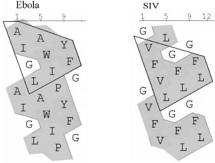
We have analyzed the Ebola virus fusion peptide sequence with respect to its hydrophobicity with the hydrophobic cluster analysis. This method is based on an analysis of the shape and position of hydrophobic clusters in a given protein sequence (13). The 25 to 35 fragment (AIGLAWIPYFG) shows a hydrophobicity profile similar to that of the SIV tilted peptide, well known for its fusogenic properties (22, 26), although, as shown on Fig. 1, the two stretches have no sequence identity.
FIG. 1.
Hydrophobic cluster analysis of the tilted peptide of SIV (residues 1 to 11) and the similar domain in Ebola virus (residues 25 to 35). In this view, the sequence is written on a cylinder that is unrolled and duplicated (11). The hydrophobic amino acids are shaded.
The three-dimensional structure of the 25 to 35 Ebola virus peptide was calculated as previously described, assuming a helical secondary structure (3, 6, 7, 28). Small peptides can adopt α and β type structures (12, 22, 33). It was shown that the ability of the Ebola virus peptide to induce lipid destabilization correlates with an α-helical structure, in the absence of Ca2+ (33). Furthermore, the fusogenic activity of the tilted fragments of apolipoprotein A-II and SIV fusion protein increases with the helical content (22, 28), stressing the importance of the helix structure for lipid destabilizing capacities. Moreover, nuclear magnetic resonance studies have indicated that the fragments of the fusogenic domain of GP41 and of HA2, which correspond to tilted peptides in terms of hydrophobic/hydrophilic properties, are predominantly α-helical in a lipid-like environment (15, 16). In addition, we have tested the properties of a β strand conformation for the Ebola virus peptide and other tilted peptides (23) and we found no correlation between the orientation of the peptides towards the interface and the experimental results on liposome fusion. All these data support our assumption that the Ebola virus tilted peptide has a helical structure in the lipid bilayer.
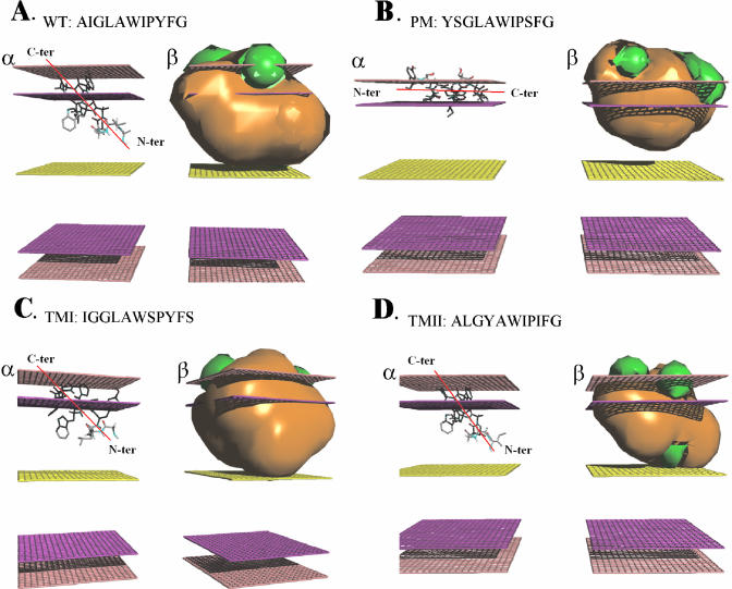
The behavior of the peptide was analyzed by integral membrane protein and lipid association (IMPALA). This method allows the study of a peptide or protein interaction with a modeled membrane (10). The most stable position of the Ebola virus fusion peptide in the simplified bilayer corresponds to a tilt of around 47° towards the interface plane (Fig. 2Aα). The mass center is about 11 Å from the bilayer center. As predicted by de Planque et al., the tryptophan residue is close to the level of the lipid carbonyl region (9). An analysis of restraint profiles shows angle variations of more than 25° with no significant influence on the energy of the system (data not shown), indicating that the peptide could adopt metastable positions in the bilayer, as has already been observed for other viral tilted fragments. It was suggested that this behavior is related to the destabilizing properties of tilted peptides (24).
FIG. 2.
Lowest energy position of the different peptides after IMPALA calculations (α) and representation of the molecular hydrophobicity potential (β). Hydrophobic and hydrophilic envelopes are orange and green, respectively. Mid plane, bilayer center (z = 0); first upper (bottom), lipid acyl chain/polar headgroups interface at 13.5Å from the center; second upper (bottom) plane, lipid/water interface (Z = 18 Å). The N to C orientation was shown. (A) Wild type; (B) PM; (C) TMI; (D) TMII.
Calculations of the molecular hydrophobicity potentials allow for visualization of the hydrophilic/hydrophobic environment of a molecule (3). In Fig. 2Aβ we observe that the hydrophilic/hydrophobic envelopes are not split as for an amphipathic or a transmembrane peptide (3). The Ebola virus peptide presents an asymmetric distribution of hydrophobicity, similar to what is seen for tilted peptides like those of SIV or bovine leukemia virus (18, 35).
Since most tilted peptides induce liposome fusion in vitro (22, 23, 26, 27, 30), the peptide was tested for fusogenic properties. Large unilamellar vesicles were prepared by the extrusion technique of Hope et al. (17). Peptides were synthesized by conventional solid phase peptide synthesis, by Fmoc for transient NH2-terminal protection, and were characterized by mass spectrometry (1). Peptide purity varies between 80% and 85%, as indicated by analytical high-pressure liquid chromatography.
Lipid mixing assays were carried out. Mixing of liposome membranes was followed by measuring the fluorescence increase of octadecyl rhodamine chloride (R18), in the same conditions as previously described (23). A series of assays were first conducted to optimize the fusion conditions of the peptide. Fusion was optimal when noncharged lipid films (30.2% phosphatidylcholine, 28.6% sphingomyelin, 29.6% phosphatidylethanolamine, and 11.6% cholesterol [wt/wt]) at pH 6 were used with peptides dissolved in 100% trifluoroethanol (TFE) (Table 1). These experimental conditions were thereafter used in all lipid destabilization assays.
TABLE 1.
Determination of optimal lipid fusion conditionsa
| Liposome compositionb | pH | Relative fluorescence (%) |
|---|---|---|
| PC(30.2%); PE(29.6%); SM(28.6%); Chol(11.6%) | 4 | 82 |
| 6 | 100 | |
| 7.4 | 83 | |
| PC(34%); PE(34%); SM(6%); Chol(13%); PI(3%); PS(11%) | 4 | 7 |
| 6 | 11 | |
| 7.4 | 40 |
Relative fluorescence is measured after 900 s, and 100% fluorescence corresponds to the highest level obtained in the different assays.
PC, phosphatidylcholine; PE, phosphatidylethanolamine; SM, sphingomyelin; Chol, cholesterol; PI, phosphatidylinositol; PS, phosphatidylserine.
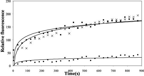
At a 1:5 peptide to lipid molar ratio, the Ebola virus wild-type peptide induced a significant increase of fluorescence similar to what is observed for the SIV peptide used as positive control, at a molar ratio of 1:50 (Fig. 3). Indeed, the SIV peptide has been extensively characterized by molecular modeling and by in vitro and in vivo approaches (2, 3, 18, 26). Both peptides have a similar mean hydrophobicity (<H>SIV = 0.93, <H>Ebola virus = 0.77) but Ebola virus peptide has to be 10 times more concentrated in order to induce an equivalent fusion level. This difference could be due to a higher aggregation and/or a lower α conformation propensity of the Ebola virus peptide, as it contains a α-helix-breaking proline residue. It should be noted that other tilted peptides, like the bovine leukemia virus fusion peptide, also contain a proline residue (35).
FIG. 3.
Time span of lipid mixing of phosphatidylcholine, phosphatidylethanolamine, sphingomyelin, and cholesterol large unilamellar vesicles (pH 6) induced by wild-type peptide (▪) and SIV peptide (×). The peptide/lipid molar ratio is 0.2 for Ebola virus peptide and 0.02 for the SIV peptide. Peptides, dissolved in TFE, were added to a mixture of R18-labeled and unlabeled large unilamellar vesicles (1:4, wt/wt). The increase in R18 relative fluorescence due to probe dilution is monitored at room temperature. In a control experiment, the same volume of TFE alone (•) is added to the liposome mixture.
Increasing amounts of Ebola virus wild-type peptide were added to liposomes. The R18 fluorescence intensity recorded after 900 s showed that the peptide causes lipid mixing in a concentration-dependent way (data not shown). When the unlabeled large unilamellar vesicle population was omitted, no change in fluorescence was observed. In the same way, mixing labeled and unlabeled liposomes in the absence of the peptide (and in the presence of TFE) had little effect on the R18 fluorescence, suggesting that there is no significant spontaneous vesicle instability. This also suggests that the effects observed with the peptide are not due to a simple exchange of the fluorescence probe between vesicles and/or a modification of the shape of vesicles. However, aggregation of large unilamellar vesicles with diffusion of the probe in the absence of true membrane fusion could not be completely ruled out at this stage. For that reason vesicle leakage and core mixing experiments were performed.
The HPTS (8-aminonaphtalene-1,3,6-trisulfonic acid)/DPX (p-xylylenebis[pyridinium] bromide) assay of Ellens et al. (11) was used to monitor vesicle leakage (23). Results indicate that the Ebola virus wild-type peptide induces an increase in the HPTS fluorescence at a ratio of 0.2 (0.02 for SIV) and pH 6, with LUV composition identical to that used in the lipid mixing assays. After 15 min, the wild-type, PM, TMI, TMII, and SIV peptides had a relative fluorescence of 41.5, 18.5, 31, 50, and 51%, respectively; 100% is established by lysing the vesicles with 0.5% Triton X-100. As for the lipid mixing assays, the Ebola virus wild-type peptide has to be ten times more concentrated than the SIV fusion peptide in order to induce a similar increase in fluorescence. Leakage also appears to be correlated to the concentration of Ebola virus peptide (data not shown).
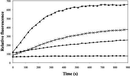
The mixing of liposome contents was monitored as described by Kendall and McDonald (21). In these assays, liposomes containing calcein and Co2+ ions are mixed with vesicles containing EDTA. Incubation of labeled and unlabeled vesicles in buffer alone (i.e., no peptide nor TFE) did not modify the calcein fluorescence intensity. Co2+ ions are present in the medium to avoid fluorescence due to leakage of vesicle contents. The maximum fluorescence was determined in the presence of 0.5% Triton X-100 (EDTA, 10 mM). When the Ebola virus wild-type peptide was added to a mixture of large unilamellar vesicles containing calcein, Co2+, EDTA, phosphatidylcholine, phosphatidylethanolamine, sphingomyelin, and cholesterol, a significant increase in calcein fluorescence was observed during the first minute (Fig. 4). Since Co2+ ions were present in the medium, the effects are probably due to the mixing of the liposome-aqueous phases. As for the lipid mixing assays, the Ebola virus wild-type peptide had to be more concentrated than the fusion peptide of SIV to induce a similar increase in fluorescence.
FIG. 4.
Core mixing of phosphatidylcholine, phosphatidylethanolamine, sphingomyelin, and cholesterol large unilamellar vesicles (pH 6) induced by 0.5% Triton X-100 and 10 mM EDTA (▴), TFE alone (▪), wild-type peptide (×; R = 1.2); SIV peptide (•; R = 0.2). R is the peptide/lipid molar ratio. Peptides dissolved in TFE are added to a mixture of calcein-, Co2+-, and EDTA-containing vesicles mixed at a 1:1 weight ratio. The calcein fluorescence is monitored at 520 nm at room temperature as a function of time.
From those results, the 25 to 35 Ebola virus peptide can therefore be considered as the active fusion peptide of the N-terminal part of GP2, since this fragment, smaller than the fusion peptide predicted by Gallaher (14), induces lipid destabilization and fusion.
Mutants were then designed via molecular modeling to assess the role of the hydrophobicity gradient in the orientation of the peptide in a membrane and in the fusion process, as has previously been done for viral and neurotoxic tilted peptides (5, 26, 28). By molecular modeling we designed a mutant with no asymmetric hydrophobicity gradient and with a predicted orientation parallel to the lipid bilayer surface (PM: YSGLAWIPSFG), as well as two mutants with an asymmetric hydrophobicity gradient and predicted as tilted by the IMPALA simulations (TMI: IGGLAWSPYFS, and TMII: ALGYAWIPIFG), but with a sequence different from that of the wild-type peptide (Fig. 2).
Due to the high hydrophobicity of the peptide, the parallel mutant (PM) could not be obtained by simple permutation of residues. Serine residues were inserted because serine was shown to be one of the best candidates for decreasing the mean hydrophobicity without creating steric clashes (20). Similar mutants (with a different mean hydrophobicity compared to the Ebola virus wild-type peptide) were already designed for the SIV tilted peptide (18, 26). TMI was designed to rule out that the differences in fusogenic and destabilizing activities between the wild-type and the PM are not due to a hydrophobicity decrease. TMII was designed to test the importance of the sequence versus the hydrophobicity gradient.
IMPALA simulations were carried out for all the mutants. For PM, the most stable position corresponds to an angle of 0°. The mass center is 16 Å from the bilayer center, similar to what is observed for amphipathic peptides (24) (Fig. 2Bα). TMI and II peptides adopt an angle of 55° and 45° towards the interfacial plane, respectively. Their mass centers are at 12 Å (Fig. 2Cα, 2Dα). Their properties resemble those of the Ebola virus wild-type peptide, showing a wide restraint minimum, and allowing metastable positions (not shown). From this analysis, we can assume that TMI and TMII behave like tilted peptides and should induce lipid destabilization, whereas PM should have no significant effect on liposomes.
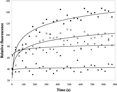
Under the same conditions as for the Ebola virus wild-type peptide, lipid mixing experiments clearly showed that the PM mutant does not induce significant liposome fusion. In contrast, TMI and TMII are almost as fusogenic as the Ebola virus wild-type peptide, reaching the same fluorescence intensity after 900 s (Fig. 5).
FIG. 5.
Time course of lipid mixing of phosphatidylcholine, phosphatidylethanolamine, sphingomyelin, and cholesterol large unilamellar vesicles (pH 6) induced by wild-type peptide (•), PM peptide (▪), TMI peptide (×), and TMII peptide (▴). The peptide/lipid molar ratio is 0.2. Peptides dissolved in TFE were added to a mixture of R18-labeled and unlabeled vesicles (1:4 wt/wt). The increase in R18 relative fluorescence due to probe dilution is monitored at room temperature. In a control experiment, the same volume of TFE alone is added to the liposome mixture; this curve has been subtracted from that of the peptides.
In the leakage assays, the mutants induced an HPTS fluorescence increase with different levels of efficiency. TMI and TMII were more lipid-destabilizing than PM. After 900 s, vesicle leakage reaches 50% for TMII, 30% for TMI, and less than 20% for PM.
Results obtained with the mutant peptides indicate that lipid destabilization is related to the hydrophobicity gradient, rather than to a specific amino acid sequence or a mean hydrophobicity. Thus, there is a relationship between the theoretical orientation of the peptide in a modeled membrane predicted by computational methods, and its fusogenic capacities, as already shown for other tilted peptides (18, 23, 26, 31, 36). We can therefore consider that the Ebola virus 25 to 35 fragment is a tilted peptide, as defined by Brasseur (5).
It should be noted that this tilted domain is not located at the N terminus of the fusion protein, as for the SIV or HIV peptides. This is also the case for viruses such as the vesicular stomatitis virus G, the Semliki Forest virus E1, the avian leucosis and sarcoma virus TM subunits, and the bovine leukemia virus gp30 (8). For the last, we have shown that the hydrophobic gradient and the tilted orientation also play a role in the fusion process (35). We assume that the presence of Pro and Gly residues allow the chain to be kinked or sufficiently flexible to allow the fusion domain to enter into the membrane. Pro and Gly residues are found close to the 25 to 35 fragment in the Ebola virus GP2 sequence and should permit lipid insertion of this domain.
In conclusion, we have shown that the Ebola virus fusion domain located on the N-terminal part of the GP2 sequence is shorter than predicted by Gallaher (14), going from residues 25 to 35 instead of 23 to 38. The 25 to 35 fragment is able to induce liposome destabilization and fusion. We propose a mechanism of action for the fusion peptide of the Ebola virus, predicting that the peptide penetrates the host membrane, adopts an oblique orientation, and destabilizes the lipid bilayer in a manner consistent with that of a tilted peptide.
Acknowledgments
R.B. and L.L. are Research Director and Research Associate at the Belgian Fund for Scientific Research (FNRS). A.T. is Research Director of the French National Institute for Medical Research (INSERM). A.B. was supported by the Ministère de la Region Wallone (DGTRE; Grant 215.370). This work was supported by “Protmem Project” from the Ministère de la Région Wallone (DGTRE; Grants 14540 and 3.4568.03).
REFERENCES
- 1.Atherton, E., C. J. Logan, and R. C. Sheppard. 1981. Peptide synthesis. II. Procedures for solid-phase synthesis by N-fluorenylmethoxycarbonylamino-acids on polyamide supports: synthesis of substance P and of acyl carrier protein 65-74 decapeptide. J. Chem. Soc. Perkin Trans. I 1:538. [Google Scholar]
- 2.Bradshaw, J. P., M. J. Darkes, T. A. Harroun, J. Katsaras, and R. M. Epand. 2000. Oblique membrane insertion of viral fusion peptide probed by neutron diffraction. Biochemistry 39:6581-6585. [DOI] [PubMed] [Google Scholar]
- 3.Brasseur, R. 1991. Differentiation of lipid-associating helices by use of three-dimensional molecular hydrophobicity potential calculations. J. Biol. Chem. 266:16120-16127. [PubMed] [Google Scholar]
- 4.Brasseur, R., T. Pillot, L. Lins, J. Vandekerckhove, and M. Rosseneu. 1997. Peptides in membranes: tipping the balance of membrane stability. Trends Biochem. Sci. 22:167-171. [DOI] [PubMed] [Google Scholar]
- 5.Brasseur, R. 2000. Tilted peptides: a motif for membrane destabilization (hypothesis). Mol. Membr. Biol. 17:31-40. [DOI] [PubMed] [Google Scholar]
- 6.Brasseur, R., L. Lins, B. Vanloo, J. M. Ruysschaert, and M. Rosseneu. 1992. Molecular modeling of the amphipathic helices of the plasma apolipoproteins. Proteins 13:246-257. [DOI] [PubMed] [Google Scholar]
- 7.Brasseur, R. (ed.). 1990. Molecular description of biological membrane components by computer-aided conformational analysis, p. 209-219. CRC Press, Boca Raton, Fla.
- 8.Delos, S. E., J. M. Gilbert, and J. M. White. 2000. The central proline of an internal viral fusion peptide serves two important roles. J. Virol. 74:1686-1693. [DOI] [PMC free article] [PubMed] [Google Scholar]
- 9.de Planque, M. R., J. A. Kruijtzer, R. M. Liskamp, D. Marsh, D. V. Greathouse, R. E. Koeppe, B. de Kruijff, and J. A. Killian. 1999. Different membrane anchoring positions of tryptophan and lysine in synthetic transmembrane alpha-helical peptides. J. Biol. Chem. 274:20839-20846. [DOI] [PubMed] [Google Scholar]
- 10.Ducarme, P., M. Rahman, and R. Brasseur. 1998. IMPALA: a simple restraint field to simulate the biological membrane in molecular structure studies. Proteins 30:357-371. [PubMed] [Google Scholar]
- 11.Ellens, H., J. Bentz, and F. C. Szoka. 1985. H+- and Ca2+-induced fusion and destabilization of liposomes. Biochemistry 24:3099-3106. [DOI] [PubMed] [Google Scholar]
- 12.Epand, R. M. 2003. Fusion peptides and the mechanism of viral fusion, Biochim. Biophys. Acta 1614:116-121. [DOI] [PubMed] [Google Scholar]
- 13.Gaboriaud, C., V. Bissery, T. Benchetrit, and J. P. Mornon. 1987. Hydrophobic cluster analysis: an efficient new way to compare and analyze amino acid sequences. FEBS Lett. 224:149-155. [DOI] [PubMed] [Google Scholar]
- 14.Gallaher, W. R. 1996. Similar structural models of the transmembrane proteins of Ebola virus and avian sarcoma viruses. Cell 85:477-478. [DOI] [PubMed] [Google Scholar]
- 15.Gordon, L. M., P. W. Mobley, R. Pilpa, M. A. Sherman, and A. J. Waring. 2002. Conformational mapping of the N-terminal peptide of HIV-1 gp41 in membrane environments by (13)C-enhanced Fourier transform infrared spectroscopy. Biochim. Biophys. Acta 1559:96-120. [DOI] [PubMed] [Google Scholar]
- 16.Han, X., J. H. Bushweller, D. S. Cafiso, and L. K. Tamm. 2001. Membrane structure and fusion-triggering conformational change of the fusion domain from influenza hemagglutinin. Nat. Struct. Biol. 8:715-720. [DOI] [PubMed] [Google Scholar]
- 17.Hope, M. J., M. G. Bally, G. Webb, and P. R. Cullis. 1985. Production of large unilamellar vesicles by a rapid extrusion procedure: characteriza-tion of size, trapped volume and ability to maintain a membrane potential. Biochim. Biophys. Acta 812:55-65. [DOI] [PubMed] [Google Scholar]
- 18.Horth, M., B. Lambrecht, M. C. Khim, F. Bex, C. Thiriart, J. M. Ruysschaert, A. Burny, and R. Brasseur. 1991. Theoretical and functional analysis of the SIV fusion peptide. EMBO J. 10:2747-2755. [DOI] [PMC free article] [PubMed] [Google Scholar]
- 19.Ito, H., S. Watanabe, A. Sanchez, M. A. Whitt, and Y. Kawaoka. 1999. Mutational analysis of the putative fusion domain of Ebola virus glycoprotein. J. Virol. 73:8907-8912. [DOI] [PMC free article] [PubMed] [Google Scholar]
- 20.Jonson, P. H., and S. B. Petersen. 2001. A critical view on conservative mutations. Protein Eng. 14:397-402. [DOI] [PubMed] [Google Scholar]
- 21.Kendall, D. A., and R. C. MacDonald. 1982. A fluorescence assay to monitor vesicle fusion and lysis. J. Biol. Chem. 257:13892-13895. [PubMed] [Google Scholar]
- 22.Lambert, G., A. Decout, B. Vanloo, D. Rouy, N. Duverger, A. Kalopissis, J. Vandekerckhove, J. Chambaz, R. Brasseur, and M. Rosseneu. 1998. The C-terminal helix of human apolipoprotein AII promotes the fusion of unilamellar liposomes and displaces apolipoprotein AI from high-density lipoproteins. Eur. J. Biochem. 253:328-338. [DOI] [PubMed] [Google Scholar]
- 23.Lins, L., C. Flore, L. Chapelle, P. J. Talmud, A. Thomas, and R. Brasseur. 2002. Lipid-interacting properties of the N-terminal domain of human apolipoprotein C-III. Protein Eng. 15:513-520. [DOI] [PubMed] [Google Scholar]
- 24.Lins, L., B. Charloteaux, A. Thomas, and R. Brasseur. 2001. Computational study of lipid-destabilizing protein fragments: towards a comprehensive view of tilted peptides. Proteins 44:435-447. [DOI] [PubMed] [Google Scholar]
- 25.Malashkevich, V. N., B. J. Schneider, M. L. McNally, et al. 1999. Core structure of the envelope glycoprotein GP2 from Ebola virus at 1.9-A resolution. Proc. Natl. Acad. Sci. USA 96:2662-2667. [DOI] [PMC free article] [PubMed] [Google Scholar]
- 26.Martin, I., F. Defrise-Quertain, V. Mandieau, N. M. Nielsen, T. Saermark, A. Burny, R. Brasseur, J. M. Ruysschaert, and M. Vandenbranden. 1991. Fusogenic activity of SIV (simian immunodeficiency virus) peptides located in the GP32 NH2 terminal domain. Biochem. Biophys. Res. Commun. 175:872-879. [DOI] [PubMed] [Google Scholar]
- 27.Martin, I., M. C. Dubois, F. Defrise-Quertain, T. Saermark, A. Burny, R. Brasseur, and J. M. Ruysschaert. 1994. Correlation between fusogenicity of synthetic modified peptides corresponding to the NH2-terminal extremity of simian immunodeficiency virus gp32 and their mode of insertion into the lipid bilayer: an infrared spectroscopy study. J. Virol. 68:1139-1148. [DOI] [PMC free article] [PubMed] [Google Scholar]
- 28.Perez-Mendez, O., B. Vanloo, A. Decout, M. Goethals, F. Peelman, J. Vandekerckhove, R. Brasseur, and M. Rosseneu. 1998. Contribution of the hydrophobicity gradient of an amphipathic peptide to its mode of association with lipids. Eur. J. Biochem. 256:570-579. [DOI] [PubMed] [Google Scholar]
- 29.Peuvot, J., A. Schanck, L. Lins, and R. Brasseur. 1999. Are the fusion processes involved in birth, life and death of the cell depending on tilted insertion of peptides into membranes? J. Theor. Biol. 198:173-181. [DOI] [PubMed] [Google Scholar]
- 30.Pillot, T., M. Goethals, B. Vanloo, C. Talussot, R. Brasseur, J. Vandekerckhove, M. Rosseneu, and L. Lins. 1996. Fusogenic properties of the C-terminal domain of the Alzheimer beta-amyloid peptide. J. Biol. Chem. 271:28757-28765. [DOI] [PubMed] [Google Scholar]
- 31.Pillot, T., L. Lins, M. Goethals, B. Vanloo, J. Baert, J. Vandekerckhove, M. Rosseneu, and R. Brasseur. 1997. The 118-135 peptide of the human prion protein forms amyloid fibrils and induces liposome fusion. J. Mol. Biol. 274:381-393. [DOI] [PubMed] [Google Scholar]
- 32.Ruiz-Arguello, M. B., F. M. Goni, F. B. Pereira, and J. L. Nieva. 1998. Phosphatidylinositol-dependent membrane fusion induced by a putative fusogenic sequence of Ebola virus. J. Virol. 72:1775-1781. [DOI] [PMC free article] [PubMed] [Google Scholar]
- 33.Suarez, T., M. J. Gomara, F. M. Goni, I. Mingarro, A. Muga, E. Perez-Paya, and J. L. Nieva. 2003. Calcium-dependent conformational changes of membrane-bound Ebola virus fusion peptide drive vesicle fusion. FEBS Lett. 535:23-28. [DOI] [PubMed] [Google Scholar]
- 34.Talmud, P., L. Lins, and R. Brasseur. 1996. Prediction of signal peptide functional properties: a study of the orientation and angle of insertion of yeast invertase mutants and human apolipoprotein B signal peptide variants. Protein Eng. 9:317-321. [DOI] [PubMed] [Google Scholar]
- 35.Voneche, V., D. Portetelle, R. Kettmann, L. Willems, K. Limbach, E. Paoletti, J. M. Ruysschaert, A. Burny, and R. Brasseur. 1992. Fusogenic segments of bovine leukemia virus and simian immunodeficiency virus are interchangeable and mediate fusion by means of oblique insertion in the lipid bilayer of their target cells. Proc. Natl. Acad. Sci. USA 89:3810-3814. [DOI] [PMC free article] [PubMed] [Google Scholar]


