Abstract
Influenza A viruses are the cause of annual epidemics of human disease with occasional outbreaks of pandemic proportions. The zoonotic nature of the disease and the vast viral reservoirs in the aquatic birds of the world mean that influenza will not easily be eradicated and that vaccines will continue to be needed. Recent technological advances in reverse genetics methods and limitations of the conventional production of vaccines by using eggs have led to a push to develop cell-based strategies to produce influenza vaccine. Although cell-based systems are being developed, barriers remain that need to be overcome if the potential of these systems is to be fully realized. These barriers include, but are not limited to, potentially poor reproducibility of viral rescue with reverse genetics systems and poor growth kinetics and yields. In this study we present a modified A/Puerto Rico/8/34 (PR8) influenza virus master strain that has improved viral rescue and growth properties in the African green monkey kidney cell line, Vero. The improved properties were mediated by the substitution of the PR8 NS gene for that of a Vero-adapted reassortant virus. The Vero growth kinetics of viruses with H1N1, H3N2, H6N1, and H9N2 hemagglutinin and neuraminidase combinations rescued on the new master strain were significantly enhanced in comparison to those of viruses with the same combinations rescued on the standard PR8 master strain. These improvements pave the way for the reproducible generation of high-yielding human and animal influenza vaccines by reverse genetics methods. Such a means of production has particular relevance to epidemic and pandemic use.
The potential of influenza A viruses to generate new human pathogenic strains from a vast natural reservoir in aquatic birds means that eradication of influenza is not feasible. Correspondingly, disease control requires the monitoring of virus reservoirs and the development of improved antiviral therapies and vaccines. The most widely used human influenza vaccines are those made from subunits of inactivated viruses propagated in embryonated chickens' eggs. These influenza vaccines are essentially genetic modifications of those generated in the mid-1970s, when production was improved by reassorting the circulating strain with A/Puerto Rico/8/34 (PR8), an H1N1 virus adapted for high growth in eggs (49). Since the mid-1970s, the influenza vaccine has been a so-called 6 + 2 reassortant containing the surface hemagglutinin (HA) and neuraminidase (NA) genes from the vaccine target strain and the remaining genes from PR8. Such reassortants are made by coinfecting eggs with both viruses and screening progeny for the desired 6 + 2 configuration.
Although routinely used to prepare human influenza virus vaccines and diagnostic reagents, embryonated chickens' eggs have potentially serious limitations as a host system, not least of which is that the cultivation of influenza viruses in eggs can lead to the selection of variants characterized by antigenic and structural changes in HA (19, 36, 40). Other problems include the lack of reliable year-round supplies of high-quality eggs, the possible presence of adventitious pathogens, and the low susceptibility of summer eggs to infection with influenza virus (27). The current cycle of interpandemic influenza vaccine production requires detailed planning up to 6 months before vaccine manufacture to ensure an adequate supply of embryonated eggs (10). Because a pandemic event cannot be predicted and a 6-month delay in vaccine production is unacceptable, there is an urgent need to develop improved cell culture systems for vaccine production. Such an improvement is a priority for the World Health Organization (WHO). As part of their Global Agenda on Influenza, the WHO has urged the development of novel vaccines and production strategies or technologies (44).
An additional need for improved cell-based protocols for the production of influenza vaccines has emerged with the development of reverse genetics, which enables the production of influenza vaccines from cloned viral cDNA (7, 16, 30). The ability to custom make influenza viruses by using this technology may dramatically improve the speed with which we can respond to pandemic emergencies. Advantages include the ability to attenuate pathogenic strains (45) and the elimination of the need to screen reassortant viruses for the 6 + 2 configuration, a procedure that can be time-consuming. The potential of reverse genetics to generate vaccine candidates has already been described (15, 39). The main drawback of this methodology is the need to use vaccine-approved cell lines; those commonly used to obtain influenza viruses from cDNA are 293T and Madin-Darby canine kidney (MDCK) cells. The 293T cell line is a transformed cell line and is therefore unlikely to be used for human vaccine production, and there are lingering concerns over the tumorigenic potential of MDCK cells (12). In addition, the use of host-specific RNA polymerase I promoters in reverse genetics systems limits usable cell lines to those of primate origin.
As an alternative cell-based system, the well-characterized African green monkey kidney (Vero) cell line has potential. Vero cells are suitable for the production of several human virus vaccines, including those against poliomyelitis and rabies (26). Despite earlier findings that influenza viruses do not replicate well in Vero cells (24, 28), the repeated addition of trypsin to the culture medium improves virus yields (20), and preliminary studies with a limited number of strains have indicated that Vero cells support the primary isolation and replication of influenza A viruses (11). Influenza virus vaccines derived from Vero cells have been produced and evaluated for immunogenicity, and their production has been scaled up to commercial levels (2, 23). These Vero-derived vaccines elicit humoral responses comparable to those elicited by the egg-grown vaccines but are more potent stimulators of the cellular response (2). These Vero-based vaccines have, however, consisted of unreassorted viruses rather than the accepted 6 + 2 PR8 reassortants.
The aim of the present study was to develop a reproducible, high-yielding Vero-based reverse genetics system to produce influenza vaccine. Govorkova and colleagues (11) previously derived a high-yielding influenza virus by passaging an H1N1 reassortant isolate, A/England/1/53 [HG], several times in Vero cells. This reassortant virus was reported to contain the surface glycoprotein genes from A/England/1/53 and the remaining genes from PR8, the standard vaccine master strain. Our aims were to identify the molecular changes responsible for this high-yielding phenotype and to produce an altered PR8 vaccine master strain adapted for optimal efficiency of viral rescue from cDNA in the eight-plasmid reverse genetics system and for growth in Vero cells.
MATERIALS AND METHODS
Viruses, cells, and plasmids.
Influenza viruses were obtained from the repository at St. Jude Children's Research Hospital, Memphis, Tenn., and propagated in 10-day-old embryonated chickens' eggs. MDCK cells and Vero cells approved by the WHO for use in vaccines were obtained from the American Type Culture Collection and maintained in minimal essential medium (MEM; Invitrogen) containing 10% fetal bovine serum. Three variants of A/England/1/53 were used in this study. These viruses were A/England/1/53, which is the wild-type virus, A/England/1/53 [HG], which is a reassortant between A/England/1/53 and PR8 with high growth in eggs, and Vero-adapted A/England/1/53 (Eng53/v-a), which is a Vero-adapted version of A/England/1/53 [HG].
The plasmids carrying the eight gene segments of the high-growth PR8 (H1N1) virus (pHW191 to pHW198) and the HA and NA genes of A/Panama/2007/99 (H3N2) (pHW444 and pHW446, respectively), A/New Caledonia/20/99 (H1N1) (pHW244 and pHW246, respectively), and A/quail/Hong Kong/G1/97 (H9N2) (pHW409 and pHW422, respectively) have been described previously (15). The plasmids carrying the eight gene segments of A/teal/Hong Kong/W312/97 (H6N1) were the same as those used by Hoffmann et al. (16).
Viral RNA extraction, reverse transcriptase PCR, and DNA sequencing.
Total RNA was extracted from virus-infected allantoic fluid by using the RNeasy kit (Qiagen). Production of cDNA and PCR were carried out under standard conditions with previously described primers (17). The Hartwell Center for Bioinformatics and Biotechnology at St. Jude Children's Research Hospital determined the sequence of template DNA by using synthetic oligonucleotides and rhodamine or dRhodamine dye terminator cycle sequencing ready reaction kits with AmpliTaq DNA polymerase FS (Perkin-Elmer Applied Biosystems Inc., Foster City, Calif.). Samples were subjected to electrophoresis, detection, and analysis on Perkin-Elmer Applied Biosystems model 373, model 373 stretch, or model 377 DNA sequencers.
Viral gene cloning.
Full-length cDNA copies of viral genes were amplified by reverse transcriptase PCR as described above. The PCR fragments were cloned into the vector pCRII-TOPO (Invitrogen) according to the manufacturer's instructions. After transformation of TOP10 cells (Invitrogen) and purification of the plasmid by using a plasmid midi kit (Qiagen), the plasmid was digested with the restriction enzyme BsmBI (New England Biolabs) and ligated into the vector pHW2000 (16). All clones were confirmed by full-length sequencing.
Virus rescue from cloned cDNA.
Vero cells were grown to 70% confluency in a 75-cm2 flask and then trypsinized with trypsin-EDTA (Invitrogen) and resuspended in 10 ml of Opti-MEM I (Invitrogen). Twenty milliliters of fresh Opti-MEM I was added to 2 ml of cell suspension, and 3 ml of this suspension was seeded into each well of a 6-well tissue culture plate (approximately 106 cells per well). The plates were incubated at 37°C overnight. The following day, 1 μg of each plasmid and 16 μl of TransIT LT-1 (Panvera) transfection reagent were added to Opti-MEM I to a final volume of 200 μl and the mixture was incubated at room temperature for 45 min. After incubation, the medium was removed from one well of the 6-well plate, 800 μl of Opti-MEM I was added to the transfection mix, and this mixture was added dropwise to the cells. Six hours later, the DNA-transfection mixture was replaced by Opti-MEM I. Twenty-four hours after transfection, 1 ml of Opti-MEM I containing l-(tosylamido-2-phenyl) ethyl chloromethyl ketone (TPCK)-treated trypsin (0.8 μg/ml) was added to the cells.
The efficiency of virus rescue was calculated by determining the numbers of PFU in the culture supernatants at various intervals after transfection. The number of PFU in MDCK cells was determined as previously described (24).
Viral growth kinetics in Vero cells.
The ability of viruses of different genotypes to grow in Vero cells was determined by analyzing multiple replication cycles. Before infection of Vero cells, the rescued viruses were amplified in 10-day-old embryonated chickens' eggs. Confluent Vero cell monolayers grown on 25-cm2 plates were washed with phosphate-buffered saline, overlaid with 0.5 ml of diluted virus suspension (to achieve a multiplicity of infection [MOI] of 0.01 PFU/cell), and incubated at room temperature for 60 min. The virus suspension was then removed by aspiration, and 3 ml of MEM containing 0.4 μg of TPCK-trypsin/ml was added. Twenty-four hours later, another 0.4 μg of TPCK-trypsin/ml was added. Sample supernatants were collected at 12-h intervals until 72 h after inoculation. The virus titer of the sample was determined by plaque assay on MDCK cells or by hemagglutination assay with 0.5% chicken erythrocytes. Antigenic analysis was performed with polyclonal antisera against PR8 (H1N1) (goat sera), A/New Caledonia/20/99 (H1N1) (sheep sera), A/Panama/2007/99 (H3N2) (sheep sera), A/teal/Hong Kong/W312/97 (H6N1) (chicken sera), and A/quail/Hong Kong/G1/97 (H9N2) (chicken sera) in hemagglutination inhibition (HI) assays (22). HI assays were performed on three independently rescued virus stocks of each recombinant.
Statistical evaluation of virus replication.
The viral growth curves were fitted with logistic function, a nonlinear regression model commonly used for modeling the sigmoid growth curves in biology and chemistry (5, 31, 33). The logistic function in this analysis is given by the following: ln(virus titer) = ø1/{1 + exp[(ø2 − t)/ø3]}, where t is the time postinoculation (in hours), ø1 is the peak titer (in ln[number of PFU per milliliter]), ø2 is the half time to peak titer, and ø3 is the time from half to 73% peak titer. The model was fitted using a nonlinear least-square procedure (1) implemented with statistical software S-PLUS (33). Each growth curve was fitted with data from three replicate experiments. Based on the estimates of parameters and their standard errors from fitted models, we compared the parameters for different strains of viruses (PR8, PR8/Eng-NS, and A/teal/Hong Kong/W312/97) by using t tests. Titers of PR8 and PR8/Eng-NS were also compared using the two-way analysis of variance (ANOVA) method separately at 12, 24, 36, and 48 h postinoculation. The two factors of the two-way ANOVA are virus type and master strain backbone.
RESULTS
Viral growth characteristics in Vero cells.
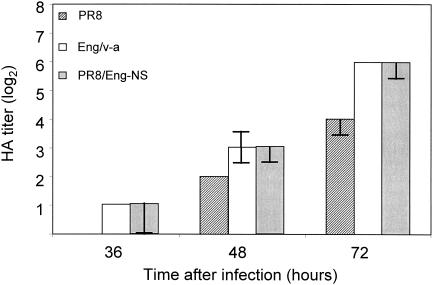
Upon multiple passaging of A/England/1/53 [HG] in Vero cells, Govorkova and colleagues (11) produced a high-yielding virus, Eng53/v-a. This reassortant virus was reported to contain the surface glycoprotein genes from A/England/1/53 and the six remaining genes from PR8. To confirm this strain's high-growth phenotype in Vero cells, we evaluated the growth kinetics of the three strains of viruses in Vero cells over 72 h by using hemagglutination activity in culture supernatants as a marker of viral growth. We confirmed the results of Govorkova et al. (11) by showing that Eng53/v-a grew faster and to higher titers than did the unpassaged parental A/England/1/53 or PR8.
Genetic characterization of the high-growth phenotype.
The Eng53/v-a strain had better growth characteristics in Vero cells than did PR8. To compare the replicative genes of Eng53/v-a and PR8 and thereby identify the molecular differences responsible for the high-growth phenotype, we first sequenced the complete genome of Eng53/v-a. Eng53/v-a contained not only the HA and NA genes from A/England/1/53 but also the nonstructural (NS) gene segment. The genes encoding the remaining Eng53/v-a proteins (PB2, PB1, PA, NP, and M) had more than 99% nucleotide identity to those of PR8. The NS genes from Eng53/v-a had only 90% identity to the corresponding genes in PR8, the HA gene had 98% identity, and the NA gene had 97% identity.
Effect of the NS genes of Eng53/v-a on the efficiency of virus rescue from cDNA in Vero cells.
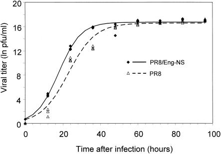
The greatest number of genetic differences between Eng53/v-a and PR8 were in the NS genes. This finding suggested that the NS genes are involved in Vero adaptation. To determine the effect of the NS genes on virus rescue from Vero cells, we cloned the NS gene segment of Eng53/v-a into the plasmid pHW2000 and set up two simultaneous virus rescue experiments. In one we used the plasmids necessary to rescue PR8, and in the other we used plasmids necessary to rescue a reassortant virus containing seven gene segments from PR8 and the NS gene segment of Eng53/v-a (PR8/Eng-NS). Supernatants from each experiment were assayed for virus titers every 12 h. The rescue efficiency of the PR8/Eng-NS virus was superior to that of PR8 12, 24, and 36 h after transfection (P < 0.001 [ANOVA]) (Fig. 1), demonstrating that the gene product(s) of the NS genes can influence viral growth characteristics in Vero cells. There was no significant difference between virus titers at times over 36 h posttransfection. The half time to peak titer was significantly (P < 0.0001) reduced for PR8/Eng-NS compared to that for PR8 (17.8 ± 0.6 and 22.9 ± 0.7 h, respectively). As a comparison of the virus rescue efficiency of PR8/Eng-NS in different culture systems, we also rescued this virus on 293T cells alone and on 293T cells in coculture with MDCK cells. At 24, 48, and 72 h after the addition of trypsin, the titers of virus in the supernatant of the coculture system were comparable to those in Vero (within a fivefold difference), whereas the titers in 293T cells alone were substantially reduced (103- to 104-fold lower). To assess the suitability of the PR8/Eng-NS virus as a master strain for vaccine purposes, we performed experiments in which we rescued the HA and NA of contemporary H1N1 (A/New Caledonia/20/99), H3N2 (A/Panama/2007/99), H6N1 (A/teal/Hong Kong/W312/97), and H9N2 (A/quail/Hong Kong/G1/97) viruses on both the PR8 and PR8/Eng-NS backbones in Vero cells. We were able to rescue all HA and NA combinations on both backbones, although the kinetics of virus rescue (half time to peak titer) were significantly faster with the PR8/Eng-NS viruses (P, <0.01 for all virus subtypes).
FIG. 1.
The efficiency of rescue of PR8 from Vero cells is improved by replacement of the NS gene segment with that from Eng53/v-a. We used the eight-plasmid reverse genetics system to transfect Vero cells and rescue PR8 and PR8 containing the NS gene segment of Eng53/v-a (PR8/Eng-NS). The numbers of PFU in culture supernatants were determined at 12-h intervals. The results shown are from three independent experiments conducted with each virus.
Effects of the NS genes from Eng53/v-a on growth characteristics of PR8 in Vero cells.
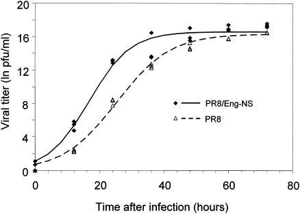
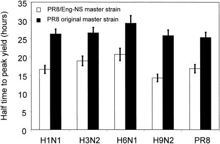
To assess the effect of the replacement of the NS gene segment in PR8 on subsequent virus amplification in Vero cells, we infected triplicate samples of near-confluent Vero cells with PR8 and PR8/Eng-NS at a low MOI (0.01) and monitored virus replication by assaying culture supernatants every 12 h. PR8/Eng-NS had a significantly (P < 0.0001) lower half time to peak titer than did PR8 (16.8 ± 0.6 and 25.3 ± 0.7 h, respectively), although there was no significant difference in peak titers (1.6 × 10⁷ and 1.2 × 10⁷ PFU/ml, respectively) (Fig. 2). We conducted similar experiments with recombinant PR8 and PR8/Eng-NS viruses carrying the surface glycoproteins of A/New Caledonia/20/99 (H1N1), A/Panama/2007/99 (H3N2), A/teal/Hong Kong/W312/97 (H6N1), and A/quail/Hong Kong/G1/97 (H9N2). The half times to peak virus yield were significantly (P < 0.0001) lower in all PR8/Eng-NS viruses than in their PR8 counterparts (Fig. 3). The peak titers of the PR8/Eng-NS viruses carrying the surface glycoproteins of the A/New Caledonia/20/99 and A/Panama/2007/99 viruses were significantly higher (P < 0.005) than those of the PR8 viruses (Table 1). The increase in growth kinetics in Vero cells did not affect the ability of the reassortant viruses to grow in eggs. The PR8/Eng-NS variants all grew in eggs to HA titers equivalent to those of their PR8 counterparts (data not shown).
FIG. 2.
The growth characteristics of PR8 in Vero cells are improved by inclusion of the NS gene segment of Eng53/v-a. Vero cell monolayers were infected with PR8 or PR8 containing the NS gene segment of Eng53/v-a (PR8/Eng-NS) at a MOI of 0.01. The numbers of PFU in culture supernatants were determined at 12-h intervals. The results shown are from three independent experiments conducted with each virus.
FIG. 3.
The half time to peak yield of different subtypes of influenza virus in Vero cells is reduced by inclusion of the NS gene segment of Eng53/v-a. The HA and NA genes of contemporary human H1N1 and H3N2 viruses, contemporary avian H6N1 and H9N2 viruses, and PR8 were rescued on the master strains PR8 and PR8 containing the NS gene segment of Eng53/v-a (PR8/Eng-NS). Vero cell monolayers were infected with each virus at a MOI of 0.01, and the numbers of PFU in culture supernatants were determined at 12-h intervals. The average half times to peak titer shown were calculated from three independent experiments conducted with each virus; the error bars indicate the 95% confidence intervals.
TABLE 1.
Estimated peak titers of viruses with HA and NA combinations rescued on different vaccine backbones in Vero cells
| Source of HA and NA | Backbone | Estimated peak titera | 95% Confidence interval | P value |
|---|---|---|---|---|
| PR8 | PR8/Eng-NS | 16.59 | 16.15-17.03 | 0.4854 |
| PR8 | 16.33 | 15.76-16.90 | ||
| A/New Caledonia/20/99 | PR8/Eng-NS | 16.87 | 16.44-17.30 | 0.0041b |
| PR8 | 15.81 | 15.27-16.34 | ||
| A/Panama/2007/99 | PR8/Eng-NS | 16.48 | 15.99-16.96 | 0.0001b |
| PR8 | 14.90 | 14.34-15.46 | ||
| A/quail/Hong Kong/G1/97 | PR8/Eng-NS | 16.29 | 15.88-16.70 | 0.2589 |
| PR8 | 15.87 | 15.28-16.46 | ||
| A/teal/Hong Kong/W312/97 | PR8/Eng-NS | 13.66 | 13.18-14.14 | 0.0011b |
| PR8 | 12.18 | 11.59-12.78 |
Expressed as natural log of number of PFU per milliliter.
Indicates subtypes for which the peak titer of the PR8/Eng-NS variant was statistically higher than that of the PR8 variant.
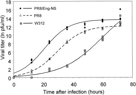
In a related experiment, we compared the growth characteristics, in Vero cells, of two reverse genetics-derived viruses containing the HA and NA genes of A/teal/Hong Kong/W312/97 with those of the reverse genetics-derived wild-type A/teal/Hong Kong/W312/97. The remaining gene segments of the two variants were from PR8 or PR8/Eng-NS. Although the peak titers of all three viruses at 72 h were similar (Fig. 4), the half times to peak titer were significantly (P < 0.0001) shorter for the PR8 and PR8/Eng-NS variants (29.3 ± 1.0 and 20.7 ± 0.8 h, respectively) than for A/teal/Hong Kong/W312/97 (60.3 ± 4.5 h) (Fig. 4).
FIG. 4.
Growth kinetics of an H6N1 virus are significantly improved by substituting all but the HA and NA genes of the H6N1 virus with genes from a master strain (PR8 or PR8/Eng-NS). We determined the growth kinetics, in Vero cells, of A/teal/Hong Kong/W312/97 (W312) and of reassortant viruses carrying the HA and NA genes of A/teal/Hong Kong/W312/97 and the remaining genes of the PR8 or PR8/Eng-NS master strain. Vero cell monolayers were infected with each virus at a MOI of 0.01, and the numbers of PFU in culture supernatants were determined at 12-h intervals.
To test whether changes in genes other than NS could lead to further improvements in the growth characteristics of PR8/Eng-NS, we determined the growth characteristics of PR8, Eng53/v-a, and PR8/Eng-NS in Vero cells. If further improvements were possible, they would be indicated by Eng53/v-a's having growth characteristics superior to those of PR8/Eng-NS. The growth characteristics of Eng53/v-a and PR8/Eng-NS were substantially better than those of PR8 but indistinguishable from each another (Fig. 5). This result suggests that the high-yielding phenotype of Eng53/v-a is due solely to the NS genes.
FIG. 5.
The NS gene segmet of Eng53/v-a is alone sufficient to confer a high-growth phenotype on PR8. Vero cell monolayers were infected with PR8, Eng53/v-a, or PR8 containing the NS gene segment of Eng53v-a (PR8/Eng-NS) at a MOI of 0.01. Virus titers in culture supernatants were estimated by using hemagglutination assays at 12-h intervals. The data represent median titers from three independent experiments, and the error bars indicate the range of values.
Stability of viruses rescued from Vero cells.
Vaccine production procedures must allow the virus produced to retain the antigenic properties of the parent viruses. Any benefit from an increase in yield would be negated by changes in the antigenicity of a vaccine. To determine whether antigenic changes occurred upon rescue of viruses in Vero cells, we used HI assays to compare the antigenic profiles of the rescued PR8/Eng-NS viruses with those of their wild-type counterparts. HI assays were performed using polyclonal antisera against the corresponding wild-type virus. All PR8/Eng-NS rescued viruses were antigenically indistinguishable from the corresponding wild-type viruses. To confirm the genetic stability of the PR8/Eng-NS rescued viruses, we compared the HA gene sequences of the rescued and wild-type viruses. We found no changes in any of the viruses. This result demonstrates that introduction of the NS gene segment from Eng53/v-a into the PR8 vaccine master strain does not lead to changes in the HA gene.
DISCUSSION
Recent discoveries in influenza pathogenicity and reverse genetics have the potential to revolutionize the way we prepare and manufacture pandemic and interpandemic influenza vaccines. Much of this technology has, however, been confined to experimental protocols; the realization of this potential awaits refinements of the methods. The results we report here go a long way toward helping to fulfill the potential of these technologies. By incorporating the NS gene segment of Eng53/v-a into the standard PR8 vaccine master strain, we have shown a reproducible improvement in vaccine virus rescue and growth in Vero cells with no drop in virus titers in eggs. Although we were able to rescue these viruses with the standard PR8 vaccine strain, the improved efficiency of rescue with our system may be crucial with other HA and NA combinations that are poorly infective in Vero cells.
With each of the HA and NA subtypes we tested, the PR8/Eng-NS viruses reached peak titers significantly faster than did the corresponding PR8 virus (Fig. 3). The peak titers of the PR8/Eng-NS variants containing the surface glycoproteins of the two contemporary vaccine strains (A/New Caledonia/20/99 and A/Panama/2007/99) and of A/teal/Hong Kong/W312/97, a virus implicated in the genesis of the 1997 H5N1 human viruses (18), were also significantly higher than those of the PR8 viruses (Table 1). In contrast, there was no difference in the peak titers of the PR8/Eng-NS and PR8 viruses carrying the surface glycoproteins of A/quail/Hong Kong/G1/97 or PR8 itself. However, by the time these peak titers were reached with the PR8 variants, the cells infected with the PR8/Eng-NS variants had been completely destroyed by cytopathic effects, and a manufacturing process in which cells and fresh media can be continually added or replenished is likely to produce higher yields for those viruses on the PR8/Eng-NS backbone.
Our data also provide support for the continued use of 6 + 2 high-growth reassortants for vaccine production in Vero cells. For example, the PR8/Eng-NS variant of the H6N1 virus had growth characteristics significantly superior to those of the wild-type H6N1 virus; half times to peak titers were three times longer in the wild-type virus. The use of 6 + 2 reassortants also reduces the risks of growing adventitious agents with influenza viruses isolated directly from clinical samples.
One of the benefits of cell-based production of vaccines is that it appears to allow the amino acid sequences of influenza virus HA molecules to remain unchanged; these molecules are altered during the adaptation of viruses to eggs (19, 36, 40). It should, however, be noted that changes in the biologic activity of a virus can occur in the absence of sequence changes. It has been shown that differences in the abilities of the same human H3N2 strain to agglutinate human and chicken red blood cells were associated with the type of cells from which the strain was isolated (MDCK or Vero) (13). Romanova and colleagues have recently shown that the inability of the Vero-grown variant to grow in eggs and agglutinate chicken red blood cells is related to the higher proportion of oligosaccharides of high mannose type in this variant than in the corresponding MDCK-derived isolates. This observation was made in the absence of any amino acid differences between the variants (37). We found that the HA genes of the PR8/Eng-NS-derived viruses were the same before and after rescue and propagation from Vero cells. This stability is an advantage in a vaccine that derives much of its protective qualities from the production of neutralizing antibody directed against the HA molecule. However, as discussed by Kemble and Greenberg (21), such benefits will be lost unless candidate vaccine viruses are first isolated in approved cell lines rather than in the widely used MDCK cells or eggs. The retention of these benefits of cell-based vaccine production requires a commitment from many agencies, including the WHO influenza network.
Host range in influenza viruses is a polygenic trait for which many influenza virus proteins have been implicated (14, 41, 43, 46). Likewise, many genes, including the NS genes, have been implicated in the attenuation of influenza viruses in different hosts (4, 25, 43). However, in our system, the transfer of the NS gene segment from Eng53/v-a to PR8 was alone sufficient to confer the high-growth phenotype (Fig. 5). Although many factors have been reported to determine the efficiency of growth of influenza viruses, the NS1 protein (one of the two proteins expressed by the NS gene segment) is thought to play a significant role in translation and replication. For example, NS1 and its interaction with host proteins have been reported to play central roles in inhibiting the nuclear export and splicing of host mRNA (8, 29, 34, 35, 47, 48). Furthermore, NS1 protein plays an important role in regulating interferon activity (42). It is unlikely, however, that the interferon pathway is involved in viral growth in Vero cells, because Vero cells do not produce interferon (6). Another study failed to identify major changes in the shutoff of host protein synthesis or in viral protein expression in Vero cells after infection with NS1 deletion mutant viruses (38). Garcia-Sastre et al. showed, however, that a mutant virus that did not express NS1 protein (delNS1) grew approximately 10 times slower on Vero cells than did the wild-type strain (9). The second protein encoded by the NS gene segment is the NS2 protein, or nuclear export protein (NEP). Although little is known about the function of NEP, this polypeptide interacts with nucleoprotein and contributes to the nuclear export of the viral ribonucleoproteins (32). Additionally, amino acid residues of the NEP have been shown to be crucial for viral replication (3). Studies are ongoing to identify the NS gene products responsible for increased viral replication in Vero cells and the mechanisms involved.
The applicability of reverse genetics to the production of influenza vaccines depends on the use of a suitable cell culture system. Technical constraints and the limited number of cells licensed for vaccine production severely limit the options available. It is therefore likely that timely improvements in reverse genetics-derived vaccines will arise from optimization of current protocols rather than identification of alternative systems. The data presented in this paper show that use of the Vero cell system with an improved master strain virus is a viable option for the rapid manufacture of influenza vaccines in pandemic emergencies and for the production of vaccines during annual epidemics. Although the technologies are now available, use of the vaccines created by them requires approval by regulatory agencies, which must be prepared and equipped to rapidly give such approval.
Acknowledgments
These studies were supported by grants AI29680 and AI95357 from the National Institute of Allergy and Infectious Diseases, by Cancer Center Support (CORE) grant CA-21765 from the National Institutes of Health, and by the American Lebanese Syrian Associated Charities (ALSAC).
We thank Janet Davies for editorial assistance.
REFERENCES
- 1.Bates, D. M., and D. Watts. 1988. Nonlinear regression analysis and its application. Wiley, New York, N.Y.
- 2.Bruhl, P., A. Kerschbaum, O. Kistner, N. Barrett, F. Dorner, and M. Gerencer. 2000. Humoral and cell-mediated immunity to vero cell-derived influenza vaccine. Vaccine 19:1149-1158. [DOI] [PubMed] [Google Scholar]
- 3.Bullido, R., P. Gomez-Puertas, M. J. Saiz, and A. Portela. 2001. Influenza A virus NEP (NS2 protein) downregulates RNA synthesis of model template RNAs. J. Virol. 75:4912-4917. [DOI] [PMC free article] [PubMed] [Google Scholar]
- 4.Clements, M. L., E. K. Subbarao, L. F. Fries, R. A. Karron, W. T. London, and B. R. Murphy. 1992. Use of single-gene reassortant viruses to study the role of avian influenza A virus genes in attenuation of wild-type human influenza A virus for squirrel monkeys and adult human volunteers. J. Clin. Microbiol. 30:655-662. [DOI] [PMC free article] [PubMed] [Google Scholar]
- 5.Davidian, M., and D. M. Gilitinan. 1996. Nonlinear models for repeated measurement data. Chapman & Hall, New York, N.Y.
- 6.Diaz, M. O., S. Ziemin, M. M. Le Beau, P. Pitha, S. D. Smith, R. R. Chilcote, and J. D. Rowley. 1988. Homozygous deletion of the alpha- and beta 1-interferon genes in human leukemia and derived cell lines. Proc. Natl. Acad. Sci. USA 85:5259-5263. [DOI] [PMC free article] [PubMed] [Google Scholar]
- 7.Fodor, E., L. Devenish, O. G. Engelhardt, P. Palese, G. G. Brownlee, and A. Garcia-Sastre. 1999. Rescue of influenza A virus from recombinant DNA. J. Virol. 73:9679-9682. [DOI] [PMC free article] [PubMed] [Google Scholar]
- 8.Fortes, P., A. Beloso, and J. Ortin. 1994. Influenza virus NS1 protein inhibits pre-mRNA splicing and blocks mRNA nucleocytoplasmic transport. EMBO J. 13:704-712. [DOI] [PMC free article] [PubMed] [Google Scholar]
- 9.Garcia-Sastre, A., A. Egorov, D. Matassov, S. Brandt, D. E. Levy, J. E. Durbin, P. Palese, and T. Muster. 1998. Influenza A virus lacking the NS1 gene replicates in interferon-deficient systems. Virology 252:324-330. [DOI] [PubMed] [Google Scholar]
- 10.Gerdil, C. 2003. The annual production cycle for influenza vaccine. Vaccine 21:1776-1779. [DOI] [PubMed] [Google Scholar]
- 11.Govorkova, E. A., N. V. Kaverin, L. V. Gubareva, B. Meignier, and R. G. Webster. 1995. Replication of influenza A viruses in a green monkey kidney continuous cell line (Vero). J. Infect. Dis. 172:250-253. [DOI] [PubMed] [Google Scholar]
- 12.Govorkova, E. A., G. Murti, B. Meignier, C. de Taisne, and R. G. Webster. 1996. African green monkey kidney (Vero) cells provide an alternative host cell system for influenza A and B viruses. J. Virol. 70:5519-5524. [DOI] [PMC free article] [PubMed] [Google Scholar]
- 13.Grassauer, A., A. Y. Egorov, B. Ferko, I. Romanova, H. Katinger, and T. Muster. 1998. A host restriction-based selection system for influenza haemagglutinin transfectant viruses. J. Gen. Virol. 79:1405-1409. [DOI] [PubMed] [Google Scholar]
- 14.Hatta, M., P. Halfmann, K. Wells, and Y. Kawaoka. 2002. Human influenza a viral genes responsible for the restriction of its replication in duck intestine. Virology 295:250-255. [DOI] [PubMed] [Google Scholar]
- 15.Hoffmann, E., S. Krauss, D. Perez, R. Webby, and R. Webster. 2002. Eight-plasmid system for rapid generation of influenza virus vaccines. Vaccine 20:3165-3170. [DOI] [PubMed] [Google Scholar]
- 16.Hoffmann, E., G. Neumann, Y. Kawaoka, G. Hobom, and R. G. Webster. 2000. A DNA transfection system for generation of influenza A virus from eight plasmids. Proc. Natl. Acad. Sci. USA 97:6108-6113. [DOI] [PMC free article] [PubMed] [Google Scholar]
- 17.Hoffmann, E., J. Stech, Y. Guan, R. G. Webster, and D. R. Perez. 2001. Universal primer set for the full-length amplification of all influenza A viruses. Arch. Virol. 146:2275-2289. [DOI] [PubMed] [Google Scholar]
- 18.Hoffmann, E., J. Stech, I. Leneva, S. Krauss, C. Scholtissek, P. S. Chin, M. Peiris, K. F. Shortridge, and R. G. Webster. 2000. Characterization of the influenza A virus gene pool in avian species in southern China: was H6N1 a derivative or a precursor of H5N1? J. Virol. 74:6309-6315. [DOI] [PMC free article] [PubMed] [Google Scholar]
- 19.Katz, J. M., C. W. Naeve, and R. G. Webster. 1987. Host cell-mediated variation in H3N2 influenza viruses. Virology 156:386-395. [DOI] [PubMed] [Google Scholar]
- 20.Kaverin, N. V., and R. G. Webster. 1995. Impairment of multicycle influenza virus growth in Vero (W. H. O.) cells by loss of trypsin activity. J. Virol. 69:2700-2703. [DOI] [PMC free article] [PubMed] [Google Scholar]
- 21.Kemble, G., and H. Greenberg. 2003. Novel generations of influenza vaccines. Vaccine 21:1789-1795. [DOI] [PubMed] [Google Scholar]
- 22.Kida, H., L. E. Brown, and R. G. Webster. 1982. Biological activity of monoclonal antibodies to operationally defined antigenic regions on the hemagglutinin molecule of A/Seal/Massachusetts/1/80 (H7N7) influenza virus. Virology 122:38-47. [DOI] [PubMed] [Google Scholar]
- 23.Kistner, O., P. N. Barrett, W. Mundt, M. Reiter, S. Schober-Bendixen, G. Eder, and F. Dorner. 1999. Development of a Vero cell-derived influenza whole virus vaccine. Dev. Biol. Stand. 98:101-110. [PubMed] [Google Scholar]
- 24.Lau, S. C., and C. Scholtissek. 1995. Abortive infection of Vero cells by an influenza A virus (FPV). Virology 212:225-231. [DOI] [PubMed] [Google Scholar]
- 25.Maassab, H. F., and D. C. DeBorde. 1983. Characterization of an influenza A host range mutant. Virology 130:342-350. [DOI] [PubMed] [Google Scholar]
- 26.Montagnon, B. J., B. Fanget, and A. J. Nicolas. 1981. The large-scale cultivation of VERO cells in micro-carrier culture for virus vaccine production. Preliminary results for killed poliovirus vaccine. Dev. Biol. Stand. 47:55-64. [PubMed] [Google Scholar]
- 27.Monto, A. S., H. F. Maassab, and E. R. Bryan. 1981. Relative efficacy of embryonated eggs and cell culture for isolation of contemporary influenza viruses. J. Clin. Microbiol. 13:233-235. [DOI] [PMC free article] [PubMed] [Google Scholar]
- 28.Nakamura, K., and M. Homma. 1981. Protein synthesis in Vero cells abortively infected with influenza B virus. J. Gen. Virol. 56:199-202. [DOI] [PubMed] [Google Scholar]
- 29.Nemeroff, M. E., S. M. Barabino, Y. Li, W. Keller, and R. M. Krug. 1998. Influenza virus NS1 protein interacts with the cellular 30 kDa subunit of CPSF and inhibits 3′end formation of cellular pre-mRNAs. Mol. Cell 1:991-1000. [DOI] [PubMed] [Google Scholar]
- 30.Neumann, G., T. Watanabe, H. Ito, S. Watanabe, H. Goto, P. Gao, M. Hughes, D. R. Perez, R. Donis, E. Hoffmann, G. Hobom, and Y. Kawaoka. 1999. Generation of influenza A viruses entirely from cloned cDNAs. Proc. Natl. Acad. Sci. USA 96:9345-9350. [DOI] [PMC free article] [PubMed] [Google Scholar]
- 31.O'Connel, M. A., B. A. Belanger, and P. D. Haaland. 1993. Calibration and assay development using the four-parameter logistic model. Chemom. Intell. Lab. Syst. 20:97-114. [Google Scholar]
- 32.O'Neill, R. E., J. Talon, and P. Palese. 1998. The influenza virus NEP (NS2 protein) mediates the nuclear export of viral ribonucleoproteins. EMBO J. 17:288-296. [DOI] [PMC free article] [PubMed] [Google Scholar]
- 33.Pinheiro, J. C., and D. M. Bates. 2000. Mixed-effects model in S and S-PLUS. Springer, New York, N.Y.
- 34.Qiu, Y., and R. M. Krug. 1994. The influenza virus NS1 protein is a poly(A)-binding protein that inhibits nuclear export of mRNAs containing poly(A). J. Virol. 68:2425-2432. [DOI] [PMC free article] [PubMed] [Google Scholar]
- 35.Qiu, Y., M. Nemeroff, and R. M. Krug. 1995. The influenza virus NS1 protein binds to a specific region in human U6 snRNA and inhibits U6-U2 and U6-U4 snRNA interactions during splicing. RNA 1:304-316. [PMC free article] [PubMed] [Google Scholar]
- 36.Robertson, J. S., C. W. Naeve, R. G. Webster, J. S. Bootman, R. Newman, and G. C. Schild. 1985. Alterations in the hemagglutinin associated with adaptation of influenza B virus to growth in eggs. Virology 143:166-174. [DOI] [PubMed] [Google Scholar]
- 37.Romanova, J., D. Katinger, B. Ferko, R. Voglauer, L. Mochalova, N. Bovin, W. Lim, H. Katinger, and A. Egorov. 2003. Distinct host range of influenza H3N2 virus isolates in Vero and MDCK cells is determined by cell specific glycosylation pattern. Virology 307:90-97. [DOI] [PubMed] [Google Scholar]
- 38.Salvatore, M., C. F. Basler, J. P. Parisien, C. M. Horvath, S. Bourmakina, H. Zheng, T. Muster, P. Palese, and A. Garcia-Sastre. 2002. Effects of influenza A virus NS1 protein on protein expression: the NS1 protein enhances translation and is not required for shutoff of host protein synthesis. J. Virol. 76:1206-1212. [DOI] [PMC free article] [PubMed] [Google Scholar]
- 39.Schickli, J. H., A. Flandorfer, T. Nakaya, L. Martinez-Sobrido, A. Garcia-Sastre, and P. Palese. 2001. Plasmid-only rescue of influenza A virus vaccine candidates. Philos. Trans. R. Soc. Lond. B 356:1965-1973. [DOI] [PMC free article] [PubMed] [Google Scholar]
- 40.Schild, G. C., J. S. Oxford, J. C. de Jong, and R. G. Webster. 1983. Evidence for host-cell selection of influenza virus antigenic variants. Nature 303:706-709. [DOI] [PubMed] [Google Scholar]
- 41.Scholtissek, C., H. Burger, O. Kistner, and K. F. Shortridge. 1985. The nucleoprotein as a possible major factor in determining host specificity of influenza H3N2 viruses. Virology 147:287-294. [DOI] [PubMed] [Google Scholar]
- 42.Seo, S. H., E. Hoffmann, and R. G. Webster. 2002. Lethal H5N1 influenza viruses escape host anti-viral cytokine responses. Nat. Med. 8:950-954. [DOI] [PubMed] [Google Scholar]
- 43.Snyder, M. H., W. T. London, H. F. Maassab, R. M. Chanock, and B. R. Murphy. 1990. A 36 nucleotide deletion mutation in the coding region of the NS1 gene of an influenza A virus RNA segment 8 specifies a temperature-dependent host range phenotype. Virus Res. 15:69-83. [DOI] [PubMed] [Google Scholar]
- 44.Stohr, K. 2003. The global agenda on influenza surveillance and control. Vaccine 21:1744-1748. [DOI] [PubMed] [Google Scholar]
- 45.Subbarao, K., H. Chen, D. Swayne, L. Mingay, E. Fodor, G. Brownlee, X. Xu, X. Lu, J. Katz, N. Cox, and Y. Matsuoka. 2003. Evaluation of a genetically modified reassortant H5N1 influenza A virus vaccine candidate generated by plasmid-based reverse genetics. Virology 305:192-200. [DOI] [PubMed] [Google Scholar]
- 46.Tian, S. F., A. J. Buckler-White, W. T. London, L. J. Reck, R. M. Chanock, and B. R. Murphy. 1985. Nucleoprotein and membrane protein genes are associated with restriction of replication of influenza A/Mallard/N.Y./78 virus and its reassortants in squirrel monkey respiratory tract. J. Virol. 53:771-775. [DOI] [PMC free article] [PubMed] [Google Scholar]
- 47.Wang, W., and R. M. Krug. 1998. U6atac snRNA, the highly divergent counterpart of U6 snRNA, is the specific target that mediates inhibition of AT-AC splicing by the influenza virus NS1 protein. RNA 4:55-64. [PMC free article] [PubMed] [Google Scholar]
- 48.Wolff, T., R. E. O'Neill, and P. Palese. 1998. NS1-binding protein (NS1-BP): a novel human protein that interacts with the influenza A virus nonstructural NS1 protein is relocalized in the nuclei of infected cells. J. Virol. 72:7170-7180. [DOI] [PMC free article] [PubMed] [Google Scholar]
- 49.Wood, J. M., and M. S. Williams. 1998. History of inactivated influenza vaccines, p. 317-323. In K. G. Nicholson, R. G. Webster, and A. J. Hay (ed.), Textbook of influenza. Blackwell Science Ltd., Oxford, United Kingdom.


