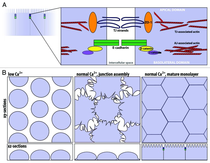
Figure 2. The apical junctional complex and calcium-switch model of junction assembly. (A) The tight junction and adherens junction, and their associated actin cytoskeleton, comprise the apical junctional complex. The tight junction strands form a semi-permeable barrier, and ZO-1 functions as a tight junction scaffolding protein. At the adherens junction, the cadherin-catenin complex contributes to cell adhesion. The apical junctional complex separates the apical and basolateral domains. (B) In the calcium-switch model, junctions are disassembled by removing calcium (left, low Ca2+). Upon calcium re-addition (center), the junctions begin to assemble, and radial actin cables are observed at early cell-cell contacts. Cells are fully polarized in a mature monolayer (right).

An official website of the United States government
Here's how you know
Official websites use .gov
A
.gov website belongs to an official
government organization in the United States.
Secure .gov websites use HTTPS
A lock (
) or https:// means you've safely
connected to the .gov website. Share sensitive
information only on official, secure websites.
