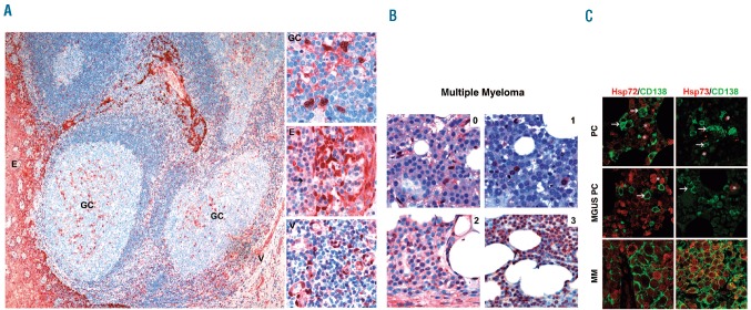
Figure 1.
Hsp72 and Hsp73 proteins are strongly expressed in MM cells as opposed to normal plasma cells (PC) or PC from patients with monoclonal gammopathy of undertermined significance (MGUS). (A) Immunohistochemical staining of Hsp72 in the normal human tonsil is shown. The overview depicts Hsp72-positive epithelial cells (E), scattered Hsp72-positive cells within the reactive germinal center (GC) and Hsp72-positive endothelial cells of small vessels (V). The insets show the corresponding regions with positive cells at higher magnification. Hsp72-negative normal PC (arrow) are interspersed between the Hsp72-positive epithelial cells. (B) Immunohistochemical staining of Hsp72 in bone marrow biopsies from MM patients representing different scoring groups (0–3; details in the Online Supplementary Design and Methods section). Of note, in the HSP72-immunonegative sample (1) stromal and endothelial cells displayed nuclear Hsp72-positivity, and thus served as a positive control. (C) Immunofluorescence analyses of Hsp72 and Hsp73 protein expression in bone marrow biopsies. Merged immunofluorescence images (CD138 and either Hsp72 or Hsp73) of bone marrow biopsies either from a patient without a plasma cell disorder (PC), from a patient with MGUS and a patient with MM. Of note, Hsp72 was strongly expressed in myeloid precursor cells (*) and Hsp73 was expressed in a few megakaryocytes (*).

